The latest survey from the European Centre for Disease Prevention and Control (ECDC), the fourth of its kind, confirms that Candidozyma auris (formerly Candida auris) continues to spread quickly across European hospitals, posing a serious threat to patients and healthcare systems. Case numbers are rising, outbreaks are growing in scale, and several countries report ongoing local transmission. The findings highlight the importance of early detection and control of transmission to avoid widespread rapid dissemination.
Candidozyma auris (C. auris) is a fungus that usually spreads within healthcare facilities, is often resistant to antifungal drugs, and can cause severe infections in seriously ill patients. Its ability to persist on different surfaces and medical equipment and to spread between patients makes it particularly challenging to control. Between 2013 and 2023, EU/EEA countries reported over 4 000 cases, with a significant jump to 1 346 cases reported by 18 countries in 2023 alone. Five countries – Spain, Greece, Italy, Romania, and Germany – have accounted for most of the cases over the decade.
“C. auris has spread within only a few years – from isolated cases to becoming widespread in some countries. This shows how rapidly it can establish itself in hospitals,” said Dr Diamantis Plachouras, Head of ECDC’s Antimicrobial Resistance and Healthcare-Associated Infections Section. “But this is not inevitable,” he added. “Early detection and rapid, coordinated infection control can still prevent further transmission.”
Recent outbreaks have been reported in Cyprus, France and Germany, while Greece, Italy, Romania and Spain have indicated they can no longer distinguish specific outbreaks due to widespread regional or national dissemination. In several of these countries, sustained local transmission has occurred within only a few years after the first documented case, highlighting a critical window for early interventions to stop its spread.
While some countries have showed positive results in limiting C. auris outbreaks, many are facing key gaps. Despite rising case numbers, only 17 of 36 participating countries currently have a national surveillance system in place for C. auris. Only 15 countries have developed specific national infection prevention and control guidance. Laboratory capacity is comparatively stronger, with 29 countries reporting access to a mycology reference or expert laboratory and 23 offering reference testing for hospitals.
While the number of C. auris infections is clearly rising, without systematic surveillance and mandatory reporting, the true scale of the problem is likely under-reported.
ECDC has regularly assessed the epidemiological situation, laboratory capacity and preparedness for C. auris in four surveys since 2018 and published rapid risk assessments including options for infection prevention and control. This is to support Member States in improving their preparedness and early response capacities to prevent or contain C. auris outbreaks in a timely manner and prevent further transmission.
Excessive amounts of visceral fat, the hidden fat surrounding organs, is linked with faster ageing of the heart, a new study has found. Ageing is the biggest risk factor for heart disease but why some people age faster than others isn’t fully understood.
The scientists leading the research, which is published in the European Heart Journal, say that visceral body fat could play an important role in accelerating ageing of the heart and blood vessels. This type of fat is known to be harmful to health and this study now links it to faster heart ageing.
Sex differences
The study, led by scientists from the Medical Research Council (MRC) Laboratory of Medical Sciences, also found differences between men and women. They discovered that fat around the hips and thighs could potentially slow heart ageing in women. The scientists analysed data from 21 241 participants in UK Biobank, which includes whole body imaging to map the amount of fat and where it is located in the body. The study was
Determining an individual’s ‘heart age’
The UK Biobank data also includes detailed imaging of the heart and blood vessels. Artificial intelligence (AI) was used to analyse these images to capture signs of organ ageing such as tissues becoming stiff and inflamed. An individual was given a ‘heart age’ which can be compared to their actual age at the time of the scan.
The risks of ‘hidden’ fat
The researchers found that faster heart ageing was linked to having more visceral adipose tissue. Visceral adipose tissue is fat found deep inside the abdomen around organs such as the stomach, intestines, and liver. This type of fat cannot be seen from the outside, and some people can have large amounts of visceral fat despite having a healthy weight.
Premature ageing
The researchers found signs on blood tests that visceral fat is linked to increased inflammation in the body, which is a potential cause of premature ageing. They also found differences between the sexes. Male-type fat distribution, which is fat around the belly and often called ‘apple-shaped’, was particularly predictive of early ageing in men.
The role of hormones
In contrast, a genetic predisposition to female-type fat, primarily fat on the hips and thighs, often called ‘pear-shaped’, was protective against heart ageing in women. The researchers also found a link between higher oestrogen levels in pre-menopausal women and a slowing of heart ageing. They suggested that this could indicate a role for hormones in protecting against heart ageing.
Increasing healthy lifespan
Professor Declan O’Regan, who led the research at the MRC Laboratory of Medical Sciences and Imperial College London, said:
We have known about the apple and pear distinction in body fat, but it hasn’t been clear how it leads to poor health outcomes. Our research shows that ‘bad’ fat, hidden deep around the organs, accelerates ageing of the heart.
But some types of fat could protect against ageing, specifically fat around the hips and thighs in women.
We also showed that body mass index wasn’t a good way of predicting heart age which underscores the importance of knowing where fat is stored in the body and not just total body weight.
The goal of our research is to find ways to increase healthy lifespan. While being active is important, we found that hidden fat could still be harmful even in fit people.
In the future, we plan to investigate how drug therapies, such as GLP-1 inhibitors (for example, Ozempic) could improve not just diabetes and obesity but target the ageing effects of hidden visceral fat.
Could a keto diet affect males differently from females? A study from The University of Texas Health Science at San Antonio (UT Health San Antonio) suggests so, and oestrogen could promote different protections against adverse effects of the diet like the accumulation of cells expressing markers of age, or senescence.
The study, published Aug. 26 in the journal Cell Reports, found that male, but not female, mice on a ketogenic diet showed the accumulation of cells in organs expressing markers of cellular senescence. A keto diet is a popular low-carbohydrate, high-fat regimen that can help some Type 2 diabetes patients control blood sugar and those with epilepsy manage seizures. Cells expressing senescence markers can contribute to age-related declines in overall bodily function.
“These results suggest sex specificity alters the effects of a ketogenic diet, with important clinical implications,” said David Gius, MD, PhD, assistant dean of research and professor with the Department of Radiation Oncology at UT Health San Antonio, associate cancer director for translational research at the institution’s Mays Cancer Center and investigator for its Barshop Institute for Longevity and Aging Studies.
He is lead author of the study, titled, “Divergent sex-specific effects on a ketogenic diet: Male, but not female, mice exhibit oxidative stress and cellular senescence.”
Ketogenic diets induce ketogenesis, the generation of ketone bodies or water-soluble molecules from fat for use as fuel in place of glucose. They have shown benefits in controlling refractory epilepsy and are being investigated as potential therapies for other health conditions.
In the past decades, keto diets also have become popular in North America and Europe for weight loss.
While the diets can improve certain health parameters, evidence from mice and clinical studies suggest the effects may be dependent on multiple variables, including adherence, metabolism and, importantly, sex, suggesting that hormone status may impact response.
Gius says the role of gender in the response to keto diets has been understudied. One reason is that male mice have been used extensively for in vivo basic and translational research because it was assumed that females would give less consistent results due to variability from the oestrous cycle. Recent studies, however, suggest that largely is unfounded.
In the new study, Gius’ team observed a keto-diet-induced increase in cellular senescence only in male mice, except when they were given the female hormone oestrogen. Male mice on a keto diet also exhibited an increase in markers of oxidative stress, which is known to contribute to senescence in cells.
Notably, the researchers found, estrogen or estradiol treatment prevented increases in cell senescence and oxidated stress in male mice on a keto diet, as did several established antioxidants.
They also observed that when females were administered tamoxifen, a “selective oestrogen receptor inhibitor” that blocks the effects of oestrogen, they then exhibited an increase in oxidative stress and cells expressing senescence markers, the same as male mice. “These results strongly suggest that oestrogen is an important variable in the response to a ketogenic diet,” Gius said.
The researchers also found that a high-fat diet – comprising more carbohydrates than a keto diet – also induces cellular senescence in male, but not female, mice.
Sceletiumtortuosum is a little succulent plant that grows in the semi-arid Karoo and Namaqualand regions of South Africa. It has a long history of traditional use among the hunter-gatherers of the region.
The plant, known as kanna or kougoed by the San and Khoikhoi people, was mainly chewed or smoked to stay alert and suppress appetite during long hunts. The San were traditionally hunter-gatherers, while the Khoikhoi were pastoralists who herded livestock.
The name kanna (meaning “eland” in the click language of the San), has a symbolic reference to this large antelope, as the “trance animal”, which was called upon during religious and spiritual gatherings. Kougoed is Afrikaans for “something to chew”. The plant can be chewed after being dried and fermented, which is believed to intensify its effects.
The first colonial governor of the Cape colony, Simon van der Stel, in 1685 wrote about kanna in his journal:
They chew mostly a certain plant which they call Canna and which they bruise, roots as well as the stem, between the stones and store and preserve in sewn-up sheepskins.
I’m part of a group of scientists from different disciplines with an interest in this plant and we pooled our expertise to understand its effects on neurochemical concentrations in different parts of the brain.
Our studies were done in mice, so there is caution about establishing effectiveness on humans. Still, the results are striking.
As a chemist with an interest in natural products, I wanted to know which alkaloids in the plant were important in bringing about these effects.
Our latest study explored the effects of Sceletiumtortuosum extracts on mouse brain chemistry.
We found that Sceletium increased the levels of certain brain chemicals which may balance mood and reduce stress. These findings lend support to the calming and mood-enhancing use of this plant in traditional medicine.
Plant chemistry
Our study examined how extracts from different chemotypes of Sceletiumtortuosum can have different effects on brain chemistry. Chemotypes are groups of the same plant species that differ in the alkaloids they produce. This is because plants often produce alkaloids in response to external cues such as the weather or the presence of a plant-eating animal or pathogen.
Alkaloids are carbon-based compounds produced by plants. They are often toxic or taste bitter, making the plants less appealing or even harmful to the predators or invaders that want to eat or inhabit them. Alkaloids generally have physiological effects of use to humans. Some commonly used ones include caffeine, morphine and quinine.
We harvested two chemotypes of kanna from the Touwsrivier and De Rust regions of South Africa. These areas were chosen because of their interesting and unusual alkaloid profiles. The chemotypes were given to healthy mice as a supplement once a day for one month. The mice were monitored every day for behavioural or unexpected adverse reactions but none were noted.
At the end of the month, the levels of chemicals in the mouse brain were measured. Both the chemotypes were found to cause a marked increase in noradrenaline and a decrease in GABA in all brain regions studied. Both molecules are neurotransmitters that transmit nerve signals in the brain affecting memory, mood, attention and sleep.
This effect on noradrenaline supports kanna’s traditional use as an appetite suppressing drug. Increased noradrenergic stimulation is also the basis of many anti-depressants as well as drugs that improve attention and alertness.
We also found an impact on the brain chemicals serotonin and dopamine which may act together to balance mood and reduce stress. Serotonin affects emotional well-being and mood; dopamine motivates feelings of pleasure and satisfaction. These findings lend support to the calming and mood-enhancing use of this plant in traditional medicine.
Importantly, the control kanna extracts that did not have the interesting alkaloid profiles did not cause any of these chemical changes in the mouse brain.
Most studies on kanna have focused on the alkaloid mesembrine. The two specific chemotypes of kanna harvested from the Touwsrivier and De Rust regions of South Africa do have the mesembrine, but they are also packed with some other lesser-known or “minor” alkaloids. These differences in alkaloids may arise from a combination of geographic, environmental and inherent genetic factors found in a particular subset of plants.
Both the Touwsrivier and De Rust plants contained higher levels of alkaloids called mesembrine alcohols, which are different from mesembrine, and were barely present in the control extract. Another minor alkaloid, known as sceletium A4, was also identified as possibly being important. Mesembrine alcohols and sceletium A4 may be the ones responsible for the activity.
This suggests that the source of the plant, and the area in which it is grown, can influence its potential as a natural treatment for mood disorders and sleep.
What the results tell us
Stress, anxiety and depression pose a risk to the ability to lead a meaningful life. The World Health Organization has reported a 25% increase in anxiety and depression worldwide since the emergence of COVID-19.
Our study showed that the plant extracts had a broad noradrenergic effect in mice. But we have to be careful about making connections between results in mice and in humans. We need to explore the behavioural impact of these extracts in both mice and humans, especially in relation to sleep, alertness and mood.
The results also highlighted that without understanding the complex chemical composition of these plants, we risk overgeneralising their benefits, or worse, using them inappropriately.
Our findings have two implications.
First, they point towards a future of precision phytotherapy (use of plants for medicinal purposes), where natural remedies are tailored not just to individuals but to selecting certain plant chemotypes that produce certain combinations of alkaloids. Manipulating the growing conditions and genetic make-up of plants to optimise for alkaloid content is an age-old art.
Second, they remind us of the enormous, still largely untapped potential of African medicinal plants in global health innovation if we invest in research that honours both indigenous knowledge and scientific rigour.
As the world searches for safer, more sustainable ways to treat mental health conditions, South Africa’s kanna plant may hold secrets worth rediscovering.
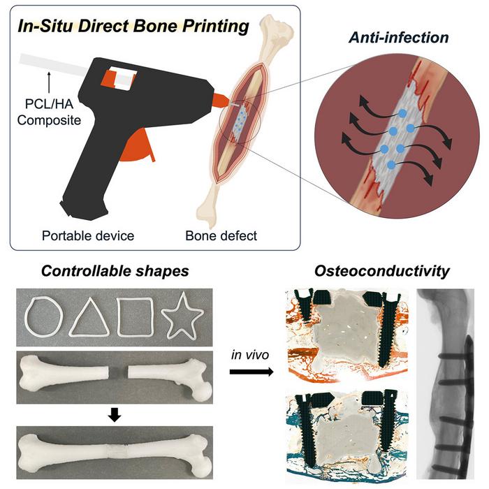
Graphical abstract. Credit: Device / Jeon et al. CC BY-SA
Scientists have developed a tool made from a modified glue gun that can 3D print bone grafts directly onto fractures and defects during surgery. The tool, described September 5th in the Cell Press journal Device, has been tested in rabbits to quickly create complex bone implants without the need for prefabricating in advance. What’s more, the team optimised the 3D-printed grafts for high structural flexibility, release of anti-inflammatory antibiotics, and promotion of natural bone regrowth at the grafting site.
Historically, bone implants have been made of metal, donor bone, or even more recently 3D-printed material. However, in cases involving irregular bone breaks, these implants must be designed and produced prior to surgery to allow for appropriate fitting.
“Our proposed technology offers a distinct approach by developing an in situ printing system that enables a real-time fabrication and application of a scaffold directly at the surgical site,” says Jung Seung Lee, co-author and associate professor of biomedical engineering at Sungkyunkwan University. “This allows for highly accurate anatomical matching even in irregular or complex defects without the need for preoperative preparation such as imaging, modelling, and trimming processes.”
The material fed into the glue gun is a filament comprised of two major components: a feature of natural bone known to promote healing called hydroxyapatite (HA) and a biocompatible thermoplastic called polycaprolactone (PCL). PCL can liquify in temperatures as low as 60°C, which when applied with a heat-modified glue gun, is cool enough to prevent tissue damage during surgical application while being able to conform to the jagged grooves of fractured bone. By adjusting the proportion of HA to PCL within the filament, the team can customise the hardness and strength of the grafts to fit different anatomical needs.
“Because the device is compact and manually operated, the surgeon can adjust the printing direction, angle, and depth during the procedure in real time,” says Lee. “Also, we demonstrated that this process could be completed in a matter of minutes. This highlights a significant advantage in terms of reducing operative time and improving procedural efficiency under real surgical conditions.”
Since infection is a common concern with surgical implants, the researchers incorporated vancomycin and gentamicin, two anti-bacterial compounds, into the filament. In both petri dish culture and liquid medium, the filament scaffold successfully inhibited the growth of E. coli and S. aureas, two common bacteria prone to cause infection post-surgery. Due to physical properties of HA and PCL within the filament, the drugs are released slowly and are able to diffuse directly onto the surgical site over several weeks.
“This localised delivery approach offers meaningful clinical advantages over systemic antibiotic administration by potentially reducing side effects and limiting the development of antibiotic resistance, while still effectively protecting against postoperative infection,” says Lee.
As a proof of concept, the device was tested on severe femoral bone fractures in rabbits. Within 12 weeks after surgery, the team found no signs of infection or necrosis and greater bone regeneration outcomes when compared to rabbits grafted with bone cement – a sealing compound commonly used for treating bone defects.
“The scaffold was designed not only to integrate biologically with surrounding bone tissue but also to gradually degrade over time and be replaced by newly formed bone,” says Lee. “The results showed that the printing group exhibited superior outcomes in key structural parameters such as bone surface area, cortical thickness, and polar moment of inertia, suggesting more effective bone healing and integration.”
Next, the team is setting their sights on optimising the anti-bacterial potential of the scaffold even further and preparing the procedure for human trials.
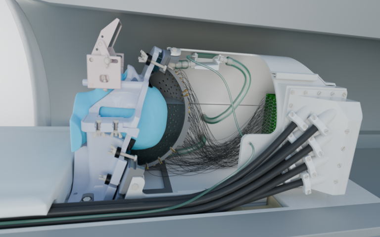
Illustration of new ultrasound device. Credit: Morgan Roberts.
Scientists have long been looking for a way to modulate brain function, which could improve our understanding of how the brain works and help to treat neurological diseases, using non-invasive methods that don’t involve surgery.
One technology that could help is transcranial ultrasound stimulation (TUS), which was recently discovered to be able to modulate the activity of neurons (the brain’s key communication cells) by delivering gentle mechanical pulses that influence how these cells send signals.
But to date current systems have struggled to reach deeper areas of the brain with sufficient precision to target specific brain structures. Conventional TUS systems often affect broader regions than intended, limiting their utility for targeted neuromodulation.
The study, published in Nature Communications, introduces a new ultrasound device capable of influencing deep brain regions without surgery for the first time, targeting areas around 1000 times smaller than conventional ultrasound devices can pinpoint and 30 times smaller than previous deep brain ultrasound devices.
The new technology features 256 elements configured within a special helmet to send focused beams of ultrasound to specific parts of the brain in order to turn neuronal activity up or down. It also includes a soft plastic face mask which helps to target the ultrasound waves more precisely by keeping the head still.
The research team demonstrated the system’s capabilities on seven human volunteers by targeting a part of the thalamus, a small structure in the centre of the brain that helps to relay sensory and motor information, called the lateral geniculate nucleus (LGN). The LGN is involved in processing visual information.
In the first experiment, participants looked at a flashing checkerboard, which sent signals to the brain through the eyes. During stimulation with the ultrasound device, a functional magnetic resonance imaging (fMRI) scan showed significantly increased activity in the participants’ visual cortex, confirming precise targeting of the LGN.
A second experiment revealed sustained decreases in visual cortex activity for at least 40 minutes after ultrasound stimulation, highlighting the system’s potential for inducing lasting changes in brain function.
Though participants did not consciously perceive any changes in what they were seeing during the experiments, the brain scans revealed significant changes in neural activity. The ultimate goal is to harness these effects to produce clinically beneficial outcomes, such as stopping hand tremors.
Professor Bradley Treeby, senior author of the study from UCL Medical Physics and Biomedical Engineering, said: “This advance opens up opportunities for both neuroscience research and clinical treatment. For the first time, scientists can non-invasively study causal relationships in deep brain circuits that were previously only accessible through surgery.
“Clinically, this new technology could transform treatment of neurological and psychiatric disorders like Parkinson’s disease, depression, and essential tremor, offering unprecedented precision in targeting specific brain circuits that play key roles in these conditions.
“The ability to precisely modulate deep brain structures without surgery represents a paradigm shift in neuroscience, offering a safe, reversible, and repeatable method for both understanding brain function and developing targeted therapies.”
In addition to its research applications, the system could pave the way for new clinical interventions. Deep brain stimulation (DBS), currently used to treat conditions like Parkinson’s disease, requires invasive surgery and carries associated risks. The new ultrasound system offers a non-invasive alternative with comparable precision, potentially allowing clinicians to test areas of the brain that could be used to treat disease before surgery or even replace surgical approaches altogether.
Recognising this clinical potential, several members of the research team have recently founded NeuroHarmonics, a UCL spinout company developing a portable, wearable version of the system. The company aims to make precise, non-invasive deep brain therapy accessible for both clinical treatment and broader therapeutic applications.
Dr Eleanor Martin, first author of the study from UCL Medical Physics and Biomedical Engineering, said: “We designed the system to be compatible with simultaneous fMRI, enabling us to monitor the effects of stimulation in real time. This opens up exciting possibilities for closed-loop neuromodulation and personalised therapies.”
The researchers emphasise that further studies are needed to fully understand the mechanisms underlying TUS-induced neuromodulation. However, the results mark a significant milestone in the development of safe, effective, and targeted brain stimulation technologies.
Excessive alcohol consumption can disrupt the liver’s unique regenerative abilities by trapping cells in limbo between their functional and regenerative states, even after a patient stops drinking, as described in a new study from researchers at University of Illinois Urbana-Champaign and collaborators.
This in-between state is a result of inflammation disrupting how RNA is spliced during the protein-making process, the researchers found, providing scientists with new treatment pathways to explore for the deadly disease. The researchers published their findings in the journal Nature Communications.
The liver has a remarkable ability to regenerate itself after damage or partial removal. However, it loses that ability in patients with alcohol-associated liver disease – the leading cause of liver-related mortality worldwide, resulting in roughly 3 million deaths annually.
“We knew that the liver stops functioning and stops regenerating in patients with alcohol-related hepatitis and cirrhosis, even when a patient has discontinued consuming alcohol, but we didn’t know why,” said U. of I. biochemistry professor Auinash Kalsotra, who co-led the study with Duke University School of Medicine professor Anna Mae Diehl. “The only real life-saving treatment option once a patient reaches the liver failure stage in those diseases is transplantation. But if we understood why these livers were failing, maybe we could intervene.”
Both the Kalsotra and Diehl labs havestudied the molecular and cellular underpinnings of liver regeneration. Over the last five years, they found that in order to regenerate, liver cells reprogram their gene expression to revert to fetal-like progenitor cells, multiply and then reverse the process back to become mature functioning cells again. Armed with this knowledge, the group turned to the question of how those mechanisms were disrupted in alcohol-associated liver disease.
The researchers compared samples of healthy livers and samples of livers with alcohol-associated hepatitis or cirrhosis obtained from Johns Hopkins University Hospital through an initiative supported by the National Institute for Alcohol Abuse and Alcoholism, part of the National Institutes of Health.
The first thing the researchers noticed in diseased livers was that, although damaged cells had begun the process of reverting to the regenerative state, they did not complete the process and instead remained in transitional limbo.
“They are neither functional adult cells nor proliferative progenitor cells. Since they are not functioning, more pressure builds on the remaining cells. So they try to regenerate, and they’re all ending up in this unproductive quasi-progenitor state, and that’s what is causing liver failure,” said U. of I. graduate students Ullas Chembazhi and Sushant Bangru, the co-first authors of the study.
To figure out why the cells were getting stuck in this state, the team investigated which proteins were being made by the liver cells and, in turn, the RNA molecules carrying the instructions for those proteins from the DNA to the cell’s protein-building machinery.
While most studies focus only on the total amounts of RNA or protein in a cell, Kalsotra’s team used deep RNA sequencing technology and computational analyses to zoom in on the splicing of RNA fragments, a key step in stitching together different parts of genetic instructions to make proteins.
“In comparing the samples, we saw RNA was getting mis-spliced broadly in alcohol-related liver disease, across thousands of genes, and it was affecting major functions of proteins,” said Kalsotra.
The researchers found a possible driver of the RNA mis-splicing: Alcohol-damaged liver cells had a deficiency of the protein ESRP2, which binds to RNA to splice it properly.
“Proteins function at a very specific place in the cell, and that is directed by sequences within the protein that take the protein to that particular spot. We found that, in many cases, the sequence that dictates where the protein localizes within a cell was mis-spliced. That’s why it was important that we did the multiple analyses we did,” said Kalsotra. “There was the same amount of RNA and protein, but the protein was not at the right place to function. Due to mis-splicing, key proteins that are required for productive liver regeneration were getting stuck in the cytoplasm, when they needed to be in the nucleus.”
To verify that ESRP2 deficiency was a likely culprit, the researchers studied mice without the gene that produces ESRP2. They displayed similar liver damage and regeneration failure to that seen in patients with advanced alcohol-related hepatitis.
But why was ESRP2 missing from liver cells from patients with alcohol-related hepatitis? Upon investigation, the researchers found that liver support cells and immune cells, drawn to the liver tissue damaged by alcohol processing, released high amounts of inflammatory and growth factors. Those factors suppress ESRP2 production and activity.
To verify this finding, the researchers treated liver cell cultures with a molecule that inhibits the receptor for one of the inflammation-promoting factors. ESRP2 levels recovered and splicing activity was corrected, pointing to the pathway as a possible treatment target.
“I’m hopeful these findings will become a launching pad for future clinical studies. We can use these mis-spliced RNAs as diagnostic markers or develop treatments that can curb the inflammation. And if we can correct the splicing defects, then maybe we can improve recovery and restore damaged livers,” Kalsotra said.
The brain has its own waste disposal system – known as the glymphatic system – that’s thought to be more active when we sleep.
But disrupted sleep might hinder this waste disposal system and slow the clearance of waste products or toxins from the brain. And researchers are proposing a build-up of these toxins due to lost sleep could increase someone’s risk of dementia.
There is still some debate about how this glymphatic system works in humans, with most research so far in mice.
But it raises the possibility that better sleep might boost clearance of these toxins from the human brain and so reduce the risk of dementia.
Here’s what we know so far about this emerging area of research.
Why waste matters
All cells in the body create waste. Outside the brain, the lymphatic system carries this waste from the spaces between cells to the blood via a network of lymphatic vessels.
But the brain has no lymphatic vessels. And until about 12 years ago, how the brain clears its waste was a mystery. That’s when scientists discovered the “glymphatic system” and described how it “flushes out” brain toxins.
Let’s start with cerebrospinal fluid, the fluid that surrounds the brain and spinal cord. This fluid flows in the areas surrounding the brain’s blood vessels. It then enters the spaces between the brain cells, collecting waste, then carries it out of the brain via large draining veins.
Scientists then showed in mice that this glymphatic system was most active – with increased flushing of waste products – during sleep.
One such waste product is amyloid beta (Aβ) protein. Aβ that accumulates in the brain can form clumps called plaques. These, along with tangles of tau protein found in neurons (brain cells), are a hallmark of Alzheimer’s disease, the most common type of dementia.
In humans and mice, studies have shown that levels of Aβ detected in the cerebrospinal fluid increase when awake and then rapidly fall during sleep.
But more recently, another study (in mice) showed pretty much the opposite – suggesting the glymphatic system is more active in the daytime. Researchers are debating what might explain the findings.
So we still have some way to go before we can say exactly how the glymphatic system works – in mice or humans – to clear the brain of toxins that might otherwise increase the risk of dementia.
In one experiment, a single night of complete sleep deprivation in healthy adults increased the amount of Aβ in the hippocampus, an area of the brain implicated in Alzheimer’s disease. This suggests sleep can influence the clearance of Aβ from the human brain, supporting the idea that the human glymphatic system is more active while we sleep.
This also raises the question of whether good sleep might lead to better clearance of toxins such as Aβ from the brain, and so be a potential target to prevent dementia.
How about sleep apnoea or insomnia?
What is less clear is what long-term disrupted sleep, for instance if someone has a sleep disorder, means for the body’s ability to clear Aβ from the brain.
Sleep apnoea is a common sleep disorder when someone’s breathing stops multiple times as they sleep. This can lead to chronic (long-term) sleep deprivation, and reduced oxygen in the blood. Both may be implicated in the accumulation of toxins in the brain.
Insomnia is when someone has difficulty falling asleep and/or staying asleep. When this happens in the long term, there’s also an increased risk of dementia. However, we don’t know the effect of treating insomnia on toxins associated with dementia.
So again, it’s still too early to say for sure that treating a sleep disorder reduces your risk of dementia because of reduced levels of toxins in the brain.
So where does this leave us?
Collectively, these studies suggest enough good quality sleep is important for a healthy brain, and in particular for clearing toxins associated with dementia from the brain.
But we still don’t know if treating a sleep disorder or improving sleep more broadly affects the brain’s ability to remove toxins, and whether this reduces the risk of dementia. It’s an area researchers, including us, are actively working on.
For instance, we’re investigating the concentration of Aβ and tau measured in blood across the 24-hour sleep-wake cycle in people with sleep apnoea, on and off treatment, to better understand how sleep apnoea affects brain cleaning.
This is an emerging field and we don’t yet have all the answers about the link between disrupted sleep and dementia, or whether better sleep can boost the glymphatic system and so prevent cognitive decline.
So if you are concerned about your sleep or cognition, please see your doctor.
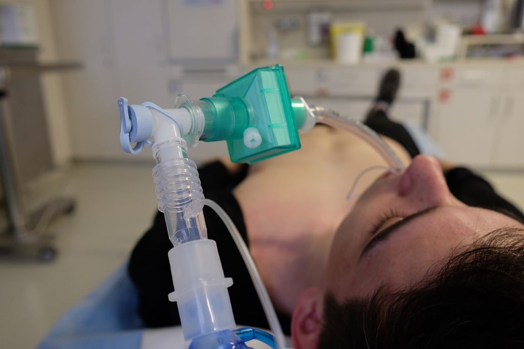
Endotracheal intubation is a difficult task for highly trained individuals and under the best of circumstances. In the field and in the ER, where seconds matter, emergency medical personnel face many unknowns and wildly challenging conditions which lower their chances of success.
But what if successful endotracheal intubation could be less reliant on ideal conditions and years of specialised training? In a paper published in Science Translational Medicine, UC Santa Barbara researchers David Haggerty, Elliot Hawkes and collaborators demonstrate a non-electronic soft robotic device that quickly and autonomously guides a soft tube into the trachea. Initial device testing with highly trained users yielded a 100% success rate, and a 96% overall success rate with prehospital medical providers (EMTs and paramedics).
“Current intubation tools require extensive anatomical knowledge, training, skill and ideal conditions to be highly successful,” said recent UC Santa Barbara Ph.D. graduate David Haggerty, a former researcher in the lab of mechanical engineering professor Elliot Hawkes. Current technology calls for the rescuer to first visualize the tracheal opening then manually direct a tube through the serpentine anatomy of the airway into the trachea. The challenge of this procedure increases in prehospital settings due to various factors including inadequate light and nonideal body position in addition to potential injuries and fluid in the airway.
This project is supported in part with funds from the National Science Foundation
From rigid tools to soft robotics
One of the main challenges to successful intubation is the body itself, and the mechanisms it has evolved to prevent food and foreign bodies from entering the lungs. The epiglottis is a small fleshy flap that closes over the trachea and guides food and liquid into the adjacent esophagus with each swallow. Conventional practice typically requires the rescuer to push a metal laryngoscope into the back of the mouth behind the tongue to lift the epiglottis out of the way in order to make room for the breathing tube.
But even with the epiglottis out of the way, the path the endotracheal tube must take is a twisted one, as it has to bend toward the front of the neck where the trachea is located, otherwise air could be delivered to the stomach via the esophagus, instead of to the lungs.
“Traditional tools, which you push from the base, are fundamentally limited in navigating delicate, tortuous anatomy,” Hawkes explained. “They must be relatively stiff so you can push them, and can only get around bends by deflecting off the sensitive tissue.”
The researchers’ device upends that paradigm with a soft, inflatable tube that everts from its tip. Called the soft robotic intubation system (SRIS), it consists of a curved “introducer” that slides into place at the back of the throat and stops at the esophagus. With that in place, a soft, pre-inflated tube is threaded through the introducer, emerging near its tip at the opening of the trachea. As the user advances the tube, it everts from its tip, carrying inside it a soft breathing tube as it enters the trachea. “So instead of trying to push this tube and bend it to get into this complex configuration, we can just mechanically create that complex configuration as we go,” Haggerty said. Once the endotracheal tube is at its destination, the user can inflate a cuff at its distal end to seal the opening and begin ventilation. The introducer can be removed, leaving the breathing tube in place.
Introducing a soft, growing tube eliminates friction with the surrounding tissue and minimizes injury due to excessive or misplaced force. It also automatically conforms to its environment, one of the major benefits of soft robotics. “This growing paradigm naturally accounts for minor variations in anatomical placement, size, shape or configuration,” Haggerty said, and because of this, users need not have extensive skill or a perfect understanding of the environment in order to navigate it.
In tests with mannequins and cadavers, the SRIS proved itself both effective, and, importantly, rapid, with a 100% success rate at a procedure duration of just seconds for expert users. For nonexpert users – the primary target of this technology – a five-minute training session was all that was needed to deliver an 87% success rate for first-pass attempts, translating into an 96% overall success rate, with a significantly lower procedure duration – 21 seconds versus 44 seconds – than state-of-the-art video laryngoscope intubation.
The next step for the researchers is to conduct clinical trials in order to get approvals from the Food and Drug Administration for clinical use. “We have good reason to believe it’s efficacious based on the data, but cadavers don’t say ‘ow’,” Haggerty said. They need to complete more testing to ascertain the device’s safety and effectiveness in a variety of airways and external conditions, he added. If successful, this device could benefit the millions of emergency intubations that occur each year in the US, and find application in military medical care. That’s in addition to its potential to save lives in global populations that have poor or no access to essential health services.
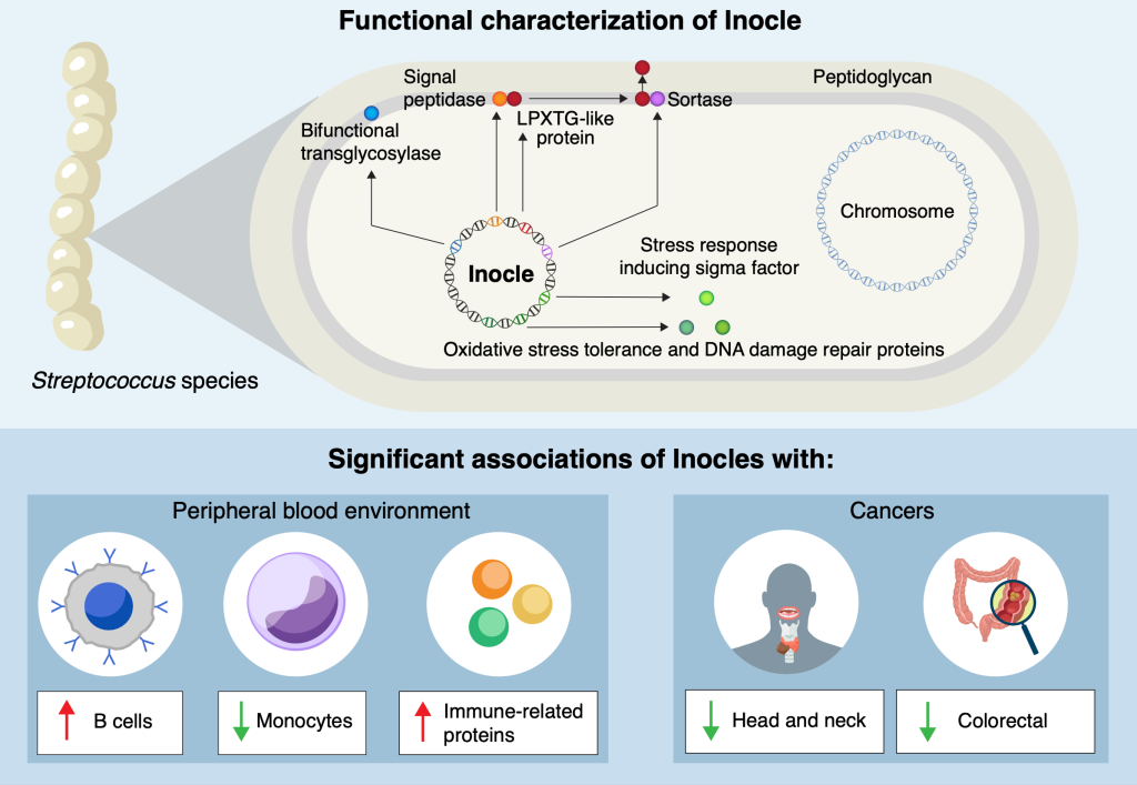
Researchers at the University of Tokyo and other institutions have made a surprising discovery hiding in people’s mouths: Inocles, giant DNA elements that had previously escaped detection. These appear to play a central role in helping bacteria adapt to the constantly changing environment of the mouth. The findings provide fresh insight into how oral bacteria colonise and persist in humans, with potential implications for health, disease and microbiome research.
You might think that modern medical science knows everything there is to know about the human body. But even within the last decade, small, previously unknown organs have been discovered, and there’s one area of human biology that is currently going through a research renaissance, the microbiome. This includes familiar areas such as the gut microbiome, but also the oral microbiome. Inspired in part by recent discoveries of extraneous DNA in the microbiome of soil, Project Research Associate Yuya Kiguchi and his team turned their sights to a large set of saliva samples collected by the Yutaka Suzuki Lab of the Graduate School of Frontier Sciences at the University of Tokyo. They wondered if they might find something similar in human saliva.
“We know there are a lot of different kinds of bacteria in the oral microbiome, but many of their functions and means of carrying out those functions are still unknown,” said Kiguchi. “By exploring this, we discovered Inocles, an example of extrachromosomal DNA – chunks of DNA that exist in cells, in this case bacteria, but outside their main DNA. It’s like finding a book with extra footnotes stapled to it, and we’re just starting to read them to find out what they do.”
Detecting Inocles was not easy, as conventional sequencing methods fragment genetic data, making it impossible to reconstruct large elements. To overcome this, the team applied advanced long-read sequencing techniques, which can capture much longer stretches of DNA. A key breakthrough came from co-first author Nagisa Hamamoto, who developed a method called preNuc to selectively remove human DNA from saliva samples, greatly improving the quality of sequencing long sections of other DNA. This allowed the researchers to assemble for the first time complete Inocle genomes, which turned out were hosted by the bacteria Streptococcus salivarius, though identifying the host itself was a difficult matter.
“The average genome size of Inocle is 350 kilobase pairs, a measure of length for genetic sequences, so it is one of the largest extrachromosomal genetic elements in the human microbiome. Plasmids, other forms of extrachromosomal DNA, are at most a few tens of kilobase pairs,” said Kiguchi. “This long length endows Inocles with genes for various functions, including resistance to oxidative stress, DNA damage repair and cell wall-related genes, possibly involved in adapting to extracellular stress response.”
The team aims to develop stable methods for culturing Inocle-containing bacteria. This will allow them to investigate how Inocles function, whether they can spread between individuals, and how they might influence oral health conditions such as cavities and gum disease. Since many Inocle genes remain uncharacterised, researchers will use a mixture of laboratory experiments and also computational simulations such as AlphaFold to predict and model the roles Inocles may play.
“What’s remarkable is that, given the range of the human population the saliva samples represent, we think 74% of all human beings may possess Inocles. And even though the oral microbiome has long been studied, Inocles remained hidden all this time because of technological limitations,” said Kiguchi. “Now that we know they exist, we can begin to explore how they shape the relationship between humans, their resident microbes and our oral health. And there’s even some hints that Inocles might serve as markers for serious diseases like cancer.”