Category: Metabolic Disorders

Do Lifetime Body Weight Patterns Affect the Risk of Kidney Cancer?

Study links higher body mass index at various ages across adulthood with greater risks of developing different types of kidney cancer.

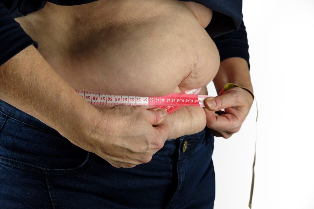
Photo by I Yunmai on Unsplash

Excess weight in mid-life is a known risk factor for kidney cancer, but new research indicates that weight patterns throughout life may also affect an individual’s likelihood of developing this malignancy. The findings are published by Wiley online in CANCER, a peer-reviewed journal of the American Cancer Society.

To assess weight patterns and their associations with kidney cancer and its different subtypes, investigators analysed data from 204 364 individuals from the NIH-AARP Diet and Health Study, including body mass index (BMI) data when participants entered the study (an average age of 61.6 years), and prior BMI recordings at 18, 35, and 50 years of age. The team noted that there were 1,425 cases of kidney cancer, or renal cell carcinoma (RCC), among the study’s participants, with 583 having aggressive RCC and 339 having fatal RCC. The researchers also recorded the different subtypes of RCC, including clear cell RCC (541 patients), papillary RCC (146 patients), and chromophobe RCC (64 patients).

Higher BMI at any of the ages assessed was linked with higher risks of overall RCC and all subtypes (except chromophobe RCC), with a 10-40% higher risk for each 5-unit increase in BMI. Similar increased risks were linked to weight gain during adulthood that resulted in overweight or obesity, compared with maintaining normal BMI.

Also, long-term excess weight was associated with higher risks of overall RCC, aggressive RCC, fatal RCC, and clear cell RCC, but not papillary RCC and chromophobe RCC. Weight loss in which BMI was reduced by at least 10%, particularly later in life, was associated with a lower risk of RCC. Specifically, weight loss from age 18–35 years and after age 50 years was associated with 21% and 28% reductions in RCC incidence, respectively.

“These findings emphasise that maintaining a healthy weight across one’s lifetime is important for reducing RCC risk. More importantly, weight loss, even later in life, may offer protective benefits,” said lead author Zhengyi Deng, PhD, of Stanford University School of Medicine. “We should support initiatives that promote healthy weight maintenance and weight loss strategies. Some of these include lifestyle interventions, weight-loss programs, and emerging medical treatments for obesity; however, individuals should consult with their healthcare providers prior to initiation of any plan.” 

Source: Wiley

Radiology’s Role in the Diagnosis and Management of Diabetic Complications

SCP -Using modern CT technology, radiologists can search for narrowed arteries in various parts of the body, including the neck and brain. This process is called CT angiography.

Radiology provides crucial insights into the complications caused by diabetes, allowing for timely diagnosis, effective management and monitoring of disease progression. Early detection of these complications can significantly improve patient outcome and quality of life.

What is diabetes?

Diabetes is known as a ‘silent killer’ because it is quite often asymptomatic at the onset. Diabetes, a major lifestyle disorder, has become one of the most dangerous and common diseases in the world. It is a chronic disease that causes high blood sugar levels and occurs when the body doesn’t produce enough insulin or use insulin properly.

Types of diabetes

  • Type 1 diabetes: The body’s immune system destroys the cells that produce insulin
  • Type 2 diabetes: The body doesn’t produce enough insulin or the body’s cells don’t react to insulin as they should
  • Gestational diabetes: Sometimes occurs during pregnancy when the placenta releases hormones that cause insulin resistance. This tampers with the expectant mom’s blood sugar level, changing the amount of glucose in the blood

Around 4.2 million people in South Africa have diabetes – 90% of whom have type 2 diabetes, a lifestyle disease exacerbated by dietary factors, coupled with too little physical activity and high levels of obesity.

Dr Jean de Villiers, senior partner and radiologist at SCP Radiology, discusses the imaging techniques used to identify and manage complications of diabetes.

Cardiovascular Disease: People with diabetes are at higher risk of developing heart disease and other cardiovascular problems. Imaging techniques such as CT angiography can be used to assess the heart’s blood vessels and detect issues such as atherosclerosis, coronary artery narrowing or blockage of the arteries. CT angiography is also used for the neck, arm and leg arteries, as well as the arteries to the gut.

Stroke: Diabetes increases the risk of stroke by damaging blood vessels through high blood sugar levels, leading to the formation of fatty deposits and clots within the arteries. This can increase the chance of clot formation and block blood flow to the brain and cause a stroke. Imaging techniques such as MRI, CT scans, and ultrasound may be able to detect these fatty deposits in the arteries. The deposits are generally seen as areas of narrowing in the involved arteries or calcification of the walls of the arteries.

Blood vessel damage: Chronic high blood sugar levels can directly damage the lining of blood vessels, making them more susceptible to inflammation and clot formation. Essentially, the excess glucose in the blood weakens and stiffens the blood vessel walls, making them more prone to blockages. CT or MRI scans can be critical in identifying and assessing strokes, transient ischemic attacks (TIAs) or other cerebrovascular issues in diabetic patients.

High blood pressure association: People with diabetes often also have high blood pressure, which can exacerbate the damage to blood vessels and increases stroke risk.  A CT of the coronary arteries is used to visualise blockages in the coronary blood vessels and assess the severity of atherosclerosis in diabetic patients. This helps in planning for interventions like stent placement or bypass surgery.

Kidney disease: Diabetes affects your kidneys by potentially damaging the blood vessels within the kidneys due to high blood sugar levels. This can lead to impaired kidney function, causing the kidneys to leak protein into the urine and eventually progressing to chronic kidney disease if left uncontrolled. This condition is often referred to as ‘diabetic nephropathy.’

Diabetic nephropathy can lead to kidney damage and radiology plays a role in assessing kidney size, structure and function. Renal ultrasound can help assess kidney size and detect signs of chronic kidney disease (CKD). In advanced cases, a CT scan or MRI can be used to further evaluate the kidneys for the presence of complications such as renal artery stenosis or renal scarring.

Diabetic neuropathy: Diabetic neuropathy is a complication of diabetes where high blood sugar levels damage nerves throughout the body.  Most commonly affected are the nerves in the legs and feet, leading to symptoms like numbness, tingling, pain and sometimes muscle weakness.  It can also impact internal organs, depending on which nerves are affected and is considered a serious diabetes complication that can affect up to 50% of diabetics.

While radiology is not typically used for direct diagnosis of diabetic neuropathy, it can help rule out other causes of neuropathy. MRI and CT scans can assess for structural issues, such as spinal problems or other nerve impingements that may be contributing to symptoms.

Infections: Diabetic patients have a higher susceptibility to infections due to impaired immune response.

Diabetic foot ulcers and infections: Over time, high blood sugar levels damage nerves, blood vessels and skin in the feet. Damaged nerves can cause loss of feeling in the feet, while damaged blood vessels slow blood flow to the feet, preventing the healing of injuries.

Imaging techniques like CT, MRI and ultrasound are useful for detecting and monitoring bone and soft tissue infections. These can be critical for determining the appropriate course of antibiotic treatment or surgical intervention. X-rays, CT and MRI can be used to assess for infection in diabetic foot, such as ulcers, osteomyelitis or abscesses that may progress to amputation if left untreated.

Liver disease: Non-alcoholic fatty liver disease (NAFLD) is commonly seen in diabetic patients. Ultrasound is the primary tool for detecting fatty liver, while CT and MRI may offer further details on liver fat content or cirrhosis. Regular monitoring through imaging can help prevent more severe liver damage.

Osteoporosis: Long-term diabetes, especially type 1, can increase the risk of osteoporosis due to lower bone density. A DEXA scan helps assess bone mineral density (BMD), aiding in the early detection of osteoporosis and providing information on fracture risk.

‘As with any lifestyle disease, prevention is best. However, second to this is early detection and timely diagnosis, effective management and monitoring of the disease,’ says Dr de Villiers. ‘In the case of diabetes, we work with physicians and patients to detect possible complications early enough to help improve medical care, monitor treatment response and ultimately, improve quality of life.’

Fatty Liver Linked to Increased Mortality Risk From Several Diseases

Human liver. Credit: NIH

People with fatty liver disease have almost twice the mortality rate of the general population, according to a comprehensive study from Karolinska Institutet. They have an increased mortality risk from both liver diseases and common diseases such as cancer and cardiovascular disease, according to the study published in The Journal of Hepatology.

In Sweden, it is estimated that one in five people has fatty liver disease known as MASLD (metabolic dysfunction-associated steatotic liver disease), and globally it may be as many as one in four. The disease is caused by overweight or obesity and is characterised by an excessive accumulation of fat in the liver, which can lead to severe liver damage and liver cancer.

Hidden health condition

“Many people are not aware that they have fatty liver disease because it rarely causes any symptoms in the earlier stages,” says Axel Wester, assistant professor at the Department of Medicine, Huddinge, Karolinska Institutet and physician at Karolinska University Hospital. “Our study shows that people diagnosed with MASLD have an increased risk of dying from many different diseases, not just liver disease.”

The researchers identified all patients diagnosed with MASLD in Sweden between 2002 and 2020, more than 13,000 patients in total, and analysed their risk of death from different causes compared to the general population.

The overall mortality rate for people with MASLD was almost twice as high. The risk was elevated for nearly all causes of death studied, but especially for death from liver disease (27 times higher mortality) and liver cancer (35 times higher mortality). However, the most common causes of death were cardiovascular disease and non-liver cancer, with mortality rates 54 and 47 per cent higher, respectively.

A holistic approach

People with MASLD also had an increased risk of dying from infections, gastrointestinal diseases, respiratory diseases, endocrine diseases or external causes, but not from mental illness.

“It is important that we do not only focus on the liver when treating patients with fatty liver disease,” says Hannes Hagström, adjunct professor at the Department of Medicine, Huddinge, Karolinska Institutet and senior physician at Karolinska University Hospital. “A holistic approach and early intervention involving different medical specialities can be crucial to improve the prognosis for these patients.”

Source: Karolinska Institutet

Time of Injury Matters: Circadian Rhythms Affect Muscle Repair

Photo by Mat Napo on Unsplash

Circadian rhythms doesn’t just dictate when we sleep — it also determines how quickly our muscles heal. A new Northwestern Medicine study in mice, published in Science Advances, suggests that muscle injuries heal faster when they occur during the body’s natural waking hours.

The findings could have implications for shift workers and may also prove useful in understanding the effects of aging and obesity, said senior author Clara Peek, assistant professor of biochemistry and molecular genetics at Northwestern University Feinberg School of Medicine.

The study also may help explain how disruptions like jetlag and daylight saving time changes impact circadian rhythms and muscle recovery.

“In each of our cells, we have genes that form the molecular circadian clock,” Peek said. “These clock genes encode a set of transcription factors that regulate many processes throughout the body and align them with the appropriate time of day. Things like sleep/wake behaviour, metabolism, body temperature and hormones — all these are circadian.”

How the study was conducted

Previous research from the Peek laboratory found that mice regenerated muscle tissues faster when the damage occurred during their normal waking hours. When mice experienced muscle damage during their usual sleeping hours, healing was slowed.

In the current study, Peek and her collaborators sought to better understand how circadian clocks within muscle stem cells govern regeneration depending on the time of day.

For the study, Peek and her collaborators performed single-cell sequencing of injured and uninjured muscles in mice at different times of the day. They found that the time of day influenced inflammatory response levels in stem cells, which signal to neutrophils — the “first responder” innate immune cells in muscle regeneration.

“We discovered that the cells’ signalling to each other was much stronger right after injury when mice were injured during their wake period,” Peek said. “That was an exciting finding and is further evidence that the circadian regulation of muscle regeneration is dictated by this stem cell-immune cell crosstalk.”

The scientists found that the muscle stem cell clock also affected the post-injury production of NAD+, a coenzyme found in all cells that is essential to creating energy in the body and is involved in hundreds of metabolic processes.

Next, using a genetically manipulated mouse model, which boosted NAD+ production specifically in muscle stem cells, the team of scientists found that NAD+ induces inflammatory responses and neutrophil recruitment, promoting muscle regeneration.  

Why it matters

The findings may be especially relevant to understanding the circadian rhythm disruptions that occur in aging and obesity, Peek said.

“Circadian disruptions linked to aging and metabolic syndromes like obesity and diabetes are also associated with diminished muscle regeneration,” Peek said. “Now, we are able to ask: do these circadian disruptions contribute to poorer muscle regeneration capacity in these conditions? How does that interact with the immune system?”

What’s next

Moving forward, Peek and her collaborators hope to identify exactly how NAD+ induces immune responses and how these responses are altered in disease.

“A lot of circadian biology focuses on molecular clocks in individual cell types and in the absence of stress,” Peek said. “We haven’t had the technology to sufficiently look at cell-cell interactions until recently. Trying to understand how different circadian clocks interact in conditions of stress and regeneration, is really an exciting new frontier.”

Source: Northwestern University

A Third of Children Worldwide Forecast to be Obese or Overweight by 2050

AI image created with Gencraft

Obesity rates are set to skyrocket, with one in six children and adolescents worldwide forecast to be obese by 2050, according to a new study. But with significant increases predicted within the next five years, the researchers stress urgent action now could turn the tide on the public health crisis.

The research, led by Murdoch Children’s Research Institute (MCRI) and published in The Lancet, found a third of children and adolescents will be overweight (385 million) or obese (360 million) within the next 25 years. The forecast equates to 356 million children aged 5–14 years and 390 million aged 15–24 years with one in six facing obesity.

The global obesity rate for those between 5-24 years old tripled from 1990 to 2021, rising by 244 per cent to 174 million, suggesting that current approaches to curbing increases in obesity have failed a generation of young people. As of 2021, 493 million children and adolescents were overweight or obese.

MCRI Dr Jessica Kerr said if immediate five-year action plans were not developed, the future was bleak for our youth. 

“Children and adolescents remain a vulnerable population within the obesity epidemic,” she said, adding that obesity drives a whole range of diseases. Prevention is key as obesity rarely resolves after adolescence.

“Despite these findings indicating monumental societal failures and a lack of coordinated global action across the entire developmental window to reduce obesity, our results provide optimism that this trajectory can be avoided if action comes before 2030.”

The analysis, released on World Obesity Day, used the 2021 Global Burden of Diseases, Injuries, and Risk Factors Study to estimate the latest overweight and obesity levels and forecasts in 204 countries and territories.

The United Arab Emirates, Cook Islands, Nauru and Tonga are forecast to have the highest prevalence while China, Egypt, India and the US will have the greatest number of children and adolescents with obesity by 2050.

In Australia, children and adolescents have experienced some of the fastest transitions to obesity in the world. Girls are already more likely to be obese than overweight. Overall, by 2050 for those aged 5-24 years, 2.2 million are forecasted to be obese and 1.6 million overweight.

Globally, there will be more boys, 5–14 years, with obesity than being overweight by 2050.

“Without urgent policy reform, the transition to obesity will be particularly rapid in north Africa, the Middle East, Latin America and in the Caribbean, where the rise is concurrent with high population numbers and limited resources,” Dr Kerr said.

“Many regions have historically had to focus on preventing undernutrition and stunting in children. To prevent a public health emergency from this newer threat, an immediate imperative should be creating national surveillance surveys of obesity in children and adolescents in every country.”

Dr Kerr said older adolescent girls, aged 15-24 years entering their reproductive years, were a priority population for intervention.

“Adolescent girls who are obese are a main focus if we are to avoid intergenerational transmission of obesity, chronic conditions and the dire financial and societal costs across future generations,” she said.

“With this age group increasingly being out of school and cared for by adult services, we need to focus interventions at the community and commercial level.”

Source: Murdoch Childrens Research Institute

Mitochondria may Hold the Key to Curing Diabetes

Cells with nuclei in blue, energy factories in green and the actin cytoskeleton in red. Credit: NIH

A new study has revealed that abnormalities in mitochondria, the powerhouses of the cell, can affect the development and maturation of pancreatic beta cells, eventually leading to them no longer being able to produced. This opens to the door to possibly restoring their function – and reversing the course of type 2 diabetes.

Mitochondrial defects are associated with the development of diseases such as type 2 diabetes. Several studies have shown that insulin-producing pancreatic β-cells of patients with diabetes have abnormal mitochondria and are unable to generate energy. Yet, these studies were unable to explain why the cells behaved this way.

In a study published in Science, University of Michigan researchers used mice to show that dysfunctional mitochondria trigger a response that affects the maturation and function of β-cells.

“We wanted to determine which pathways are important for maintaining proper mitochondrial function,” said first author Emily M. Walker, PhD, a research assistant professor of internal medicine.

To do so, the team damaged three components that are essential for mitochondrial function: their DNA, a pathway used to get rid of damaged mitochondria, and one that maintains a healthy pool of mitochondria in the cell.

“In all three cases, the exact same stress response was turned on, which caused β-cells to become immature, stop making enough insulin, and essentially stop being β-cells,” Walker said. 

“Our results demonstrate that the mitochondria can send signals to the nucleus and change the fate of the cell.”

The researchers also confirmed their findings in human pancreatic islet cells.

Mitochondrial dysfunction affects several types of cells

Their results prompted the team to expand their search into other cells that are affected during diabetes.  

Losing your β-cells is the most direct path to getting type 2 diabetes. Through our study we now have an explanation for what might be happening and how we can intervene and fix the root cause.”

-Scott A. Soleimanpour, M.D.

“Diabetes is a multi-system disease – you gain weight, your liver produces too much sugar and your muscles are affected. That’s why we wanted to look at other tissues as well,” said Scott A. Soleimanpour, M.D., director of the Michigan Diabetes Research Center and senior author of the study.

The team repeated their mouse experiments in liver cells and lipid cells and saw that the same stress response was turned on. Both cell types were unable to mature and function properly.

“Although we haven’t tested all possible cell types, we believe that our results could be applicable to all the different tissues that are affected by diabetes,” Soleimanpour said.

Reversing mitochondrial damage could help cure diabetes

Regardless of the cell type, the researchers found that damage to the mitochondria did not cause cell death. 

This observation brought up the possibility that if they could reverse the damage, the cells would function normally.

To do so, they used a drug called ISRIB that blocked the stress response. They found that after four weeks, the β-cells regained their ability to control glucose levels in mice.

“Losing your β-cells is the most direct path to getting type 2 diabetes. Through our study we now have an explanation for what might be happening and how we can intervene and fix the root cause,” Soleimanpour said.

The team is working on further dissecting the cellular pathways that are disrupted and hope that they will be able to replicate their results in cell samples from diabetic patients.

Source: Michigan Medicine – University of Michigan

Diabetes Can Drive the Evolution of Antibiotic Resistance

Photo by CDC on Unsplash

Staphylococcus aureus is a leading cause of antibiotic resistance associated infections and deaths. It is also the most prevalent bacterial infection among those with diabetes mellitus, a chronic condition that affects blood sugar control and reduces the body’s ability to fight infections.

Microbiologists at the UNC School of Medicine have just shown that people with diabetes are more likely to develop antibiotic-resistant strains of Staph, too. Their results, which were published in Science Advances, show how the diabetic microbial environment produces resistant mutations, while hinting at ways antibiotic resistance can be combatted in this patient population.

“We found that antibiotic resistance emerges much more rapidly in diabetic models than in non-diabetic models of disease,” said Brian Conlon, PhD, associate professor of immunology. “This interplay between bacteria and diabetes could be a major driver of the rapid evolution and spread of antibiotic resistance that we are seeing.”

 Staph feeds off the high levels of blood glucose in diabetes, allowing it to reproduce more rapidly. The bacterium can also grow without consequence, as diabetes also impairs the immune system’s ability to destroy cells and control infection.

As the numbers of bacteria increase in a diabetic infection, so does the likelihood of resistance. Random mutations appear and some build up resistance to external stressors, like antibiotics. Once a resistant mutant is present in a diabetic infection, it rapidly takes over the population, using the excess glucose to drive its rapid growth.

Staphylococcus aureus is uniquely suited to take advantage of this diabetic environment,” said Lance Thurlow, PhD, assistant professor of microbiology and immunology. “Once that resistant mutation happens, you have excess glucose and you don’t have the immune system to clear the mutant and it takes over the entire bacterial population in a matter of days.”

Conlon, an expert on antibiotic treatment failure, and Thurlow, an expert on Staph pathogenesis in diabetes, have long been interested in comparing the effectiveness of antibiotics in a model with and without diabetes. Using their connections within the Department of Microbiology and Immunology, the researchers brought their labs together to perform a study with antibiotics in a diabetic mouse model of S. aureus infection.

First, the team prepared a mouse model with bacterial infection in the skin and soft tissue. The mouse models were divided into two groups: one half was given a compound that selectively kills cells in the pancreas, rendering them diabetic, and the other half was not given the compound. Researchers then infected both diabetic and non-diabetic models with S. aureus and treated them with rifampicin, an antibiotic where resistance evolves at a high rate.

After five days of infection, it was time to observe the results.

Conlon and Thurlow were quick to notice that the rifampicin had practically no effect in diabetic models. So, they took some samples to investigate. Researchers were shocked to find that the bacteria had evolved to become resistant to rifampicin, with the infection harboring over a hundred million rifampicin resistant bacteria. There were no rifampicin resistant bacteria in the non-diabetic models.

Their new findings have left Conlon and Thurlow with many questions; however, they are certain that the evolution of antibiotic resistance in people with diabetes could spell trouble for the population at large.

And, even more surprisingly, the mutation had taken over the entire infection in just four days. They next inoculated diabetic and non-diabetic models with Staphylococcus aureus as before, but this time supplemented with a known number of rifampicin resistant bacteria. Again, these bacteria rapidly took over the diabetic infection, but remained as only a sub-population in non-diabetic models after 4 days rifampicin treatment.

Their new findings have left Conlon and Thurlow with many questions; however, they are certain that the evolution of antibiotic resistance in people with diabetes could spell trouble for the population at large. Antibiotic-resistant strains of bacteria spread from person to person in the same ways as other bacteria and viruses do – in the air, on doorknobs, and the food that we eat – which makes preventing these types of infections a major priority.

So, what can be done to prevent it? Well, the Conlon and Thurlow labs showed that reducing blood sugar levels in diabetic models (through administration of insulin) deprived bacteria of their fuel, keeping their numbers at bay, and reducing the chances of antibiotic-resistant mutations from occurring. Their findings suggest that controlling blood sugar through insulin use could be key in preventing antibiotic resistance.

“Resistance and its spread are not only associated with the prescription of drugs, but also the health status of those that are taking antibiotics,” said Conlon. “Controlling blood glucose then becomes really important. When we gave our mice insulin, we were able to bring their blood sugar back to normal and we didn’t get this rapid proliferation of resistant bacteria.”

Now, Conlon and Thurlow are expanding their efforts to study the evolution of resistance in humans (with and without diabetes) and other antibiotic-resistant bacteria of interest, including Enterococcus faecalisPseudomonas aeruginosa, and Streptococcus pyogenes. Recognizing how large a role the host plays a role in the evolution of antibiotic resistance, the researchers plan to perform similar studies in patients undergoing chemotherapy and recent transplant recipients to see if those populations are also prone to antibiotic resistant infections.

Source: University of North Carolina Health Care

Skeletal Muscle Health Amid Growing use of Weight Loss Medications

Photo by Andres Ayrton on Pexels

A recent commentary published in The Lancet highlights the critical importance of skeletal muscle mass in the context of medically induced weight loss, particularly with the widespread use of GLP-1 receptor agonists. These medications, celebrated for their effectiveness in treating obesity, have raised concerns regarding the potential for substantial muscle loss as part of the weight loss process. 

Dr Steven Heymsfield, professor of metabolism and body composition, and Dr M. Cristina Gonzalez, adjunct professor in metabolism-body composition, both of Pennington Biomedical Research Center joined colleagues Dr Carla Prado of the University of Alberta, and Dr Stuart Phillips of McMaster University on authoring the commentary, titled “Muscle Matters: The Effects of Medically Induced Weight Loss on Skeletal Muscle.”  

The authors emphasise that muscle loss, as measured by decreases in fat-free mass, can account for 25 to 39% of total weight lost over a period of 36 to 72 weeks. This rate of muscle decline is significantly higher than what is typically observed with non-pharmacological caloric restriction or normal aging and could lead to unintended negative health consequences. 

Despite the promising metabolic benefits associated with GLP-1 receptor agonists, including improvements in fat-to-fat-free tissue ratios, the potential adverse effects of muscle loss are gaining attention. Skeletal muscle plays critical roles not only in physical strength and function but also in metabolic health and immune system regulation.  

A decline in muscle mass has been linked to decreased immunity, increased risk of infections, poor glucose regulation, and other health risks. The authors suggest that muscle loss due to weight reduction may exacerbate conditions like sarcopenic obesity, which is prevalent among individuals with obesity and contributes to poorer health outcomes, including cardiovascular disease and higher mortality rates. 

While the short-term effects of muscle loss on physical strength and function remain unclear, the commentary calls for future research to explore how reductions in muscle mass might improve muscle composition and quality. The authors stress the need for a multimodal approach to weight loss treatment, combining GLP-1 receptor agonists with exercise and nutritional interventions to preserve muscle mass. 

“We have to be mindful of the side effects that we are seeing with the new weight loss medications, such as a person eating less while on the medications and not getting the appropriate amount of dietary vitamins and minerals,” Dr Heymsfield said. “Also, when a person loses weight, they are not only losing fat, they also lose muscle. We are looking at how that muscle loss can be better managed with consumption of an adequate amount of protein along with an optimum amount of exercise.” 

This evolving conversation underscores the importance of ensuring that weight loss interventions promote overall health, including muscle preservation, as part of a comprehensive strategy for treating obesity. 

For more information, please refer to the full commentary in The Lancet at https://www.thelancet.com/journals/landia/article/PIIS2213-8587(24)00272-9/fulltext.  

Source: Pennington Biomedical Research Center

Drug may Counteract the Muscle Loss and Osteoporosis after Rapid Weight Loss

Photo by I Yunmai on Unsplash

Weight loss medication has taken the world by storm and helped many overweight people. But for some, significant weight loss also comes with a loss of muscle mass and can lead to an increased risk of osteoporosis.

New research now suggests that the monoclonal antibody drug bimagrumab may be able to alleviate some of this risk, says PhD student Frederik Duch Bromer and postdoc Andreas Lodberg from the Department of Biomedicine at Aarhus University, who are behind the study published in the Journal of Cachexia, Sarcopenia and Muscle.

“We are the first to study how certain drugs affect bones, and the results show that bimagrumab can increase the amount of bone tissue while building muscle mass, and this could be very important for the many people currently taking weight loss medication.”

Bimagrumab was originally developed to treat muscle loss and dysfunction, but since then, it has beome apparent that it also has a fat “burning” component to it. So, if approved, it could be part of a second-generation weight loss drug on the market.

Therefore, it’s relevant to research how this particular patient group reacts to the drug,” says Andreas Lodberg.

“An estimated two billion people will be categorised as overweight by 2035, so it’s also important that we research the drugs that come on the market for this particular patient group in order to better understand their long-term impact on the body.”

Osteoporosis can prove costly for patients and society

Patients on weight loss medication often have a history of weight fluctuation, which can contribute to the development of osteoporosis. Brittle bones increase the risk of serious fractures, and this is costly for both patients and society.

Therefore, the research results could be good news for patients on weight loss medication. And according to Frederik Duch Bromer, the study shows that bimagrumab not only counteracts the breakdown of bone and muscle tissue, it actually promotes the build-up of both.

“Bimagrumab slightly increases the calcium content in bones and promotes the formation of new bone in what we call the shell (cortex) of the long bones. We also saw a significant build-up of bone tissue in the area around the femoral head, which is typically where many older people incur fractures.”

According to Frederik Duch Bromer, the results also showed that bimagrumab has no effect on the blood. Similar drugs have previously been shown to increase red blood cell production, increasing the risk of blood clots.

The study is based on mice with both osteoporosis and reduced muscle mass, and the drug is now being tested in several phase 2 clinical trials. Andreas Lodberg emphasises that more research is needed.

“Our study shows that bimagrumab has a positive effect in many areas, but we also have indications that the drug may have other side effects, and we’ll now investigate this further to get a clearer picture of the implications of using the drug for patients.”

Andreas Lodberg and Frederik Duch Bromer hope to be able to continue with further research to investigate both the positive results and possible side effects.

Source: Aarhus University

Research Reveals New Insights into how LDL Cholesterol Works in the Body

Image by Scientific Animations, CC4.0

National Institute of Health (NIH) scientists have made a significant breakthrough in understanding how “bad” cholesterol, known as low-density lipoprotein-cholesterol or LDL-C, builds up in the body. The researchers were able to show for the first time how the main structural protein of LDL binds to its receptor – a process that starts the clearing of LDL from the blood – and what happens when that process gets impaired.

The findings, published in Nature, further the understanding of how LDL contributes to heart disease, the world’s leading cause of death, and could open the door to personalising LDL-lowering treatments like statins to make them even more effective.

“LDL is one of the main drivers of cardiovascular disease which kills one person every 33 seconds, so if you want to understand your enemy, you want to know what it looks like,” said Alan Remaley, MD, PhD, co-senior author on the study who runs the Lipoprotein Metabolism Laboratory at NIH’s National Heart, Lung, and Blood Institute.

Until now scientists have been unable to visualise the structure of LDL, specifically what happens when it links up with its receptor, a protein known as LDLR. Typically, when LDL binds to LDLR, the process of clearing LDL from the blood begins. But genetic mutations can prevent that work, causing LDL to build up in the blood and get deposited into the arteries as plaque, which can lead to atherosclerosis, a precursor for heart disease.

In the new study, the researchers were able to use high-end technology to get a view of what’s happening at a critical stage of that process and see LDL in a new light.

“LDL is enormous and varies in size, making it very complex,” explained Joseph Marcotrigiano, PhD, chief of the Structural Virology Section in the Laboratory of Infectious Diseases at NIH’s National Institute of Allergy and Infectious Diseases and co-senior author on the study. “No one’s ever gotten to the resolution we have. We could see so much detail and start to tease apart how it works in the body.”

Using cryo-electron microscopy, the researchers were able to see the entirety of the structural protein of LDL when it bound to LDLR. Then, with AI-driven protein prediction software, they were able to model the structure and locate the known genetic mutations that result in increased LDL.

The researchers found that many of the mutations that mapped to the location where LDL and LDLR connected, were associated with familial hypercholesterolaemia (FH). FH is marked by defects in how the body uptakes LDL into its cells, and people with it have extremely high levels of LDL and can have heart attacks at a very young age. They found that FH-associated variants tended to cluster in particular regions on LDL.

The study findings could open new avenues to develop targeted therapies aimed at correcting these kinds of dysfunctional interactions caused by mutations. But, as importantly, the researchers said, they could also help people who do not have genetic mutations, but who have high cholesterol and are on statins, which lower LDL by increasing LDLR in cells. By knowing precisely where and how LDLR binds to LDL, the researchers say they may now be able to target those connection points to design new drugs for lowering LDL from the blood.

Source: NIH/National Heart, Lung and Blood Institute