Category: Cancer

Is it Time for the International Definition of Triple-negative Breast Cancer to be Revised?
Photo by National Cancer Institute on Unsplash

An analysis of Swedish data, where the definition of triple negative breast cancer (TNBC) differs from that used internationally, brings additional insights to on ongoing discussion in the scientific community. The study was presented at the 2023 European Society for Medical Oncology (ESMO) meeting and is now published in Lancet Regional Health – Europe.

The Swedish definition of TNBC differs from the international version in that it also includes tumours with low expression of the Oestrogen Receptor (ER) biomarker, ie in 1–9% of tumour cells. Internationally, ER-low breast cancer is classified as hormone-sensitive and treated differently from TNBC patients. This is despite previous studies demonstrating that the majority of ER-low tumours are molecularly similar to ER-zero, the latter completely without expression of ER, and meta-analyses that show no survival benefit from endocrine therapy in ER-low tumours.

The Swedish population-based study included all women diagnosed with TNBC in Sweden during 2008–2020 using the National Quality Register for Breast Cancer. Patient and tumour characteristics, treatment and survival in patients with low ER expression was compared to patients with no ER tumour expression.

The study identified and included 5655, and 560 patients (10%) were defined as ER-low and 5095 (90%) as ER-zero. The data demonstrated there are only small differences in tumour characteristics, no differences in response to neoadjuvant chemotherapy and no significant differences in prognosis.

“The international cut-off for ER-positivity and thus the definition of TNBC as only completely ER-negative is now increasingly questioned. ER-low tumours behave like ER-zero tumours and should be treated as such. On the basis of real-world data, the Swedish cutoff for hormone receptor positivity appears to be more clinically relevant. A changed international definition would give patients with ER-low expressing breast cancer the same treatment options as in TNBC, within studies and in clinical routine,” says study leader Dr Irma Fredriksson.

The study was carried out in collaboration with the pharmaceutical company MSD.

Source: Karolinska Institutet

Man’s Best Friend Shares Similarities in Genetics of Meningiomas

Photo by Pauline Loroy on Unsplash

Researchers have discovered that meningiomas – the most common type of brain tumour in humans and dogs – are extremely similar genetically. These newly discovered similarities will allow doctors to use a classification system that identifies aggressive tumours in both humans and dogs, while also opening the door for new and exciting collaborations between human and animal medicine. The researchers, from Texas A&M School of Veterinary Medicine & Biomedical Sciences (VMBS), Baylor College of Medicine and Texas Children’s Hospital, published their findings in the scientific journal Acta Neuropathologica.

Until now, the lack of reliable and viable experimental models has been a barrier to understanding the biology of and developing effective treatments for these brain tumours.

“The discovery that naturally occurring canine tumours closely resemble their human counterparts opens numerous avenues for exploring the biology of these challenging tumors,” said Dr. Akash Patel, an associate professor of neurosurgery at Baylor College of Medicine and principal investigator at the Jan and Dan Duncan Neurological Research Institute (Duncan NRI) at Texas Children’s Hospital.

“It also provides opportunities for developing and studying novel treatments applicable to both humans and dogs.”

The study was led by Patel; Dr Jonathan Levine, a VMBS professor and head of the Department of Small Animal Clinical Sciences (VSCS); and Dr Tiemo Klisch, assistant professor at Baylor College of Medicine and principal investigator at Duncan NRI. VSCS assistant professor Dr Beth Boudreau was a key collaborator.

For the project, the team analysed 62 canine meningiomas from 27 dog breeds and discovered that the tumours shared remarkable similarities to the same kinds of tumours when they occur in humans.

This is the largest study to date of the gene expression profiles of canine meningiomas.

Watching the signs

The new discovery was made possible by building on recent work conducted by Patel’s team, as well as previous work by Levine and Boudreau that explored gliomas, another type of brain tumour.

In 2019, Patel and others at Baylor College of Medicine and Texas Children’s Hospital found that they could classify meningiomas in humans into three biologically distinct subtypes – MenG A, B, and C – by analysing their RNA.

The new classification system can predict patient outcomes with greater accuracy than the standard tissue sample analysis.

“Because RNA shows how a tumour’s genes activate, it allows researchers to accurately predict how a tumour will behave – whether it will be aggressive or if it’s going to respond to certain therapies,” Levine said.

“We ended up agreeing to provide Patel with canine tumor samples we had worked years and years to archive, to see if he could isolate the RNA, which is not always easy to do,” Levine said.

“He was able to produce this very robust dataset that showed a similar pattern structure to human tumours. Our team also provided Dr Patel with key clinical outcome data, including responses to certain treatments.”

Onward to clinical trials

Now that the researchers have established a connection between tumors across the two species, they can begin preparations for clinical trials, which can take several years to plan and fund.

“We’re really interested in creating wins for both human and animal medicine,” Levine said.

“For example, we hope to give dog owners access to therapy that’s not available anywhere else in the world through clinical trials. At the same time, that information will also inform the next step of human trials.”

Incidentally, a separate group of researchers from the University of California, Davis, conducted a similar study with matching conclusions about meningiomas in dogs and people and published its work in the same journal.

The two research groups look forward to collaborating in the future to develop tumour treatments for both species.

Source: Texas A&M University

In the Breast Cancer Fight, the Next Battleground is Malignancy Hibernation

Photo by Michelle Leman on Pexels

There is a surprising dearth of research about how breast cancer cells can go dormant, spread and then resurface years or even decades later, according to a new review of in vitro breast cancer studies conducted by researchers at the University of Massachusetts Amherst.

“[Our review found that] less than 1% of all these studies that combine cells with designer environments look at dormancy,” says Shelly Peyton, Provost Professor of Chemical Engineering. “It’s not enough. We just don’t understand what’s happening – and it’s killing patients.”

Breast cancer dormancy is a phenomenon in which breast cancer cells metastasise (typically to the liver, lungs, brain or bones) but don’t grow. “They’re not detectable or symptomatic tumours,” Peyton explains. “A patient will have their primary tumour removed and appear to be disease-free for months, years, even decades. And for reasons we don’t understand, something changes about the environment that causes those cells to start regrowing, and then you have a deadly metastasis.”

Patients with metastatic breast cancer have a 30% five-year survival rate, compared to a 99% survival rate for localised breast cancer. “Early detection is key, particularly in the Western world,” says Peyton. “You can have lumpectomies, radiation, small surgeries. And women can survive. It’s when that cancer has spread that it becomes much harder to treat.”

This relapse in distant organs impacts 40% of early-stage breast cancer patients, and breast cancer dormancy is a contributing factor. But while metastasis has known biomarkers, dormant cancer cells are very hard to identify. 

“When you have a single dormant breast cancer cell that’s hiding in a distant tissue, it’s really hard to detect that,” says Nate Richbourg, lead author on the paper and postdoctoral researcher in the Peyton Lab. “And you don’t want to do an invasive biopsy or prescribe toxic chemotherapy for something that might not be a problem.”

With these challenges in mind, the review, published in Science Advances, aimed to identify gaps in the research, particularly focusing on in vitro studies, or research using benchtop-model environments instead of animal models or humans. In vitro studies allow for the precise control of the environment, which Peyton’s research group says may play a deciding role in whether a cell remains dormant or reactivates into a deadly metastatic tumor. 

“What can we control in these artificial environments that will give us insight into how breast cancer dormancy happens, and what we can do to treat it as well?” Richbourg asks, describing the importance of in vitro modelling. “When we create this artificial dormancy, we can see how many of those cells could turn back into proliferating and potentially deadly cells.”

Their review highlights just how complex the role of the environment is. “If you have a [breast cancer] cell somewhere in the bone marrow, you’re going to have other cells there, the physical factors in your environment, and the biochemical factors,” Richbourg gives as an example. “We try to use reductive models to separate the thing that is influencing this behaviour. But what we’re seeing is that everything works together to create this breast cancer dormancy effect. The better we can create models that capture all that nuance, the better we’re going to be able to understand it.”

For Peyton, their work is also a call to action. “The paper is calling out to the field that we need to do more,” she says. This includes being more creative with the materials that already exist and developing new materials; identifying ways to model the decades-long progression of dormancy that is impossible to recreate in a single study; and expanding the diversity of cell lines used for research (Richbourg points out that many of the studies they reviewed used the same cell line, MDA-MB-231, derived from one 40-to-50-year-old white woman).

Finally, the researchers have an eye to the ultimate goal: better treatments to save patients. “We see that that there are some clinical trials that are happening that are derived from some of those in vitro models,” says Ninette Irakoze, graduate student in the Peyton Lab. “The paper gives hope that, with more development of these in vitro models, eventually we could find treatments to eradicate dormant cancer.”

Source: University of Massachusetts Amherst

New Treatment Quadruples 3-year Survival for Rare and Aggressive Cancer

Photo by National Cancer Institute on Unsplash

An innovative treatment significantly increases the survival of people with malignant mesothelioma, a rare but rapidly fatal type of cancer with few effective treatment options, according to results from a clinical trial led by Queen Mary University of London and published in JAMA Oncology.

The phase 3 clinical trial, led by Professor Peter Szlosarek at Queen Mary and sponsored by Polaris Pharmaceuticals, has unveiled a breakthrough in the treatment of malignant pleural mesothelioma (MPM), a rare and often rapidly fatal form of cancer with limited therapeutic options.

The ATOMIC-meso trial, a randomised placebo-controlled study of 249 patients with MPM, found that a treatment – which combines a new drug, ADI-PEG20, with traditional chemotherapy – increased the median survival of participants by 1.6 months, and quadrupled the survival at 36 months, compared to placebo-chemotherapy.

The findings are significant, as MPM has one of the lowest 5-year survival rates of any solid cancer of around 5-10%. This innovative approach marks the first successful combination of chemotherapy with a drug that targets cancer’s metabolism developed for this disease in 20 years.

MPM is a rare, aggressive cancer that affects the lining of the lungs and is associated with exposure to asbestos. It’s usually treated with potent chemotherapy drugs, but these are seldom able to halt the progression of the disease.

The premise behind this new drug treatment is elegant in its simplicity – starving the tumour by cutting off its food supply. All cells need nutrients to grow and multiply, including amino acids like arginine. ADI-PEG20 works by depleting arginine levels in the bloodstream. For tumour cells that can’t manufacture their arginine due to a missing enzyme, this means their growth is thwarted.

The ATOMIC-meso trial is the culmination of 20 years of research at Queen Mary’s Barts Cancer Institute that began with Professor Szlosarek’s discovery that malignant mesothelioma cells lack a protein called ASS1, which enables cells to manufacture their own arginine. He and his team have since dedicated their efforts to using this knowledge to create an effective treatment for patients with MPM.

Professor Szlosarek said: “It’s truly wonderful to see the research into the arginine starvation of cancer cells come to fruition. This discovery is something I have been driving from its earliest stages in the lab, with a new treatment, ADI-PEG20, now improving patient lives affected by mesothelioma. I thank all the patients and families, investigators and their teams, and Polaris Pharmaceuticals for their commitment to defining a new cancer therapy.”

There are ongoing studies assessing ADI-PEG20 in patients who have sarcoma or glioblastoma multiforme and other cancers dependent on arginine. The success of this novel chemotherapy in MPM also suggests that the drug may be of benefit in the treatment of multiple other types of cancer. 

Source: Queen Mary University London

Dual Immunotherapy Drugs Show Promise vs a Range of Advanced Cancers

Squamous cancer cell being attacked by cytotoxic T cells. Image by National Cancer Institute on Unsplash

In an early phase clinical trial, a combination of antibody-based medications targeting the immune system generated promising safety data and anti-tumour activity in individuals with various types of advanced cancer. The findings appear online in CANCER, a peer-reviewed journal of the American Cancer Society.

Both medications tested in the trial are checkpoint inhibitors, and support immune responses against tumour cells. CS1002 increases the activation and proliferation of T immune cells by binding to a T cell receptor called CTLA-4. CS1003, also called nofazinlimab, blocks the programmed cell death protein 1 that is expressed on various types of immune cells and plays a role in suppressing the immune system.

In this first-in-human multicentre, open-label study conducted from April 26, 2018 to January 18, 2022 at 9 study sites in Australia and China, phase Ia involved monotherapy dose-escalation (Part 1), which was followed by phase Ib combination therapy dose escalation (Part 2) and expansion (Part 3). Various dosing schedules of CS1002 (0.3, 1, or 3mg/kg once every three weeks, or 3mg/kg once every 9 weeks) were evaluated with 200mg CS1003 once every three weeks.

Parts 1, 2, and 3 of the trial included 13, 18, and 61 patients, respectively, who had advanced/metastatic solid, relapsed, or refractory tumors. During treatment, investigators did not observe any dose-limiting toxicities or a maximum tolerated dose. Treatment-related side effects such as diarrhoea, fatigue, and rash were reported in 30.8%, 83.3%, and 75.0% of patients in Parts 1, 2, and 3, respectively. Serious side effects such as intestinal inflammation and severe skin reactions were experienced by 15.4%, 50.0%, and 18.3% of patients in each part.

Of 61 patients evaluable for treatment efficacy, 23 (37.7%) with different types of tumours experienced a positive response. Higher response rates occurred with conventional and high-dose CS1002 regimens (1mg/kg once every three weeks or 3mg/kg once every 9 weeks) compared with low-dose CS1002 (0.3mg/kg once every three weeks) in certain cancers such as melanoma and skin cancer.

“CS1002 in combination with CS1003 had manageable safety profile across a broad dosing range and showed promising anti-tumor activities across CS1002 dose levels when combined with CS1003,” the investigators wrote. They concluded that this warranted more testing of CS1002 in combination with CS1003 for the treatment of solid tumours.

Source: Wiley

Terahertz Biosensor can Accurately Detect Skin Cancer

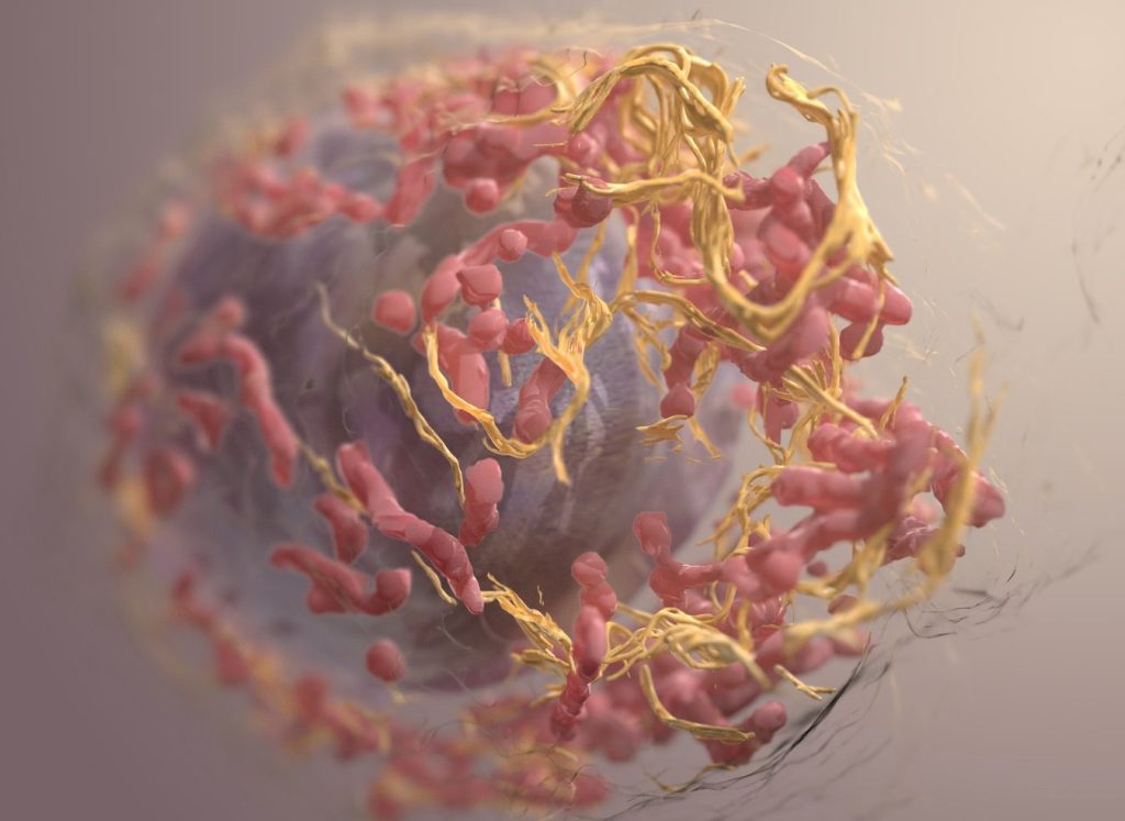
3D structure of a melanoma cell derived by ion abrasion scanning electron microscopy. Credit: Sriram Subramaniam/ National Cancer Institute

Researchers have developed a revolutionary biosensor using terahertz (THz) waves that can detect skin cancer with exceptional sensitivity, potentially paving the way for earlier and easier diagnoses. Published in the journal IEEE Transactions on Biomedical Engineering, the study presents a significant advancement in early cancer detection, thanks to a multidisciplinary collaboration of teams from Queen Mary University of London and the University of Glasgow.

“Traditional methods for detecting skin cancer often involve expensive, time-consuming, CT, PET scans and invasive higher frequencies technologies,” explains Dr Shohreh Nourinovin, Postdoctoral Research Associate at Queen Mary’s School of Electronic Engineering and Computer Science, and the study’s first author.

“Our biosensor offers a non-invasive and highly efficient solution, leveraging the unique properties of THz waves – a type of radiation with lower energy than X-rays, thus safe for humans – to detect subtle changes in cell characteristics.”

The key innovation lies in the biosensor’s design. Featuring tiny, asymmetric resonators on a flexible substrate, it can detect subtle changes in the properties of cells.

Unlike traditional methods that rely solely on refractive index, this device analyses a combination of parameters, including resonance frequency, transmission magnitude, and a value called “Full Width at Half Maximum” (FWHM). This comprehensive approach provides a richer picture of the tissue, allowing for more accurate differentiation between healthy and cancerous cells and to measure malignancy degree of the tissue.

In tests, the biosensor successfully differentiated between normal skin cells and basal cell carcinoma (BCC) cells, even at different concentrations. This ability to detect early-stage cancer holds immense potential for improving patient outcomes.

“The implications of this study extend far beyond skin cancer detection,” says Dr Nourinovin.

“This technology could be used for early detection of various cancers and other diseases, like Alzheimer’s, with potential applications in resource-limited settings due to its portability and affordability.”

Dr Nourinovin’s research journey wasn’t without its challenges.

Initially focusing on THz spectroscopy for cancer analysis, her project was temporarily halted due to the COVID pandemic. However, this setback led her to explore the potential of THz metasurfaces, a novel approach that sparked a new chapter in her research.

Source: Queen Mary University of London

Removing a Protein Lets Glioblastoma Chemo Remain Effective for Longer

Photo by Anna Shvets on Pexels

New research by the University of Sussex could help to increase life expectancy and improve treatment for glioblastoma. In the study, published in the Journal of Advanced Science, researchers have discovered that an understudied protein, called PANK4, is able to block cancer cells from responding to chemotherapeutic treatment for the highly intrusive brain cancer, glioblastoma.

Scientists at Sussex have demonstrated that if the protein is removed, cancer cells respond better to temozolomide, the main chemotherapy drug for the treatment of glioblastoma.

Prof Georgios Giamas, Professor of Cancer Cell Signalling at the University of Sussex explains: “Glioblastoma is a devastating brain cancer, and researchers are working hard to identify ways to delay progression of the disease, and tackle cell resistance to treatment. As this is the first time that PANK4 has been linked to glioblastoma, the next step is to develop a drug targeting this protein to try to reverse chemo-resistance and restore sensitivity, ensuring that patients receive the best treatment and have better outcomes.”

Glioblastoma is one of the most aggressive forms of brain cancer. Approximately 250 000 – 300 000 globally are diagnosed with it annually, with a best-case survival rate of just one to 18 months after diagnosis.

Following surgery to remove the tumour, glioblastoma patients are typically treated with radiation and the chemotherapeutic drug, temozolomide. Although patients initially respond well to the drug, the cancer cells quickly develop resistance to this treatment.

The University of Sussex scientists led an international research team to understand the possible reasons for this resistance, helping to guide future therapies to improve quality of life and increase life expectancy for those with glioblastoma.

The team identified a protein called PANK4 which, when removed from the cancer cells, can lead to the cell’s death, and saw patients better responding to temozolomide. Linked to this, the researchers found that patients expressing high levels of the PANK4 protein had lower survival rates.

Dr Viviana Vella, research fellow at the University of Sussex explains: “There are a multitude of under-investigated proteins that may hold great potential for therapeutic intervention. Our study sheds light on this understudied protein, PANK4, unveiling a protective role in temozolomide-resistant cancer cells. Ultimately, PANK4 depletion represents a vulnerability that can now be exploited to restore sensitivity to the drug and improve treatment.”

Source: University of Sussex

Extract of Sandalwood Oil Prevents Prostate Cancer Progression

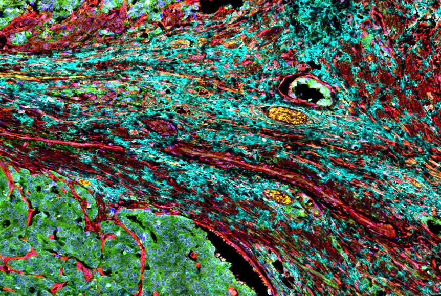
Ccancer-associated fibroplasts surrounding a prostate tumour. Credit: Moscat and Diaz Meco lab.

Extracted from the core of sandalwood trees (santalum album tree), sandalwood oil has been used for many centuries by several cultures throughout the world for perfume, soaps, incense and candles. With its earthy sweet scent, this essential oil also is used in the food industry and topically in various cosmetic preparations.

Importantly, this natural oil is known for its health benefits and medicinal applications from antibacterial to anticancer because of its phytochemical constituents. In addition to containing esters, free acids, aldehydes, ketones and santenone, sandalwood oil primarily (> 90%) constitutes santalol, equal amounts of two compounds, alpha and beta-santalol.

Now, researchers from Florida Atlantic University’s Schmidt College of Medicine and collaborators are the first to demonstrate in vivo the chemo-preventive properties of alpha-santalol against prostate cancer development using a transgenic mouse model.

Results of the study, published in the journal Phytomedicine Plus, showed that administration of alpha-santalol decreased the incidence of prostate tumours by decreasing cell proliferation and inducing apoptosis, without causing weight loss or any noticeable side effects.

Apoptosis (programmed cell death) is a method the body uses to get rid of unneeded or abnormal cells such as cancer cells. Findings revealed that the area occupied by normal tissue in alpha-santalol-treated mice was 53% compared to 12% in control mice.

This suggests that administering alpha-santalol protected the normal tissue and delayed progression from prostatic intraepithelial neoplasia, a precancerous condition, to poorly differentiated carcinoma, a high-grade form of cancer where cancer cells and tissue look very abnormal.

These results are significant because mortality in prostate cancer patients is mainly attributable to advanced stages of the disease.

In prior studies, the researchers demonstrated the efficacy of alpha-santalol in suppressing growth and inducing apoptotic cell death in cultured human prostate cancer cells.

Based on these observations, they selected a genetically engineered mouse model that resembles many features similar to human prostate cancer, eliciting different lesion grades and cancer progression.

“Although our cellular studies provided important mechanistic insights, relevant in vivo models are vital for developing novel chemo-preventive agents for clinical use and to determine if alpha-santalol offers protection against prostate cancer development,” said senior author Ajay Bommareddy, PhD, associate professor of pharmacology in the Department of Biomedical Science, FAU Schmidt College of Medicine.

“Prior to this new study, alpha-santalol’s in vivo efficacy against prostate cancer development had not yet been established.”

Additional findings of the current study showed alpha-santalol reduced the incidence of visible prostate tumors compared to control-treated mice.

Only 11% in the treated group developed prostate tumours whereas more than half in the control group developed the tumours.

The differences in urogenital and prostate weights were statistically significantly different in alpha-santalol-treated mice compared with controls.

The average wet weight of urogenital tract in alpha-santalol treated mice was about 74.28% lower compared with control mice.

Similarly, the average wet weight of the prostate gland was lower by 52.9% compared with control mice.

Current treatment methods for prostate cancer include androgen ablation, chemotherapy, radiotherapy and radical prostatectomy, but are ineffective against advanced prostate cancers.

Early detection and local therapy have resulted in improved outcomes but has been challenging with the management of advanced stages.

“Identifying agents that have the ability to selectively target cancerous cells and delay onset and progression of prostate cancer is greatly needed,” said Bommareddy.

“Additional studies are essential to systemically explore the feasibility of alpha-santalol as a promising chemo-preventive and anti-tumour agent against human prostate cancer development and to elucidate the mechanisms surrounding the role of pro-apoptotic and antiapoptotic proteins.”

Source: Florida Atlantic University

New Trial Flips the Script for Hormonal Treatment of Breast Cancer

Photo by National Cancer Institute

For decades, hormonal treatment of breast cancer has been going in one direction: blocking oestrogen. Now, a global study has discovered there may be another, less toxic way to defeat the most common form of breast cancer. The results, published in The Lancet Oncology, showed that the androgen receptor (AR) agonist enobosarm, is effective against oestrogen receptor-positive (ER+) breast cancer, which constitutes up to 80% of all breast cancer cases.

“The effectiveness of enobosarm lies in its ability to activate the AR and trigger a natural defence mechanism in breast tissue, thereby slowing the growth of ER+ breast cancer, which relies on the hormone oestrogen to grow and spread,” said senior co-author Professor Wayne Tilley, Director of the Dame Roma Mitchell Cancer Research Laboratories at the University of Adelaide.

“This clinical study is supported by our pre-clinical research, previously published in Nature Medicine, which established that the AR is a tumour suppressor in both normal breast tissue and ER+ breast cancer.”

Along with investigators from the University of Adelaide and Dana-Farber Cancer Institute (DFCI) in Boston, USA, the international study also included researchers from the University of Liverpool in the UK and other experts around the world.

The team assessed enobosarm’s efficacy and safety in 136 postmenopausal women with advanced or metastatic ER-positive, HER2-negative breast cancer.

Enobosarm showed significant anti-tumour activity and was well-tolerated by patients, without adversely affecting their quality of life or causing masculinising symptoms.

This discovery represents the first advancement in hormonal treatment of ER+ breast cancer in decades and offers a promising new oral treatment strategy for the most prevalent form of breast cancer.

The new hormonal strategy differs from the existing standard-of-care hormonal treatments, which have been around for decades and involve suppressing oestrogen activity in the body or inhibiting the ER.

Although successful initially, treatments targeting ER can cause severe side effects and treatment-resistant progression of the disease is common.

“Our findings are very promising. They demonstrate that stimulating the androgen receptor pathway with enobosarm can be beneficial,” said senior co-author and study Principal Investigator Dr Beth Overmoyer from DFCI.

“This is the first time a non-oestrogen receptor hormonal treatment approach has been shown to be clinically advantageous in ER+ breast cancer. The study supports further investigation of enobosarm in earlier stages of breast cancer as well as in combination with targeted therapies, such as ribociclib, a CDK 4/6 inhibitor.”

estrogen to grow and spread,” said senior co-author Professor Wayne Tilley, Director of the Dame Roma Mitchell Cancer Research Laboratories at the University of Adelaide.

“The data strongly encourages more clinical trials for AR-stimulating drugs in treating AR-positive and ER-positive breast cancer. The fact that this drug is well-tolerated also opens possibilities for its use in breast cancer prevention,” said co-author Dr Stephen Birrell, a clinical affiliate of the University of Adelaide.

Source: University of Adelaide

How Lung Cancer Transforms from One Type to Another

Lung cancer metastasis. Credit: National Cancer Institute

Adenocarcinomas sometimes respond to initially effective treatments by transforming into a much more aggressive small cell lung cancer (SCLC) that spreads rapidly and has few options for treatment. Researchers at Weill Cornell Medicine have developed a mouse model that illuminates this problematic process, known as histological transformation.

The researchers, whose results were published in Science, discovered that during the transition from lung adenocarcinoma to small cell lung cancer (SCLC), the mutated cells appeared to undergo a change in cell identity through an intermediate, stem cell-like state, which facilitated the transformation.

“It is very difficult to study this process in human patients. So my aim was to uncover the mechanism underlying the transformation of lung adenocarcinoma to small cell lung cancer in a mouse model,” said study lead Dr Eric Gardner, a postdoctoral fellow in the laboratory of Dr Harold Varmus.

The complex mouse model took several years to develop and characterise but has allowed the researchers to crack this difficult problem.

This study was in collaboration with Dr Ashley Laughney, assistant professor of physiology and biophysics and a member of the Meyer Cancer Center at Weill Cornell Medicine and Ethan Earlie, a graduate student in the Laughney lab and part of the Tri-Institutional Computational Biology and Medicine program.

“It is well known that cancer cells continue to evolve, especially to escape the pressure of effective treatments,” said Dr Varmus.

“This study shows how new technologies – including the detection of molecular features of single cancer cells, combined with computer-based analysis of the data – can portray dramatic, complex events in the evolution of lethal cancers, exposing new targets for therapeutic attack.”

Catching Transformation in the Act

SCLC most commonly occurs in heavy smokers, but this type of tumour also develops in a significant number of patients with lung adenocarcinomas, particularly after treatment with therapies that target a protein called Epidermal Growth Factor Receptor (EGFR), which promotes tumour growth.

The new SCLC-type tumours are resistant to anti-EGFR therapy because their growth is fuelled by a new cancer driver, high levels of Myc protein.

To unravel the interplay of these cancer pathways, the researchers engineered mice to develop a common form of lung adenocarcinoma, in which lung epithelial cells are driven by a mutated version of the EGFR gene.

They then turned the adenocarcinoma tumours into SCLC-type tumours, which generally arise from neuroendocrine cells.

They did this by shutting off EGFR in the presence of several other changes including losses of the tumour suppressor genes Rb1 and Trp53 as well as turning up the production of Myc,a known driver of SCLC.

Oncogenes, such as EGFR and Myc, are mutated forms of genes that normally control cell growth. They are known for their roles in driving the growth and spread of cancer. Tumor suppressor genes, on the other hand, normally inhibit cell proliferation and tumor development.

Context-dependent change

Surprisingly, this study showed that oncogenes act in a context-dependent manner.

While most lung cells are resistant to becoming cancerous by Myc, neuroendocrine cells, are very sensitive to the oncogenic effects of Myc. Conversely, epithelial cells, which line the air sacs of the lungs and are the precursors to lung adenocarcinomas, grow excessively in response to mutated EGFR.

“This shows that an ‘oncogene’ in the wrong cell type doesn’t act like an oncogene anymore,” Dr Laughney said.

“So, it fundamentally changes how we think about oncogenes.”

The researchers also discovered a stem cell-like intermediate that was neither adenocarcinoma nor SCLC.

Cells in this transitional state became neuroendocrine in nature only when mutations in the tumour suppressor genes RB1 and TP53 were present.

They observed that loss of another tumour suppressor called Pten accelerated this process.

At that stage, oncogenic Myc could drive these intermediate stem-like cells to form SCLC-type tumours.

This study further supports efforts seeking therapeutics that target Myc proteins, which are implicated in many types of cancers. The researchers now plan to use their new mouse model to further explore the adenocarcinoma-SCLC transition, detailing, for example, how the immune system normally responds to this transition.

Source: Weill Cornell Medicine