Tag: oestrogen

Study Proposes Safer Alternative to Hormone Therapy During Menopause
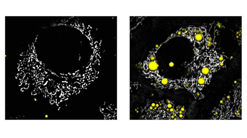
Liver cells viewed under a fluorescence microscope. On the left are healthy cells, and on the right, fat droplets stand out in yellow. The research revealed that activating the ERβ receptor induces the organ to completely oxidize these lipids to generate energy. Credit: Débora Santos Rocha et al./Comprehensive Physiology

By Maria Fernanda Ziegler  |  Agência FAPESP – When it comes to balancing hormonal changes during the transition to menopause, less can be more. Researchers at the University of São Paulo (USP) in Brazil demonstrated this when testing an alternative strategy to conventional therapy. Rather than replacing oestrogen entirely, the team activated only one type of oestrogen receptor in the cells. This approach achieved a broad and beneficial metabolic effect without stimulating the growth of reproductive tissues, such as the uterus and breasts. This is important for individuals with a genetic predisposition to developing tumours.

“It’s a more targeted strategy. By acting only on that receptor, we were able to rebalance the metabolic effects of the drop in oestrogen during menopause without affecting potentially sensitive areas of the body,” explains the researcher Débora Santos Rocha, a FAPESP postdoctoral fellow at the Institute of Chemistry (IQ-USP) and first author of the article published in the journal Comprehensive Physiology.

The study was conducted on female rats that had their ovaries removed to simulate menopause. In this model, the researchers used an experimental drug to activate only oestrogen receptor beta (ERβ), which is an important regulator of metabolism and does not stimulate reproductive tissues or increase the risk of hormone-sensitive tumours.

As Rocha explains, the significant drop in oestrogen during the transition to menopause disrupts the entire metabolism, potentially promoting the accumulation of visceral fat, insulin resistance, and cardiovascular risk. “In addition to their importance in reproduction, oestrogens play a central role in energy regulation. When their levels decrease, widespread metabolic changes also occur, increasing the risk of metabolic syndrome and diseases such as diabetes and hypercholesterolaemia [high cholesterol],” she states.

Conventional hormone replacement therapy attempts to counteract this loss by providing oestrogen and progesterone through pills, patches, or creams. However, by activating all oestrogen receptors indiscriminately, it may increase the risk of tumour growth in predisposed women. “It isn’t hormone replacement therapy that causes cancer. What happens is that if there are tumour cells in oestrogen-sensitive tissues, such as the uterus, breast, or endometrium, they may respond to the hormone and multiply more quickly,” Rocha explains.

A targeted solution with broad benefits

The results of the study show that the isolated activation of ERβ promoted profound metabolic reprogramming. “With the new approach, we restored fasting blood glucose levels, normalised the lipid profile, and reduced the size of fat cells. The pancreatic islets [cells that produce insulin and other hormones] regained their normal shape, and blood levels of cholesterol, triglycerides, and free fatty acids returned to healthy levels. All of that occurred without any effect on the uterus, which indicates the safety of the approach,” comments Alicia Kowaltowski, a professor at IQ-USP and the research coordinator.

The study primarily focused on the liver. This is because the organ begins to process fats differently during the transition to menopause and in the absence of oestrogen. “Lipid metabolism changes significantly, and the liver accumulates more fat. ‘Bad’ cholesterol [LDL] in the blood increases, and various lipid profiles are altered. However, with the activation of ERβ, those parameters returned to normal,” says Rocha.

The same effect was observed in the analysis of isolated liver cells. “The drug remodels the metabolism of liver cells [hepatocytes], causing them to oxidise fatty acids more [ie, to ‘burn’ the building blocks of lipids to generate energy]. That reduces fat accumulation and improves the overall lipid profile. By activating that specific pathway, we modulate liver metabolism and make circulating lipids healthier,” the postdoctoral fellow explains.

Kowaltowski highlights another important finding from the study. “By activating ERβ, the drug causes liver cells to fully oxidize fatty acids, something that normally doesn’t happen since the liver typically performs only partial oxidation, generating ketone bodies [alternative energy sources that the body creates to compensate for a lack of carbohydrates]. With that ‘complete oxidation,’ fat is fully converted into CO₂, significantly improving the body’s lipid profile. It’s important to note that this pathway can be activated to provide metabolic benefits – something we hadn’t imagined,” she explains.

There is no one-size-fits-all model

Although the treatment did not prevent the weight gain characteristic of menopause, the authors note in the article that it broadly improved metabolic quality and the functioning of the liver, pancreas, and blood.

“Menopause is a phase characterised by a wide variability of symptoms among women, which makes it important to have a variety of therapeutic approaches. If we can modulate specific pathways, we pave the way for more personalized treatments. Perhaps the approach we’re presenting won’t be ideal for one person, but it could be decisive for another. The important thing is to have treatment options,” Kowaltowski emphasizes.


“Perhaps the approach we’re presenting won’t be ideal for one person, but it could be decisive for another. The important thing is to have treatment options,” emphasises Alicia Kowaltowski, a professor at IQ-USP (photo: Cecília Bastos/USP Imagens)

The research continues in a new project funded by FAPESP that is now focused on the transition to menopause (perimenopause), using an experimental model that more closely mimics human reality.

Rather than abruptly removing the ovaries from female rats, the new study will use a model of gradual ovarian reserve decline, enabling the researchers to observe hormonal change dynamics throughout the process. “We want to understand that transition and identify new therapeutic targets that could serve as alternatives to conventional hormone replacement therapy,” Rocha concludes.

The article “Estrogen receptor beta activation coordinates liver lipid remodeling and metabolic fluxes, preventing lipotoxicity” can be read at onlinelibrary.wiley.com/doi/10.1002/cph4.70228.

Source: FAPESP

Study Identifies Oestrogen Receptor as Key Protector of the Right Heart in Pulmonary Hypertension

Human heart. Credit: Scientific Animations CC4.0

Researchers at National Jewish Health have identified a key way oestrogen receptor alpha (ERα) helps protect the right side of the heart in pulmonary hypertension. The preclinical findings reveal that ERα preserves the survival and movement of endothelial cells, which line blood vessels, allowing the stressed heart to maintain the small vessels it needs to function.

The study, published online Sept. 10 in Arteriosclerosis, Thrombosis, and Vascular Biology, may help explain an important difference between women and men with pulmonary hypertension and points to a potential path for developing therapies tailored to the right ventricle, the chamber that pumps blood through the lungs.

Pulmonary hypertension causes abnormally high pressure in the blood vessels of the lungs. That pressure forces the right ventricle to work harder, and a patient’s outlook depends heavily on how well the chamber adapts. Although women are more likely to develop certain forms of pulmonary hypertension, their right ventricles often function better than those of men. Researchers have been working to understand the biology behind that apparent paradox.

“We know that the right ventricle is a major driver of survival in pulmonary hypertension, but we still have very few treatments designed specifically to protect it,” said pulmonologist Tim Lahm, MD, senior author of the study and a researcher at National Jewish Health. “These findings show that oestrogen receptor alpha is doing important work inside the blood vessels of the right heart, particularly in females, by helping endothelial cells survive, move and build the vascular network the heart needs under stress.”

Using preclinical models of pulmonary hypertension and right ventricular pressure overload, researchers examined how ERα affects the endothelial cells that line blood vessels in the right side of the heart. When normal ERα function was disrupted, these cells were less able to migrate, form vessel-like networks and maintain the capillaries needed to support the heart under increased pressure. The effects were more pronounced in females and included increased endothelial cell death, greater enlargement of the right ventricle and reduced capillary density.

Single-nucleus RNA sequencing provided additional insight into the underlying mechanisms. In females with impaired ERα function, endothelial cells showed reduced activity in pathways involved in cell movement and increased activity in pathways associated with cell death. These changes were not observed to the same degree in males, further supporting a sex-specific role for the receptor.

“A healthy network of capillaries is essential when the right ventricle is working against increased pressure,” Dr Lahm said. “By showing how ERα supports that network, this work gives us a more precise biological target to investigate.”

The findings do not establish oestrogen or ERα-targeted therapy as a treatment for patients yet, but are an important first step. Additional research is needed to confirm the mechanism in people and determine whether it can be translated into a safe and effective therapy.

Source: National Jewish Health

‘Momnesia’ is Real – A Biological Explanation for Temporary Forgetfulness During Pregnancy

Photo by SHVETS production

Many women usually say the same thing during pregnancy: they walk into a room and forget why, misplace their keys or struggle to follow a conversation. This phenomenon, often called ‘pregnancy brain’ or ‘momnesia,’ has long lacked a clear biological explanation.

Now, a new study by researchers at Baylor College of Medicine and collaborating institutions and published in Science Bulletin, identifies a specific brain circuit in an animal model that becomes disrupted under the sustained high oestrogen levels present during pregnancy. The findings offer the first biological explanation of how exposure to high-level circulating oestrogen can temporarily impair memory.

A novel brain circuit links high oestrogen levels with memory problems

“We worked with mouse models designed to mimic the sustained, high blood-oestrogen levels of pregnancy. These models showed that elevated oestrogen caused reversible memory impairment without affecting mood or motivation, suggesting a specific cognitive effect rather than a general change in well-being,” said senior author Dr Zheng Sun, associate professor of medicine – endocrinology, diabetes and metabolism and of molecular and cellular biology at Baylor.

Digging into the underlying biology, the team found that oestrogen receptor alpha, the protein that transmits oestrogen’s signals into cells, is the dominant oestrogen receptor in the brain region called the lateral hypothalamus. Furthermore, this region has abundant GABAergic neurons – brain cells that normally send calming, inhibitory signals to other parts of the brain. Using single-nucleus RNA sequencing, the researchers discovered that high oestrogen levels suppress signaling in these neurons, leading them to fire more frequently. “When we genetically removed oestrogen receptors from these hypothalamic neurons, both oestrogen-induced and pregnancy-induced memory problems in mice were reversed,” said Sun, a member of Baylor’s Dan L Duncan Comprehensive Cancer Center.

The team also found that these overactive hypothalamic neurons project directly into a region of the hippocampus that is a hub for memory formation. Using chemogenetics, a technique that allows researchers to turn specific neurons on or off, the team showed that silencing this hypothalamus-to-hippocampus pathway protected mice from estrogen-induced memory problems, whereas artificially activating the same pathway was sufficient to impair memory on its own, even without elevated estrogen.

Reconciling mixed evidence

Oestrogen’s relationship with memory has puzzled researchers for decades. For instance, hormone replacement therapy after menopause has been linked to cognitive benefits in some studies, while high oestrogen during pregnancy or with oral contraceptive use has been linked to memory complaints in others. The new findings suggest a possible explanation – it may not simply be a matter of ‘more oestrogen is better’ or ‘worse,’ but rather where in the brain that oestrogen acts, and at what levels.

“Low-level, cyclical oestrogen exposure appears to support cognitive function, which is part of why hormone therapy can help postmenopausal women,” said senior author Dr Yanlin He, associate professor at Pennington Biomedical Research Center. “But sustained, high-level oestrogen exposure seems to engage a different pathway altogether, one centred in the hypothalamus rather than the hippocampus itself. That distinction may help reconcile a lot of conflicting data in the field.”

Confirming the link in pregnant women

To determine whether these findings translate to humans, the researchers assessed memory performance in women across different stages of pregnancy. They found task-specific memory impairments that emerged during late pregnancy, and that correlated with circulating oestrogen levels, even after accounting for other factors that might influence cognition. This human data supports the idea that the hormone-driven circuit identified in mice may underlie the memory changes many pregnant women experience.

“Momnesia is real, it has a defined biological basis and is temporary,” said senior author Dr Xianghua Zhuang, professor at the Second Qilu Hospital of Shandong University. “We hope this work helps validate what many women have described anecdotally for years, and gives researchers a concrete target for future study.”

“The memory changes observed in both mice and women were temporary and task-specific, not a sign of broader cognitive decline,” said senior author Dr Xinguo Hou, professor at the Qilu Hospital of Shandong University. “Nonetheless, understanding the underlying circuit could eventually inform how clinicians counsel patients about the cognitive side effects of pregnancy or hormonal contraceptives, and could open avenues for therapies targeting this specific pathway without disrupting oestrogen’s broader, beneficial roles in the body.”

Source: Baylor College of Medicine, EurekAlert!

How Oestrogen in the Brain Impacts Stress and Trauma Response

New research reveals how oestrogen levels in the brain influence vulnerability to stress-related memory problems, helping explain sex differences in PTSD risk.

Photo by Sherise Van Dyk on Unsplash

For some people, a single traumatic event like a shooting, a natural disaster or a violent assault, can leave an imprint that lingers long after the immediate danger has passed. Memories of that event may return with unusual intensity, shaping mood, behaviour, and mental health in ways that are difficult to predict. Others exposed to similar trauma recover without developing lasting memory problems or trauma-related symptoms.

Why those outcomes diverge is a central question in stress and trauma research. Clinicians have long observed that severe acute stress can permanently alter memory for some people but not others, and that women face roughly twice the lifetime risk of posttraumatic stress disorder (PTSD). Recent research from the University of Pennsylvania in collaboration with the University of California-Irvine suggests that part of the answer may lie in the brain’s biological state at the precise moment trauma occurs.

Elizabeth Heller, PhD, an associate professor of Pharmacology in the Perelman School of Medicine at the University of Pennsylvania, and her team in the Heller Lab, have now shed light on how the brain’s biological state at the time of stress, particularly its oestrogen levels, can shape vulnerability long after the acute stress has lifted. Heller helped uncover that oestrogen levels in the brain may play a surprising role in this vulnerability, and for both sexes. The study, published in Neuron, also provides new insight into why women are more likely than men to develop post-traumatic stress disorder (PTSD) and to face higher dementia risk later in life.

Unpacking oestrogen’s role in memory vulnerability

Oestrogen is widely known to support learning and memory. This study found that high levels of oestrogen in the hippocampus, a brain region critical for memory, help the brain’s cells change and adjust more easily. However, in the context of severe acute stress, this flexibility can increase vulnerability to stress-related memory problems.

Heller and the Penn team mapped how high levels of oestrogen interact with chromatin structure (the storage packaging up DNA inside cells) in the hippocampus to make some brains more susceptible to PTSD‑like memory changes.

The findings help explain why traumatic events such as natural disasters, mass violence, and assaults can cause long-term memory problems, and why women are roughly twice as likely as men to develop PTSD.

“A lot of what determines vulnerability is the state your brain is already in,” Heller explained. “If a traumatic event hits during a period when oestrogen is already unusually high, the resulting plasticity can amplify the impact in lasting ways, promoting vulnerability to stress. Even with these findings in hand, the word oestrogen can mislead readers into assuming the biology applies only to women. That assumption shaped public understanding for decades, but it doesn’t hold up against what this research, and years of foundational neuroscience, actually shows.

As Heller notes, oestrogen is a critical brain hormone in both sexes. It is produced locally in regions like the hippocampus where it helps regulate learning, mood, and responses to stress. Recognising that universality is essential to understanding what this study truly reveals.

“The striking thing is that oestrogen levels are actually high in both males and in females in some parts of the hormonal cycle. Thus, the effects of high oestrogen levels happen in both males and females,” Heller said. “We tend to treat oestrogen as a women’s health hormone, but the brain makes its own oestrogen, and it plays powerful roles in stress, memory, mood, and emotion across sexes.”

By Eric Horvath

Source: Penn Medicine

Do Hormones Explain Why Women Experience More Gut Pain?

UCSF researchers discover that oestrogen can turn on pain signals associated with conditions like irritable bowel syndrome.

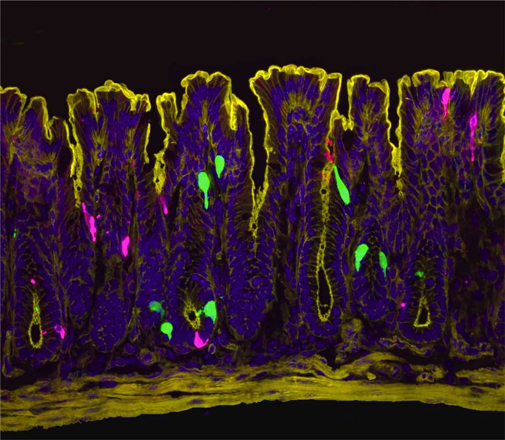
A zoomed in image of the lining of the colon. Cells that produce the hormone  PYY (peptide YY) are in green. Cells that produce the neurotransmitter serotonin are in magenta. PYY triggers the release of serotonin, which activates pain-sensing nerve fibers. Image by Archana Venkataraman/UCSF

Women are dramatically more likely than men to suffer from irritable bowel syndrome (IBS), a chronic condition causing abdominal pain, bloating, and digestive discomfort. Now, scientists at UC San Francisco have discovered why.

Oestrogen, the researchers report in Science, activates previously unknown pathways in the colon that can trigger pain and make the female gut more sensitive to certain foods and their breakdown products. When male mice were given oestrogen to mimic the levels found in females, their gut pain sensitivity increased to match that of females.

The findings not only explain the female predominance in gut pain disorders but also point to potential new ways to treat the conditions.

“Instead of just saying young women suffer from IBS, we wanted rigorous science explaining why,” said Holly Ingraham, PhD, professor UCSF and co-senior author of the study. “We’ve answered that question, and in the process identified new potential drug targets.”

The research also suggests why low-FODMAP diets – which eliminate certain fermentable foods, such as onions, garlic, honey, wheat, and beans – help some IBS patients, and why women’s gut symptoms often fluctuate with their menstrual cycles.

“We knew the gut has a sophisticated pain-sensing system, but this study reveals how hormones can dial that sensitivity up by tapping into this system through an interesting and potent cellular connection,” said co-senior author David Julius, PhD. Julius won the 2021 Nobel Prize for Physiology or Medicine for his work on pain sensation.

Search for oestrogen

Previous research had hinted that oestrogen was to blame for higher rates of IBS in females, but not why. To understand how oestrogen might be involved, Ingraham’s and Julius’s teams first needed to see exactly where the hormone was working in the gut.

“At the time I started this project, we didn’t know where and how oestrogen signalling is set up in the female intestine,” said Archana Venkataraman, PhD, a postdoc in Ingraham’s lab and co-first author of the research. “So, our initial step was to visualise the oestrogen receptor along the length of the female gut.”

The team expected to see oestrogen receptors in enterochromaffin (EC) cells, which were already known to send pain signals from the gut to the spinal cord. Instead, they got a surprise: oestrogen receptors were clustered in the lower part of the colon and in a different cell type known as L-cells.

The scientists pieced together a complex chain reaction that occurs when oestrogen binds to the L-cells. First, oestrogen causes L-cells to release a hormone called PYY (peptide YY). PYY then acts on neighbouring EC cells, triggering them to release the neurotransmitter serotonin, which activates pain-sensing nerve fibres. In female mice, removing the ovaries or blocking oestrogen, serotonin, or PYY dramatically reduced the high gut pain observed in females.

For decades, scientists believed PYY primarily suppressed appetite – drug companies even tried developing it as a weight-loss medication. But those clinical trials failed due to a troubling side effect that was never fully explained; participants experienced severe gut distress. The new findings mesh with this observation and suggest a completely new role for PYY.

“PYY had never been directly described as a pain signal in the past,” said co-first author Eric Figueroa, PhD, a postdoc in Julius’ lab. “Establishing this new role for PYY in gut pain reframes our thinking about this hormone and its local effects in the colon.”

This video shows what happens to the enterochromaffin (EC) cells in the colon when they are treated with PYY. Upon PYY treatment, calcium activity increases in the EC cell, causing it to fluoresce more brightly as it releases serotonin that is detected by nearby pain-sensing nerve fibres. Video by Eric Figueroa/UCSF

A link between IBS and diet

Increased PYY wasn’t the only way that L-cells responded to oestrogen. Levels of another molecule, called Olfr78, also went up in response to the hormone. Olfr78 detects short-chain fatty acids – metabolites produced when gut bacteria digest certain foods. With more Olfr78 receptors, L-cells become hypersensitive to these fatty acids and are more easily triggered to become active, releasing more PYY.

“It means that oestrogen is really leading to this double hit,” said Venkataraman. “First it’s increasing the baseline sensitivity of the gut by increasing PYY, and then it’s also making L-cells more sensitive to these metabolites that are floating around in the colon.”

The observation may explain why low-FODMAP diets help some IBS patients. FODMAPs (fermentable oligosaccharides, disaccharides, monosaccharides, and polyols) are carbohydrates that gut bacteria ferment into those same fatty acids sensed by Olfr78. By eating fewer FODMAPs, patients may be preventing the activation of Olfr78, and, in turn, keeping L-cells from churning out more of the pain signalling PYY.

While men have this same cellular pathway, their lower oestrogen levels keep it relatively quiet. However, the pathway could engage in men taking androgen-blocking medications, which block the effects of testosterone and can elevate oestrogen in some cases, potentially leading to digestive side-effects.

The new work suggests potential ways to treat IBS in women and men alike.

“Even for patients who see success with a low-FODMAP diet, it’s nearly impossible to stick to long term,” Ingraham said. “But the pathways we’ve identified here might be leveraged as new drug targets.”

The researchers are now studying how such drugs might work, as well as asking questions about what other hormones, such as progesterone, might play a role in gut sensitivity and how pregnancy, lactation, and normal menstrual cycles affect intestinal function.

By Sarah C.P. Williams

Source: University of California – San Francisco

Magnesium Inhibits Colorectal Cancer Development – Mostly in Females

The findings from the current study suggest that magnesium also increases the gut synthesis of vitamin D, which does not go to the blood and takes effect locally.

Photo by Danilo Alvesd on Unsplash

Researchers from Vanderbilt University Medical Center have demonstrated in a precision-based clinical trial that a magnesium supplement increases gut bacteria in humans that have been shown to synthesise vitamin D and inhibit colorectal cancer carcinogenesis.

However, the effect was observed primarily in females – an outcome that the researchers surmised may be attributable to the role that oestrogen plays in shifting magnesium from circulation into cellular uptake.

Intestinal microbiome data and colonoscopy results were analysed from participants who were randomised by whether they had the TRPM7 genotype, which plays a crucial role in regulating magnesium and calcium uptake.

Previously, the investigators showed in the same randomised trial that magnesium enhances the synthesis of vitamin D and increases the blood levels of vitamin D. The findings from the current study suggest that magnesium also increases the gut synthesis of vitamin D, which does not go to the blood and takes effect locally.

These results from the trial were published in The American Journal of Clinical Nutrition.

“Our previous study showed magnesium supplementation increased blood levels of vitamin D when vitamin D levels were low,” said Qi Dai, MD, PhD, professor of Medicine. “The current study reveals that magnesium supplementation also increases the gut microbes which have been shown to synthesise vitamin D in the gut without sunlight and locally inhibit colorectal cancer development.”

The participants were divided into two arms, one that received the magnesium supplement and another that received a placebo. Their gut microbiome was analysed from stools, rectal swabs and rectal tissues. Among participants with adequate TRPM7 function, the magnesium supplement increased Carnobacterium maltaromaticum and Faecalibacterium prausnitzii, which were previously found to work synergistically to increase vitamin D and decrease colorectal carcinogenesis. Among those with inadequate TRPM7 function, the magnesium supplement reduced the abundance of F. prausnitzii in rectal mucosa.

Among 236 participants who all had a history of colorectal polyps, 124 underwent colonoscopies after completing the trial with a 3.5-year median follow-up time. A higher abundance of F. prausnitzii in rectal mucosa was associated with an almost threefold increase in developing additional polyps.

Source: Vanderbilt University Medical Center

Oestrogen and Progesterone Stimulate the Body to Make Opioids

Source: Pixabay CC0

Female hormones can suppress pain by making immune cells near the spinal cord produce opioids, a new study from researchers at UC San Francisco has found. This stops pain signals before they get to the brain.

The discovery could help with developing new treatments for chronic pain. It may explain why some painkillers work better for women than men and why postmenopausal women, whose bodies produce less of the key hormones oestrogen and progesterone, experience more pain.

The work reveals an entirely new role for T regulatory immune cells (T-regs), which are known for their ability to reduce inflammation.

“The fact that there’s a sex-dependent influence on these cells – driven by oestrogen and progesterone – and that it’s not related at all to any immune function is very unusual,” said Elora Midavaine, PhD, a postdoctoral fellow and first author of the study, which appears in Science.

The researchers looked at T-regs in the protective layers that encase the brain and spinal cord in mice. Until now, scientists thought these tissue layers, called the meninges, only served to protect the central nervous system and eliminate waste. T-regs were only discovered there in recent years.

“What we are showing now is that the immune system actually uses the meninges to communicate with distant neurons that detect sensation on the skin,” said Sakeen Kashem, MD, PhD, an assistant professor of dermatology. “This is something we hadn’t known before.”

That communication begins when a neuron, often near the skin, receives a stimulus and sends a signal to the spinal cord.

The team found that the meninges surrounding the lower part of the spinal cord harbour an abundance of T-regs. To learn what their function was, the researchers knocked the cells out with a toxin.

The effect was striking: Without the T-regs, female mice became more sensitive to pain, while male mice did not. This sex-specific difference suggested that female mice rely more on T-regs to manage pain.

“It was both fascinating and puzzling,” said Kashem, who co-led the study with Allan Basbaum, PhD. “It actually made me sceptical initially.”

Further experiments revealed a relationship between T-regs and female hormones that no one had seen before: Estrogen and progesterone were prompting the cells to churn out enkephalin, a naturally occurring opioid.

Exactly how the hormones do this is a question the team hopes to answer in a future study. But even without that understanding, the awareness of this sex-dependent pathway is likely to lead to much-needed new approaches for treating pain.

In the short run, it may help physicians choose medications that could be more effective for a patient, depending on their sex. Certain migraine treatments, for example, are known to work better on women than men.

This could be particularly helpful for women who have gone through menopause and no longer produce oestrogen and progesterone, many of whom experience chronic pain.

The researchers have begun looking into the possibility of engineering T-regs to produce enkephalin on a constant basis in both men and women.

Source: University of California – San Francisco

Does the Brain Produce Oestrogen to Control Appetite?

Photo by Fakurian Design on Unsplash

Although a woman’s ovaries produce the most oestrogen, various types of oestrogen are also synthesised throughout different tissues in the body, including the brain’s neurons. New research in The FEBS Journal indicates that such neuro-oestrogens help suppress appetite.

Knowing that the enzyme aromatase is important for the production of oestrogens, investigators depleted or knocked out the gene encoding aromatase in mice, so that the animals were unable to synthesise oestrogens in a systemic or body-wide manner. These mice demonstrated increased food intake and body weight compared with their aromatase-expressing counterparts. Restoring aromatase expression specifically in the brain reduced food intake and increased sensitivity to leptin, the “fullness” hormone, confirming that neuro-oestrogens can influence appetite.

To further investigate the role of neuro-oestrogens independently of ovarian oestrogen involvement, the researchers removed the ovaries in female mice. The brain’s hypothalamus (the central hub for appetite signals) in ovariectomised mice showed increased expression of the gene encoding aromatase, and these mice decreased their food intake.

“Our results imply that neuro-oestrogens likely contribute to appetite regulation and may be relevant for body weight reduction” the authors wrote.

Source: Wiley

Why Does Tamoxifen Work Only for Some Patients but not Others?

Photo by Danilo Alvesd on Unsplash

A new study has shown that variation in the microbiota of the human gut impacts the pharmacokinetics of tamoxifen and thus the effectiveness of the drug. The finding, published in the journal mBio, suggests that in the future, doctors may use a simple stool test to check for certain bacteria in the gut and help predict tamoxifen’s effectiveness for them.

Tamoxifen is a selective oestrogen receptor modulator used to prevent breast cancer. It prevents breast cancer cells from being able to use oestrogen to grow.

“The key takeaway from this study is that while tamoxifen is a common and important treatment for preventing breast cancer recurrence, nearly 50% of patients don’t respond well to it,” said lead study author Yasmine Alam, a PhD candidate in the Department of Biological Chemistry, University of California Irvine. “Since tamoxifen is taken orally and passes through the gut, this difference in how patients respond may be linked to the gut microbiome – the trillions of bacteria in our intestines, which vary greatly from person to person. Our study aims to better understand how these gut bacteria influence the way tamoxifen is absorbed, broken down and recycled in the body, with the goal of improving treatment outcomes for breast cancer patients.”

In the new study, the researchers set out to define the role that gut microbes play in how tamoxifen is processed (ie, absorption, distribution, metabolism and excretion), given its significant variable efficacy across patients. The researchers provided tamoxifen to mice that had no gut microbiome and to mice with a human microbiome (introduced to the mice by a human faecal sample). They found that mice with gut bacteria had higher amounts of tamoxifen in their bloodstream. The scientists then went on to explore what part of the gut microbiome was responsible for controlling the level of drug in the bloodstream. By examining the faecal samples from people, they linked a specific enzyme in bacteria, beta-glucuronidase, as a key factor that allows the drug to enter the bloodstream.

Tamoxifen is absorbed into the bloodstream from the intestine. Tamoxifen is carried by the bloodstream to the liver, where it is converted to its cancer-fighting form. Sometimes a sugar molecule can get attached to it, which signals the body to dump the cancer-fighting form of the drug back into the intestine. This drug can only get out of the intestine by taking the sugar off the molecule – and the researchers found that beta-glucuronidase in gut bacteria can eat the sugar off the drug so it can go on to fight breast cancer.

“Specifically, we found that certain enzymes produced by gut bacteria, called β-glucuronidase, play a role in how tamoxifen is broken down. These enzymes help recycle tamoxifen back into the bloodstream, which can make the drug more effective,” Alam said. “We discovered that a particular type of bacteria, Bacteroides fragilis, was strongly linked to the ability of these enzymes to affect tamoxifen levels in the blood in a positive way. This suggests that the gut microbiome plays an important role in how tamoxifen works in the body.”

The long-term goal of the study is to pave the way for more tailored and effective therapeutic interventions in the prevention of breast cancer recurrence.

Source: American Society for Microbiology

How Oestrogen can Trigger Nerve Impulses in Milliseconds

Photo by Julian Jagtenberg on Pexels

Oestrogen, the major female ovarian hormone, can trigger nerve impulses within milliseconds to regulate a variety of physiological processes. At Baylor College of Medicine, Louisiana State University and collaborating institutions, researchers discovered that oestrogen’s fast actions are mediated by the coupling of the oestrogen receptor-alpha (ER-alpha) with an ion channel protein called Clic1.

Clic1 controls the fast flux of electrically charged chloride ions through the cell membrane, which neurons use for receiving, conducting and transmitting signals. The researchers propose that interacting with the ER-alpha-Clic1 complex enables oestrogen to trigger fast neuronal responses through Clic1 ion currents. The study appeared in Science Advances.

“Oestrogen can act in the brain to regulate a variety of physiological processes, including female fertility, sexual behaviours, mood, reward, stress response, cognition, cardiovascular activities and body weight balance. Many of these functions are mediated by oestrogen binding to one of its receptors, ER-alpha,” said co-corresponding author Dr Yong Xu, professor of pediatrics – nutrition and associate director for basic sciences at the USDA/ARS Children’s Nutrition Research Center at Baylor. 

Fast and slow

It is well known that, upon stimulation by oestrogen, ER-alpha enters the cell nucleus where it mediates the transcription of genes. This classical mode of action as a nuclear receptor takes minutes to hours.

“Oestrogen also can change the firing activity of neurons in a manner of milliseconds, but it was not clear how this happens,” Xu said. “In this case, it did not make sense to us that the minutes-long nuclear receptor function of ER-alpha was involved in such a rapid action. We explored the possibility that ion channels, proteins in the cell membrane that regulate the fast flux of ions, mediated oestrogen’s quick actions.”

In the current study, working with cell lines and animal models, the team searched for cell membrane proteins that interact with ER-alpha. They found that protein Clic1, for chloride intracellular channel protein-1, can physically interact with ER-alpha. Clic1has been implicated in the regulation of neuronal excitability, so the researchers considered it a candidate to mediate oestrogen-triggered fast actions.

“We discovered that oestrogen enhances Clic1-mediated ion currents, and eliminating oestrogen reduced such currents,” Xu said. “In addition, Clic1 currents are required for oestrogen to induce rapid responses in neurons. Also, disrupting the Clic1 gene in animal models blunted oestrogen regulation of female body weight balance.”

The findings suggest that other nuclear receptors could also interact with ion channels, a possibility the researchers look forward to studying in the future.

“This study was conducted with female mice. However, Clic1 is also present in males. We are interested in investigating its role in male physiology,” Xu said.

Chloride channels are not as well studied as other ion channels, such as potassium, sodium or calcium channels. “We are among the first to study the role Clic1 plays in female physiology,” Xu said. “We hope that our findings will inspire other groups in the field to expand these promising investigations.”

Source: Baylor College of Medicine