For some leukaemia patients, the only potential chemotherapy option is ponatinib, a drug that also carries a high risk of heart failure. This means that some patients who recover from their cancer will end up dying of heart disease brought on by the cure.
In a new study, researchers from the University of Illinois Chicago and other universities have identified mechanisms that cause ponatinib to harm the heart. They also identified a promising treatment that could reverse this process.
The paper, with senior author Sang Ging Ong, assistant professor of pharmacology and medicine at UIC, is published in Circulation Research. The study is part of a growing field called cardio-oncology that investigates drugs that shrink tumours but can also cause heart problems.
While there are three options of drugs for treating chronic myeloid leukaemia, many patients are resistant to the other two, leaving ponatinib as their only choice.
“These patients have no other options for treatment,” Ong said, despite the concerns about the drug’s side effects.
In fact, ponatinib was pulled from the market for a few months after its introduction in 2012 because of concerns about heart problems.
The researchers were interested in understanding the interaction between ponatinib and the heart cells responsible for contraction.
They discovered that ponatinib damages these cells by activating a process known as the integrated stress response.
The mechanism for this is related to the functioning of a kinase (an enzyme involved in energy transfer) called GCN2.
The researchers found that ponatinib, despite being a kinase inhibitor, actually activates GCN2, which in turn switches on the integrated stress response.
While this response isn’t always a bad thing, normally protecting cells, it can also lead to their death under prolonged stress.
To see if this response was harming the cells, the researchers studied what would happen if they used a small molecule to block the integrated stress response in both cells and in mice during ponatinib treatment.
They found that the treatment helped protect heart cells from the damaging side effects of the drug yet did not diminish ponatinib’s tumour-fighting efficacy.
“It protects the heart but at the same time, it still allows us to kill cancer cells,” Ong said.
More research is needed to know if this protective measure would work well in humans, Ong said.
The mechanisms they identified are important in other cardiac diseases, as well, which could lead to future research on how to protect cells against different conditions.
Study shows that higher levels of physical activity are linked with less pain, and to a similar extent in adults with and without a history of cancer.
Photo by Barbara Olsen on Pexels
People who have had cancer often experience ongoing pain, but a new study reveals that being physically active may help lessen its intensity. The study is published by Wiley online in CANCER, a peer-reviewed journal of the American Cancer Society.
Although physical activity has been shown to lessen various types of pain, its effects on cancer-related pain are unclear. To investigate, a team led by senior author Erika Rees-Punia, PhD, MPH, of the American Cancer Society, and first author Christopher T.V. Swain, PhD, of the University of Melbourne, in Australia, analysed information pertaining to 51 439 adults without a history of cancer and 10,651 adults with a past cancer diagnosis. Participants were asked, “How would you rate your pain on average,” with responses ranging from 0 (no pain) to 10 (worst pain imaginable). Participants were also asked about their usual physical activity.
US guidelines recommend 150 minutes (2 hours 30 minutes) to 300 minutes (5 hours) a week of moderate-intensity, or 75 minutes (1 hour 15 minutes) to 150 minutes (2 hours 30 minutes) a week of vigorous-intensity aerobic physical activity.
Based on participants’ responses, the investigators found that, for individuals who had cancer in the past as well as for those without a history of cancer, more physical activity was linked with lower pain intensity. The extent of the association was similar for both groups of individuals, indicating that exercise may reduce cancer-related pain just as it does for other types of pain that have been studied in the past.
Among participants with a past cancer diagnosis, those exceeding physical activity guidelines were 16% less likely to report moderate-to-severe pain compared to those who failed to meet physical activity guidelines. Also, compared with people who remained inactive, those who were consistently active or became active in older adulthood reported less pain.
“It may feel counterintuitive to some, but physical activity is an effective, non-pharmacologic option for reducing many types of pain. As our study suggests, this may include pain associated with cancer and its treatments,” said Dr Rees-Punia.
Receiving the news that their child has been diagnosed with cancer is devastating for any parent, but this is even worse when they hear that, after 18 months of remission, their little one will need to battle the disease all over again.
This was the case for mom of two Arthie Ishwarlal. Back in 2021, her then two-year-old daughter, Preshthi, was diagnosed with Acute Lymphoblastic Leukaemia (ALL), a type of blood cancer that affects the bone marrow, white blood cells, red blood cells, and blood platelets. But, despite undergoing inpatient treatment, Preshthi experienced a relapse earlier this year with doctors saying that her only chance for survival is a stem cell transplant from a matching donor. Unfortunately, however, there is no match for her on the country’s stem cell registry at present.
As the world observes International Childhood Cancer Day (ICCD) on 15February, Palesa Mokomele, Head of Community Engagement and Communications at DKMS Africa explains that South Africans can potentially save Preshthi’s life. While there are currently over 73 000 donors on the South African registry, each only has a 1 in 100 000 chance of being a match for a blood cancer patient in need. But exacerbating the situation for little Preshthi is the lack of Indian donors since the best chance of a match comes from within one’s own ethnic group.”
She adds that it is not just Preshthi who needs a stem cell transplant for a second chance at life. “This is often the only treatment offering children with other blood cancers, like lymphomas, any hope of a cure.”
With leukaemia and lymphomas being two of the five most common cancers among South Africa’s youth, with the former accounting for 34% of childhood cancer cases and the latter 11%, Mokomele urges South Africans aged between 17 and 55 who are in good general health to register as donors. “In doing so, you might save a child’s life.”
The UK’s King Charles has been diagnosed with cancer, though reportedly at an early stage. This follows a brief hospital stay where he underwent a procedure for a benign enlarged prostate. For the time being, he will not being public duties, but will continue his private duties.
Speaking to the BBC, UK Prime Minister Rishi Sunak said that the cancer was “caught early”. The nature of the cancer was not disclosed by Buckingham Palace, which is in line with its usual practice of sharing only basic information concerning the health of the royal family. It however noted that it was not connected to his prostate treatment, ruling out prostate cancer.
Well-wishes for the king have come in from around the globe. Mia Mottley, Barbados’ first female PM, wished wishes King Charles a “full and speedy recovery”. Barbados became a republic in 2021, ending the the role of UK monarchs as its Head of State.
Since the UK is a constitutional democracy, his private duties consist of governmental approvals. For example, the king has constitutional duties, such as approving the passing of laws and appoints new judges, ambassadors and prime ministers. Public activities such as charity events and giving honours for public or voluntary service.
It is expected that certain activities such as his weekly meetings with Prime Minister Sunak will continue unless his doctors advise otherwise. Other members of the royal family will be able to stand in for him for ceremonial duties if he is unable to perform them. Recently, 41-year-old Catherine, Princess of Wales,
It is not an unexpected medical condition to occur for the 75-year old monarch – age is a major factor for almost all cancers – just over a quarter of all cancers are diagnosed from age 75 onward. The American Cancer Society now recommends general cancer screenings start at 45.
Diagnoses of breast cancer have increased steadily in women under age 50 over the past two decades, with steeper increases in more recent years, according to a study published in JAMA Network Open. The surge is driven largely by increases in the number of women diagnosed with oestrogen-receptor positive (ER+) tumours.
While overall trends show increases, however, some decreases have occurred in specific tumour types and among specific groups of women. Such changes in disease rates in young women observed over time – analysed by age, race, tumour type, tumour stage and other factors – may offer clues to possible prevention strategies.
“For most women, regular breast cancer screening does not begin until at least age 40, so younger women diagnosed with breast cancer tend to have later-stage tumours, when the disease is more advanced and more difficult to treat,” said senior author Adetunji T. Toriola, MD, PhD, a professor of surgery and co-leader of the Cancer Prevention and Control Program at Siteman Cancer Center, based at Barnes-Jewish Hospital and Washington University School of Medicine. “This research offers a way to begin identifying the factors driving these increasing rates, with the goal of finding ways to slow or reverse them. It also could help identify young women who are at high risk of developing early-onset breast cancer, so that we can design interventions to evaluate in clinical trials to see if we can lower that risk.”
The research team analysed data from over 217 000 U.S. women diagnosed with any type of breast cancer from 2000 through 2019. In 2000, the incidence of breast cancer among women ages 20 to 49 was about 64 cases per 100 000 people. Over the next 16 years, that rate slowly went up, increasing at about 0.24% per year. By 2016, the rate had reached about 66 cases per 100 000. But after 2016, for reasons researchers do not yet understand, the trend line made a steep uphill turn, suddenly increasing at 3.76% per year. By 2019 the rate had reached 74 cases per 100 000.
An additional intriguing aspect of the data is that the increase in breast cancer incidence is due almost entirely to an increase in tumours that are ER+ according to Toriola, who is also a William H. Danforth Washington University Physician-Scientist Scholar. These tumours have proteins on their surfaces that bind to oestrogen, which fuels tumour growth. In fact, the incidence of tumours without the oestrogen receptor decreased over the 20 years of data analysed in the study.
“We need to understand what is driving the specific increase in oestrogen-receptor positive tumours,” Toriola said. “We also hope to learn from the decrease in oestrogen-receptor negative tumours. If we can understand what is driving that rate down, perhaps we can apply it in efforts to reduce or prevent other breast tumour types.”
The researchers also found higher rates of breast cancer among Black women, especially among those ages 20 to 29. Black women in this age group have a 53% increased risk of breast cancer compared with white women of the same age group. A higher risk for Black women also continues from ages 30 to 39, but the increased risk is smaller, at about 15% greater risk compared with white women of the same age range. Then, from ages 40 to 49, the rate for Black women drops below that of white women.
Toriola said his group is evaluating breast tumour tissue from cancer patients of different ages and races to see if there are molecular differences that could shed light on what is driving cancer to develop more in young Black women. Of note, Hispanic women in the study had the lowest incidence of breast cancer of any group.
The researchers also showed an increase in diagnoses of stage 1 and stage 4 tumours, and a decrease in diagnoses of stage 2 and stage 3 tumours. Toriola said such data suggest that improvements in screening over the past two decades, and perhaps greater awareness of family history and genetic risk factors for breast cancer, have led to many tumours being caught earlier. But it also suggests that when stage 1 tumours are missed in younger women, the tumours tend not to be found until they reach stage 4.
The researchers also found differences in breast cancer risk by year of birth. Toriola said the most dramatic difference was a greater than 20% increased risk of breast cancer among women born in 1990 compared with women born in 1955.
“We are hopeful this study will offer clues to prevention strategies that will be effective in younger women, especially younger Black women, who are at particularly high risk of developing breast cancer before age 40,” Toriola said.
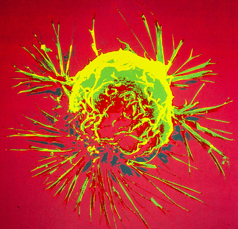
Colourised scanning electron micrograph of a breast cancer cell. Credit: NIH
Calcium ions are essential for cells, but can be toxic in higher concentrations. A team of researchers has now designed and prepared a combination drug that kills tumour cells by modulating the calcium influx into the cell. An external calcium source is not necessary because only the calcium ions already present in the tumour tissue are used, according to the study published in the journal Angewandte Chemie.
Biological cells need calcium ions, among other things, for the proper functioning of the mitochondria, the powerhouses of the cells.
However, if there is too much calcium, the mitochondrial processes become unbalanced and the cell suffocates.
A research group led by Juyoung Yoon of Ewha Womens University in Seoul, South Korea, together with teams from China, has now taken advantage of this process and developed a synergistic antitumour drug that can open calcium channels and thus trigger a deadly calcium storm inside the tumour cell.
The researchers targeted two channels, the first one in the outer membrane, and the other was a calcium channel in the endoplasmic reticulum, a cell organelle that also stores calcium ions.
The channel located in the outer membrane opens when it is exposed to a large amount of reactive oxygen species (ROS), while the channel in the endoplasmic reticulum is activated by nitric oxide molecules.
To generate the ROS that open the outer membrane calcium channel, the researchers used the dye indocyanine green.
This bioactive agent can be activated by irradiation with near-infrared light, which not only triggers reactions that lead to ROS, but it also heats up the environment.
The team explains that the high local temperature activates the other active agent, BNN-6, to release nitric oxide molecules that open the channel in the endoplasmic reticulum.
Following successful trials in tumour cell lines, the team tested an injectable formulation in tumour-implanted mice.
To create a biocompatible combined drug, the researchers loaded the active ingredients into tiny modified porous silica beads that are not harmful to the body, but can be recognized by tumour cells and transported into the cell.
After injecting the beads into the bloodstream of the mice, the researchers observed that the drug accumulated in the tumour.
Exposure to near-infrared light successfully triggered the mechanism of action, and the tumour disappeared after a few days in mice that received the preparation.
The authors emphasise that this ion influx approach may also be useful in related biomedical research areas where a similar mechanism could activate ion channels different from calcium in order to find new therapeutic approaches.
An international study has uncovered a mechanism by which cancer cells prevent the immune system from activating and attacking the cancerous invaders. The study, published in the peer-reviewed journal Cell Reports, sheds light on why immunotherapy treatments don’t work for all people or all diseases.
Certain types of cancers, such as colon, pancreatic, prostate and brain cancers, have stubbornly resisted immunotherapy.
And while breast, oesophageal and head and neck cancers often respond favourably, sometimes the treatments don’t work as planned.
Researchers still don’t understand exactly why, but the study, co-authored by University of Texas at Arlington scientists, may offer a clue.
“Immunotherapy is an incredibly promising new treatment avenue for cancer, but we still have work to do determining why it doesn’t work for all people or types of cancer,” said Jon Weidanz, UTA associate vice president for research and innovation.
He and Soroush Ghaffari, a UTA postdoctoral fellow, were co-authors of the study, along with colleagues at Leiden University in Leiden, Netherlands, and Karolinska University in Solna, Sweden.
The team determined that a key checkpoint in the immune system, called NKG2A, doesn’t engage with its specific binding molecule expressed in cancer cells until the appropriate signal is received.
“The team reasoned that monotherapy agents targeting the NKG2A receptor may not be effective without receiving an inflammatory trigger,” Ghaffari said.
“This might explain why drugs designed to bind to the NKG2A receptor to disrupt this immune checkpoint have been only effective when used in combination with other agents that can induce the necessary inflammatory signal.”
A second major finding of the study revealed how certain cancers can inhibit the immune system from activating its macrophages.
“These data give us a new molecular understanding of why some immunotherapies work and some don’t,” said Weidanz, who also is a professor kinesiology with an appointment in bioengineering and a member of the Multi-Interprofessional Center for Health Informatics.
“These results will help us identify and treat more cancers effectively with immunotherapy, helping more people live longer lives despite a cancer diagnosis.”
These findings have implications for immune system research and the development of more effective immunotherapy drugs, said Kate C. Miller, vice president of research and innovation at UTA.
“These are exciting new research results that have the potential to impact people living with cancer,” Miller said. “This is another great example of the calibre of biomedical research we’re performing both here at UTA and with our partners at other institutions.”
Using mRNA technology developed and matured for certain COVID vaccines, researchers have successfully restored the tumour-suppressing p53 protein in mouse models of advanced human ovarian cancer, significantly extending their survival. They report their results in Cancer Communications.
Ovarian cancer is often only detected at an advanced stage and metastases have already formed — usually in the intestines, abdomen or lymph nodes. At such a late stage, only 20 to 30% of all those affected survive the next five years. “Unfortunately, this situation has hardly changed at all over the past two decades,” says Professor Klaus Strebhardt, Director of the Department of Molecular Gynecology and Obstetrics at University Hospital Frankfurt.
In 96% of all ovarian cancer (high-grade) patients, the tumour suppressor gene p53 has mutated and is now non-functional. The gene contains the building instructions for an important protein that normally recognises damage in each cell’s DNA. It then prevents these abnormal cells from proliferating and activates repair mechanisms that rectify the damage.
If this fails, it induces cell death. “In this way, p53 is very effective in preventing carcinogenesis,” explains Strebhardt. “But when it is mutated, this protective mechanism is eradicated.”
If a cell wants to produce a certain protein, it first makes a transcript of the gene containing the building instructions for it. Such transcripts are called mRNAs. In women with ovarian cancer, the p53 mRNAs are just as defective as the gene from which they were copied.
“We produced an mRNA in the laboratory that contained the blueprint for a normal, non-mutated p53 protein,” says Dr Monika Raab from the Department of Molecular Gynecology and Obstetrics, who conducted many of the key experiments in the study.
“We packed it into small lipid vesicles, known as liposomes, and then tested them first in cultures of various human cancer cell lines. The cells used the artificial mRNA to produce functional p53 protein.”
In the next step, the scientists cultivated ovarian tumours – organoids – from patient cells sourced by the team led by Professor Sven Becker, Director of the Women’s Clinic at University Hospital Frankfurt.
After treatment with the artificial mRNA, the organoids shrank and began to die.
To test whether the artificial mRNA is also effective in organisms and can combat metastases in the abdomen, the researchers implanted human ovarian tumour cells into the ovaries of mice and injected the mRNA liposomes into the animals some time later.
The result was very convincing, says Strebhardt: “With the help of the artificial mRNA, cells in the animals treated produced large quantities of the functional p53 protein, and as a result both the tumours in the ovaries and the metastases disappeared almost completely.”
That the method was so successful is partly due to recent advances in mRNA technology: Normally, mRNA transcripts are very sensitive and degraded by cells within minutes.
However, it is meanwhile possible to prevent this by specifically modifying the molecules.
This extends their lifespan substantially, in this study to up to two weeks.
In addition, the chemical composition of the artificial mRNA is slightly different to that of its natural counterpart.
This prevents the immune system from intervening after the molecule has been injected and from triggering inflammatory responses.
In 2023, the Hungarian scientist Katalin Karikó and her American colleague Drew Weissman were awarded the Nobel Prize in Physiology or Medicine for this discovery.
“Thanks to the development of mRNA vaccines such as those of BioNTech and Moderna, which went into action during the SARS-CoV-2 pandemic, we now also know how to make the molecules even more effective,” explains Strebhardt.
Strebhardt, Raab and Becker are now looking for partners to join the next step of the translational project: testing on patients with ovarian cancer. “What is crucial now is the question of whether we can implement the concept and the results in clinical reality and use our method to help cancer patients,” says Strebhardt. The latest results make him very optimistic that the tide could finally turn in the treatment of ovarian carcinomas. “p53 mRNA is not a normal therapeutic that targets a specific weak point in cancer cells. Instead, we are repairing a natural mechanism that the body normally uses very effectively to suppress carcinogenesis. This is a completely different quality of cancer therapy.”
Credit: Darryl Leja National Human Genome Research Institute National Institutes Of Health
Because low-risk prostate cancer is unlikely to spread or impact survival, experts and guidelines recommend active surveillance, which involves regular monitoring and thus avoid or delay treatment like surgery or radiation therapy and their life-changing complications. A new study examined the rates of active surveillance use and evaluated the factors associated with selecting this management strategy over surgery or radiation, with a focus on underserved Black patients who have been underrepresented in prior studies. The findings are published by Wiley online in CANCER, a peer-reviewed journal of the American Cancer Society.
For the study, called the Treatment Options in Prostate Cancer Study, Jinping Xu, MD, MS, of Wayne State University, and her colleagues analysed data from metro-Detroit, Michigan, and Georgia cancer registries, focusing on patient self-reported information related to Black and White patients who were newly diagnosed with low-risk prostate cancer in 2014–2017.
Among 1688 patients, 57% chose active surveillance (51% of Black patients and 61% of White patients) over other treatments. After adjusting for other influencing factors, the strongest determinant of active surveillance uptake was a urologist’s recommendation to choose this option. Other factors linked with the decision to undergo active surveillance included a shared patient-physician treatment decision and greater knowledge about prostate cancer. Also, participants living in metro-Detroit were more likely to choose active surveillance than those living in Georgia.
Conversely, men were less likely to try active surveillance if their considerations were strongly influenced by the desire to achieve a “cure” or they expected to “live longer” with treatment, or if they perceived that their low-risk prostate cancer diagnosis was more “serious.”
Education and interventions for patients and especially urologists that address these factors may increase the use of recommended active surveillance among individuals with low-risk prostate cancer.
“I am glad to see that the majority of our study participants selected active surveillance, which indicates that acceptance has improved over the last decade; however, there is room for greater acceptance. Our study findings shed new light on potentially modifiable factors that can help further increase active surveillance use among patients with newly diagnosed low-risk prostate cancer to avoid unnecessary invasive treatment and improve their quality of life,” said Dr Xu.
Deaths from breast cancer dropped 58% between 1975 and 2019 due to a combination of screening mammography and improvements in treatment, according to a new study led by Stanford Medicine clinicians and biomedical data scientists.
Nearly one-third of the decrease (29%) is due to advances in treating metastatic breast cancer, also known as stage 4 breast cancer or recurrent cancer. Although these advanced cancers are not considered curable, women with metastatic disease are living longer than ever.
The analysis helps cancer researchers assess where to focus future efforts and resources.
“We’ve known that deaths from breast cancer have been decreasing over the past several decades, but it’s been difficult or impossible to quantify which of our interventions have been most successful, and to what extent,” said Jennifer Caswell-Jin, MD, assistant professor of medicine. “This type of study allows us to see which of our efforts are having the most impact and where we still need to improve.”
Caswell-Jin and Liyang Sun are co-first authors of the study, which was published in the Journal of the American Medical Association. Sylvia Plevritis, PhD, professor and chair of biomedical data science, and Allison Kurian, MD, MSc, professor of medicine and of epidemiology and population health, are co-senior authors.
The study was a collaborative effort by a national consortium of researchers called CISNET, or the Cancer Intervention and Surveillance Modeling Network. CISNET was established in 2000 by the National Cancer Institute to understand the impact of cancer surveillance, screening and treatment on incidence and mortality. Doing so requires sophisticated computer algorithms capable of modelling the natural course of the disease and the typical treatment paths of individual patients, then translating that information to population-level data collected by the national Surveillance, Epidemiology, and End Results Program, or SEER registry, from 1975 to 2019.
The study is the third in a trio of papers from CISNET published since 2005 that assess the relative contributions of regular screening and treatment advances on breast cancer deaths. The previous two papers informed national guidelines and helped cancer researchers focus their efforts on the most intractable problems.
“Twenty years ago, there was a question whether routine screening mammography actually decreased the number of deaths from breast cancer,” Plevritis said. But in 2005, she and other CISNET researchers published a paper in the New England Journal of Medicine that conclusively demonstrated that screening was responsible for anywhere from 28% to 65% (different models came up with varying degrees of impact) of the reduction in mortality by 2000 between 1975 and 2000.
The second paper, published in 2018 in the Journal of the American Medical Association, highlighted the differences in treatment responsiveness and survival outcomes among women with differing breast cancer subtypes from 2000 to 2012, pinpointing subgroups with poorer survival.
“We found that, while screening still had an important impact, most of the decline in annual deaths was due to improvements in treating early-stage breast cancer based on each cancer’s molecular profile,” Plevritis said.
The current study is the first to explicitly include patients with metastatic breast cancer in its models. The finding that 29% of the decrease in mortality is due to advances in treating metastatic breast cancer both surprised and gratified the researchers.
“Initially, we assumed that treatment of advanced disease was unlikely to make a significant contribution to the declines in mortality we documented in the previous two papers,” Caswell-Jin said. “But our treatments have improved, and it’s clear that they are having a significant impact on annual mortality.”
The CISNET researchers used four computer models to assess the SEER data from 1975 to 2019 — one developed at Stanford Medicine in the Plevritis Lab, one by researchers at the Dana-Farber Cancer Institute, one at MD Anderson Cancer Center, and another jointly developed by researchers at the University of Wisconsin and Harvard Medical School. The four models came up with remarkably similar estimates for the impact of each intervention: screening mammography, treatment of early-stage (stages 1, 2 or 3) breast cancer and treatment of metastatic breast cancer.
The models reproduced the decline in mortality in breast cancer known from SEER data, from 48 per 100 000 women dying of breast cancer each year in 1975 to 27 per 100,000 in 2019, a decrease of about 44%. The models arrived at a larger estimated reduction in mortality of about 58% because the incidence of breast cancer has risen during the same period and more women would have died had screening and treatments not improved.
The models concluded that about 47% of this reduction in mortality is the result of improved treatments for early-stage breast cancer, and about 25% is attributed to screening mammography. The remainder, or about 29%, is due to improvements in treating metastatic disease.
“Designing the new model, which had to account for individuals with non-metastatic cancer who underwent treatment but later progressed to metastatic cancer, and who may have been treated with multiple drugs over the course of their disease, was extremely complex,” Plevritis said. “It took about four years. But it was really satisfying when we were able to validate the model’s behaviour and see that all four models from different institutions, which used the new model inputs in different ways, delivered consistent findings. The models not only make sense, but also produce meaningful insights.”
The impact of treating metastatic disease is exemplified by the increases in median survival time after metastasis: Patients diagnosed in 2000 with metastatic disease lived an average of 1.9 years versus an average of 3.2 years for those diagnosed in 2019. Survival time varies by subgroup status, however. Patients with what are known as oestrogen receptor-positive and HER2 positive cancers saw an average increase in survival time of 2.5 years. Those with oestrogen receptor-positive and HER2-negative cancers lived an average of 1.6 years longer, but those with cancers that are oestrogen receptor-negative and HER2-negative lived about 0.5 years longer in 2019 than in 2000.
“It was meaningful as a breast oncologist to spend time with this history and see real progress over the past decades,” Caswell-Jin said. “There is much more work to be done; metastatic breast cancer isn’t yet curable. But it is rewarding to see that advances have made a difference in these numbers,” she added. “Our scientific and clinical work is helping our patients live longer, and I believe deaths from breast cancer will continue to steadily decline as innovation continues to grow.”