Category: Cancer

Study Finds no ‘Participation Effect’ Benefit for Patients in Cancer Trials

Photo by Tima Miroshnichenko on Pexels

Cancer patients who participate in clinical trials hoping for better outcomes fare no better than those who do not, when setting aside the new treatment’s effect, according to the results of a study published in the Journal of the American Medical Association. The analysis found that while overall, trials had a positive benefit, this effect diminished after accounting for various factors common to trial participants such as being younger. Evidence of publication bias was also uncovered.

Participation in a clinical trial may confer a survival benefit to cancer patients is known as a trial effect, and results from access to effective new therapies (the treatment effect), but it is also thought that a trial’s closer monitoring provides a distinct benefit as well (the participation effect). The treatment effect only applies if the treatment proves to be effective, while the participation effect should apply regardless of treatment effect. But the evidence for the participation effect has been conflicting. A pair of reviews, one conducted in 2001 and the other in 2004, found no evidence of a participation effect.

The researchers therefore sought to account for biases and confounding in differences between routine care patients and trial patients. A search was performed for studies comparing survival outcomes for the two groups between January 1 2000 and August 31 2022, which turned up 12 791 records. After screening for eligibility and duplicates, this yielded 39 studies (85 comparisons) for analysis. These comparisons involved haematologic (21%), breast (16%), lung (14%), central nervous system (7%), prostate (7%), and pancreatic cancers (5%), as well as melanoma (6%). The remaining 24% consisted of bladder, cervical, colorectal, oesophageal, gastric, head and neck, kidney, ovarian, and solid mix tumours. One-third of the comparisons involved advanced or metastatic cancer.

Initially, the meta-analysis revealed a statistically significant overall survival benefit for trial participants (HR [hazard ratio], 0.76) when all studies were pooled without regard to their design or quality. But in study subsets matching trial participants and routine care patients for eligibility criteria, the survival benefits diminished (HR, 0.85). Finally, the survival benefit disappeared when only high-quality studies were pooled (HR, 0.91). They also disappeared when estimates were adjusted for potential publication bias (HR, 0.94).

Further analysis (using funnel plots and Egger’s regression test) indicated there was a publication bias against studies which lacked a participation effect.

In an accompanying editorial, Wilson et al. note that the participation effect explains that, “Patients in trials are generally younger, fitter, have fewer comorbidities, and come from higher socioeconomic groups; this enrollment bias largely explains the participation effect. The implications of this finding are important for understanding how trials are often viewed in clinical practice. The participation effect is often used to promote the view that “a clinical trial is the best treatment option, ‘but this may be a false narrative.”

Corresponding author Jonathan Kimmelman, PhD concluded: “Our findings provide reassurance that inability to enroll in a cancer trial doesn’t disadvantage a patient, at least in terms of survival. Our findings can help patients (and physicians) focus their consent discussions on the most relevant and evidence-based benefits of trial participation: the prospects of advancing the care of future patients.”

Implantable LED Device Uses Light to Treat Deep-seated Cancers

Certain types of light have proven to be an effective, minimally invasive treatment for cancers located on or near the skin when combined with a light-activated drug. But deep-seated cancers, surrounded by tissue, blood and bone, have been beyond the reach of light’s therapeutic effects.

This miniature, implantable LED device fights cancer with light.

To bring light’s benefits to these harder-to-access cancers, engineers and scientists at the University of Notre Dame have devised a wireless LED device that can be implanted. In combination with a light-sensitive dye, the device not only destroys cancer cells, but also rallies the immune system’s cancer-targeting response. The research was published in Photodiagnosis and Photodynamic Therapy.

“Certain colours of light penetrate tissue deeper than other ones,” said Thomas O’Sullivan, associate professor of electrical engineering and co-author on the paper. “It turns out that the kind of light – in this case green – that doesn’t penetrate as deeply has the capability of producing a more robust response against the cancer cells.”

Before the light can be effective in destroying cancer cells, a dye with light-absorbing molecules must be administered to the cells. The device turns on, the dye transfers the light into energy and that energy makes the cells’ own oxygen toxic – in effect, turning the cancer cells against themselves.

While other treatments also weaponise the cells’ own oxygen, this device causes a particularly serendipitous form of cell death.

“Working together, biochemistry graduate student Hailey Sanders and electrical engineering graduate student SungHoon Rho perceptively noted that the treated cells were swelling, which is the hallmark of a kind of cell death, pyroptosis, that’s particularly good at triggering the immune response,” said Bradley Smith, the Emil T. Hofman Professor of Science and co-author on the paper.

“Our goal is to induce just a little bit of pyroptotic cell death, which will then trigger the immune system to start attacking the cancer.”

In future studies, the device will be used in mice to see whether the cancer-killing response initiated in one tumour will prompt the immune system to identify and attack another cancerous tumour on its own.

O’Sullivan noted that the device, which is the size of a grain of rice, can be injected directly into a cancerous tumour and activated remotely by an external antenna. The goal is to use the device not only to deliver treatment but also to monitor the tumour’s response, adjusting signal strength and timing as needed.

Source: University of Notre Dame

Breast Cancer Chemo Disrupts Gut Microbiome and Impacts Cognition

Photo by Tima Miroshnichenko on Pexels

Chemotherapy is known to cause behavioural side effects, including cognitive decline. Notably, the gut microbiome communicates with the brain to affect behaviour, including cognition. 

“For the first time ever, our Intelligut Study found that the gut microbiome has been implicated in cognitive side effects of chemotherapy in humans,” said senior author Leah Pyter, associate professor of psychiatry and neuroscience at Ohio State University. “The potential connection between the gut and the brain would allow us to create treatments for the gut to treat the brain.”

Study findings are published in the journal Brain, Behavior, and Immunity.

This clinical longitudinal observational study explored whether chemotherapy-induced disruption of the gut microbiome relates to cognitive decline and circulating inflammatory signals. 

Faecal samples, blood and cognitive measures were collected from 77 patients with breast cancer before, during and after chemotherapy.

“We found that patients treated with chemotherapy who showed decreases in cognitive performance also had reductions in the diversity of their gut microbiome,” said Pyter, also a researcher with Ohio State’s Institute for Behavioral Medicine Research and member of the Cancer Control Research Program at The Ohio State University Comprehensive Cancer Center – Arthur G. James Cancer Hospital and Richard J. Solove Research Institute (OSUCCC – James). 

This research builds on Pyter’s prior research in mouse models that found chemotherapy-induced shifts in the gut microbiome cause neurobiological changes and behavioural side effects.  The current study indicates that an association between gut microbiome and cognitive performance exists in humans as well. 

“Side effects of chemotherapy are common and may reduce quality of life, but these side effects can be dismissed as ‘part of chemotherapy’ and therefore overlooked and under-treated,” Pyter said. “We believe that gut microbiome-focused interventions, such as faecal microbial transplantation, may improve behavioural side effects of chemotherapy.” 

OSUCCC—James researchers are also conducting research studies on how the gut microbiome impacts cancer treatment effectiveness and its role in reducing or increasing cancer risk. 

“Chemotherapy is a very important tool for stopping many cancers and side effects should not deter patients who would benefit from this type of therapy from pursuing it, but we know the side effects of some treatment regimens can be quite challenging for patients to complete,” said David Cohn, MD, interim chief executive officer of the OSUCCC – James. “It’s a careful tightrope of walking between effective cancer control and side effect management – and our team is working every day, in the hospital clinics and the lab, to develop ways to manage the side effects of disease treatment with an eye toward quality of life.” 

Source: Ohio State University

How Metastatic Cancer Cells Gain a Foothold

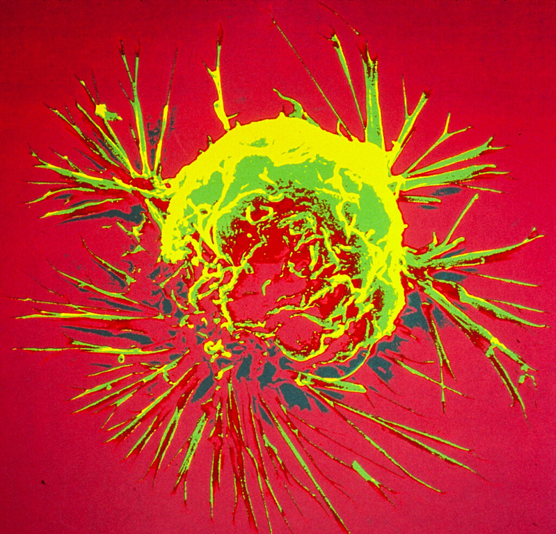
Colourised scanning electron micrograph of a breast cancer cell. Credit: NIH

Metastatic cancer cells, which cause 90% of cancer-related deaths, must overcome numerous hurdles to spread from a primary tumour through the bloodstream. Now, a new study led by investigators from the Mass General Cancer Center has identified a gene whose expression confers a growth advantage to these cells.

Mechanistically, the gene’s expression allows metastatic cancer cells to cause changes to their surrounding environment so that they can grow in new locations in the body. The findings are published in Nature Cell Biology.

“Our results point to potentially novel therapeutic avenues to specifically target metastatic cancer,” said senior author Raul Mostoslavsky, MD, PhD, who is the scientific director of the Krantz Family Center for Cancer Research at the Mass General Cancer Center.

Mostoslavsky and colleagues first compared gene expression patterns in primary versus metastatic tumours in mice with pancreatic cancer or breast cancer. After identifying various genes whose expression increased in metastatic tumour cells, the researchers silenced each gene individually.

In these experiments, silencing the Gstt1 gene had no effect on primary tumour cells from mice, but it stripped metastatic cancer cells of their ability to grow and spread. It also blocked cell growth in two metastatic-derived human pancreatic cancer cell lines.

Gstt1 encodes an enzyme that is a member of a superfamily of proteins involved with protecting cells from toxins, among other functions. Mechanistic studies indicated that the Gstt1 enzyme causes metastatic cancer cells to modify and secrete a protein called fibronectin, which is important for helping cells to attach themselves to the extracellular matrix, a large network of proteins and other molecules that surround, support, and give structure to cells and tissues in the body.

“Gstt1 alters the matrix surrounding the metastatic cells so they can grow in these foreign niches,” said Mostoslavsky. “Our results could lead to new strategies for the treatment of metastatic disease. This would be especially impactful for pancreatic cancer, in which most patients present with metastases when initially diagnosed.”

Source: Massachusetts General Hospital

New Brain Surgery Approach Targets Difficult Tumours at Skull Base

Source: CC0

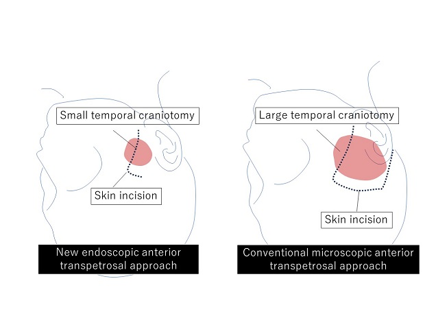
Tumours arising in the base of the skull are among the most difficult to remove in neurosurgery. The current treatment method is to perform surgical removal by what is known as the microscopic anterior transpetrosal approach (ATPA). Seeking to lessen the risk of damage and postoperative complications, as the skull base is densely packed with nerves, blood vessels, and other tissues, not to mention the brain stem, an Osaka Metropolitan University medical research team is taking a new approach.

Led by Dr Hiroki Morisako, a lecturer in the Graduate School of Medicine’s Department of Neurosurgery, and its department head Professor Takeo Goto, the team has developed a minimally invasive surgical technique called a purely endoscopic subtemporal keyhole ATPA. The team members write in The Journal of Neurosurgery that this is, to their knowledge, the first time this procedure to remove lesions in the skull base region known as the petrous apex has been described in an article.

Diagram of skin incision and extent of craniotomy. New endoscopic neurosurgery approach does not require a large craniotomy, so the result is a smaller scar. Credit: Osaka Metropolitan University

The endoscopic technique means a smaller area of the skull needs to be surgically opened compared to the microscopic approach, an average of only 11.2 cm² versus 33.9 cm². The risk of damage to the brain is also reduced.

The team performed 10 neurosurgeries using their method from 2022 to 2023 at Osaka Metropolitan University Hospital and compared the results to 13 surgeries using the microscopic ATPA from 2014 to 2021. In terms of operative time, the endoscopic approach reduced it noticeably, from an average of 410.9 minutes to 252.9 minutes. Similarly, blood loss lessened from a mean of 193 ml to 90 ml. The degree of tumour resection (surgical removal) was just as high as the microscopic method, while neurological functions were preserved at a rate equal to or higher than with the conventional approach.

“Comparison of the new endoscopic method and the conventional microscopic method showed no significant difference in tumour resection rate or in the ability to perform daily activities before and after surgery, with the new endoscopic approach resulting in shorter operative times and less blood loss,” Professor Goto stated. “The widespread use of this surgical procedure is expected to improve the treatment results of brain tumours in the base of the skull, not only in Japan but also worldwide.”

Source: Osaka Metropolitan University

New Clue in Cancer’s Obesity Paradox could Yield Immunotherapy Gains

Source: Pixabay CC0

Obesity is a well-known cancer risk – but is also known to increase the likelihood of immunotherapy success. A new discovery from Vanderbilt University Medical Center-led research team may explain why – and how this could lead to improved treatment for non-obese patients as well. The findings, published in Nature, reveal that obesity increases the frequency of macrophages in tumours and induces their expression of the immune checkpoint protein PD-1 – a target of cancer immunotherapies. 

“Obesity is the second leading modifiable risk factor for cancer, behind only smoking, and obese individuals have a greater risk for worse outcomes. But they also can respond better to immunotherapy,” said Jeffrey Rathmell, PhD, Cornelius Vanderbilt Professor of Immunobiology and director of the Vanderbilt Center for Immunobiology. “How is it that there can be this worse outcome on one hand, but better outcome on another? That’s an interesting question.” 

Postdoctoral fellow Jackie Bader, PhD, led the studies to examine the influence of obesity on cancer and to explore this “obesity paradox” – that obesity can contribute to cancer progression but also improve response to immunotherapy. 

In a mouse model, the researchers found striking differences between the macrophages isolated from tumours in obese versus lean mice. While the protein PD-1 is an immunotherapy target normally thought to act on T cells, they discovered that the macrophages in tumours from obese mice expressed higher levels of PD-1, and that PD-1 acted directly on the macrophages to suppress their function. 

In tumour samples from patients with kidney cancer, the researchers also found PD-1-expressing macrophages, and in human endometrial tumour biopsies from patients before and after 10% weight loss, they showed that PD-1 expression on tumour-associated macrophages decreased following weight loss. 

“We were very fortunate to have collaborators that provided us with samples from the same patients before and after weight loss that reinforced the findings from our mouse models,” Bader said. 

Blocking PD-1 with an immunotherapy drug in the mouse models increased tumour-associated macrophage activity, including their ability to stimulate T cells. 

‘Team macrophage’

Cancer immunotherapy studies have largely focused on T cells, because they are the immune cells that can kill cancer cells, Bader and Rathmell said. But macrophages play important roles in influencing what T cells do. 

“I’ve always been ‘team macrophage,’” Bader said. “Macrophages are thought of as being like a garbage truck: They clean up the mess. But they have a huge spectrum of activity to enhance the immune response, and they’re more plastic and manipulatable than other immune cells, which makes them really interesting.” 

The presence of more macrophages expressing PD-1 in tumours in an obese setting provides a mechanistic explanation for the obesity paradox, Bader and Rathmell said. Increased PD-1 expression suppresses immune surveillance by macrophages, subsequently suppressing the killer T cells and allowing tumours to grow (the increased cancer risk of obesity). PD-1 blockade with immunotherapy allows the increased number of PD-1-expressing macrophages to act (the enhanced response to immunotherapy). 

Currently, immune checkpoint inhibitors work in only 20–30% of patients.  

“We clearly want to find ways to make immunotherapies work better, and in the obese setting, they naturally work better,” Rathmell said. “Understanding how these processes are working biologically may give us clues about how to improve immunotherapy in general.” 

The findings also suggest that examining levels of PD-1-expressing tumour macrophages may help identify patients who will respond better to immunotherapy. 

“It could be that the greater the proportion of PD-1-expressing macrophages a tumour has, the better the response to immunotherapy will be,” Rathmell said. 

Source: Vanderbilt University

Chemo Drug may Cause Significant Hearing Loss in Longtime Cancer Survivors   

Photo by Brett Sayles

An interdisciplinary study led by researchers at the University of South Florida and Indiana University has uncovered significant findings on the long-term effects of one of the most common forms of chemotherapy on cancer survivors.

Published in JAMA Oncology, the study tracked a cohort of testicular cancer survivors who received cisplatin-based chemotherapy. The team followed the patients for an average of 14 years, revealing that 78% experience significant difficulties in everyday listening situations, negatively impacting their quality of life. This collaborative research is the first to measure real-world listening challenges and hearing loss progression in cancer survivors over a long period of time.

“It’s important that we understand the real-world effects of patients’ sensory problems and if we can understand that, then we can develop better therapeutic strategies and preventive measures to improve the long-term quality of life for cancer survivors,” said Robert Frisina, distinguished university professor and chair of the USF Department of Medical Engineering.

Cisplatin is commonly used in chemotherapy treatments for a variety of cancers, including bladder, lung, neck and testicular. It is administered intravenously and affects various parts of the body. However, the ears are particularly vulnerable as they have little ability to filter out the drug, causing it to become trapped. This leads to inflammation and the destruction of sensory cells that are critical for coding sound, causing permanent hearing loss that can progressively get worse well after cisplatin treatments are completed.

Lead author Victoria Sanchez, associate professor in the USF Health Department of Otolaryngology Head & Neck Surgery, said that despite the known risks, there’s a nationwide lack of routine hearing assessments for patients undergoing chemotherapy. “Most patients still do not get their hearing tested prior to, during or after chemotherapy. Our study highlights the need for regular auditory evaluations to manage and mitigate long-term hearing damage.”

The research team found higher doses of cisplatin led to more severe and progressing hearing loss, especially in patients with risk factors, such as high blood pressure and poor cardiovascular health. They also experienced increased difficulty hearing in common environments, such as a loud restaurant.

“It will be critically important to follow these patients for life. Their current median age is only 48 years, and eventually they will enter the years at which age-related hearing loss also begins to develop,” said Dr. Lois B. Travis, Lawrence H. Einhorn Professor of Cancer Research at Indiana University School of Medicine and a researcher at the IU Melvin and Bren Simon Comprehensive Cancer Center. This research is part of The Platinum Study, an ongoing research effort led by Dr. Travis and funded by the National Cancer Institute to study cisplatin-treated testicular cancer survivors.

The hope is that this study will inspire further investigation into alternative chemotherapeutic protocols and preventive measures, such as FDA-approved drugs to prevent or reduce hearing loss.

“This research gives oncologists the information they need to explore alternative treatment plans that could reduce the long-term side effects, such as altering the dosages and timing of the cisplatin in the treatment, when that could be an appropriate option,” Frisina said.

Innovative solutions, such as Pedmark, a new FDA-approved injection that mitigates cisplatin-induced hearing loss in children, represent promising steps forward, according to Frisina.

“We want to protect our hearing or treat a hearing loss if hearing damage occurs,” Sanchez said. “Hearing allows us to connect to the world we love. Staying connected through conversations with family and friends, enjoyment of music and entertainment, staying safe and finding pleasure in our vibrant surroundings. Promoting optimal hearing for overall wellness is essential for healthy living.”

According to the American Cancer Society, in addition to cisplatin, other platinum chemotherapy drugs, such as carboplatin, cause damage to the cochlea in the inner ear and lead to hearing loss. The risk of damage is greater with higher doses of chemotherapy.

Source: University of San Francisco

Malignant Melanoma Resists Treatment by Subverting Immune Cells

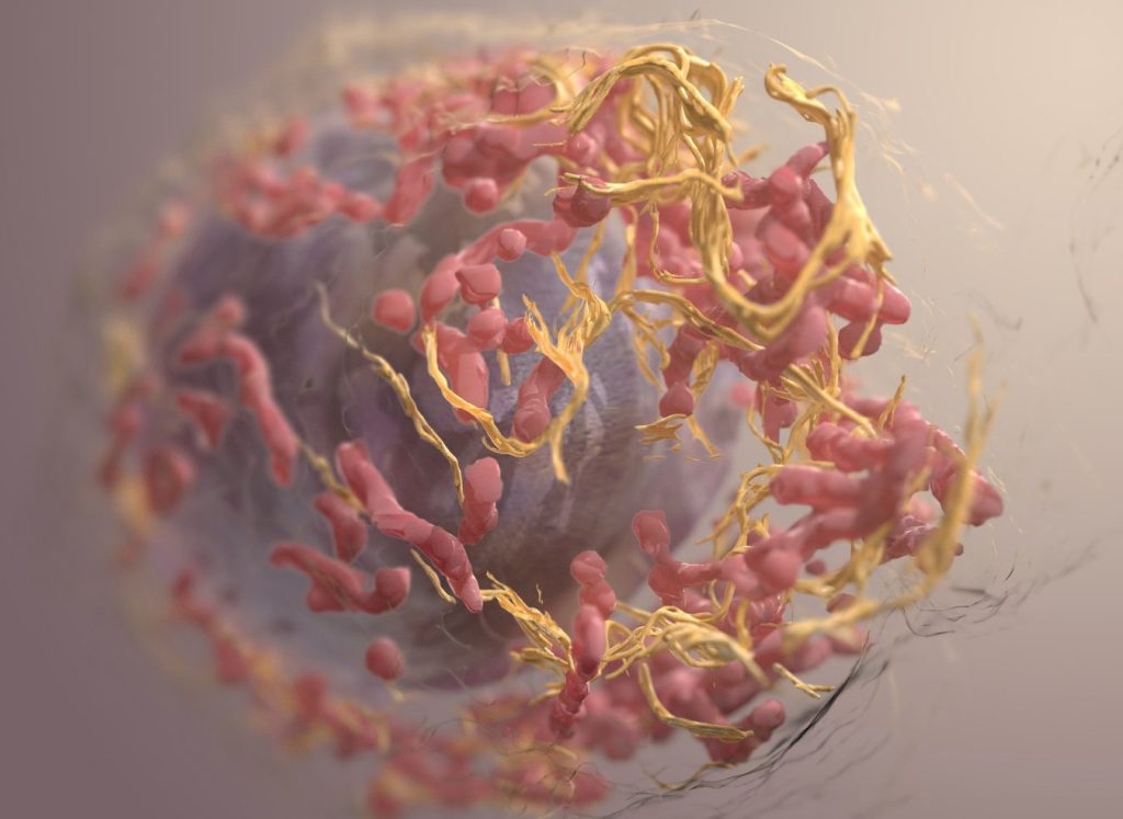
3D structure of a melanoma cell derived by ion abrasion scanning electron microscopy. Credit: Sriram Subramaniam/ National Cancer Institute

Malignant melanoma is one of the most aggressive types of cancer. Despite recent progress in effective therapies, the tumours of many patients are either resistant from the outset or become so during the course of treatment.

A University of Zurich (UZH) study published in Cell Reports Medicine has now identified a mechanism involving subverted immune cells that impedes the effectiveness of therapies. The result provides new ideas for treatments to suppress the development of resistance.

Comparing resistant and non-resistant tumour cells

For the study, the team utilised an innovative fine-needle biopsy to sample tumour cells before and during therapy. This allowed the researchers to analyse each cell individually. The patients providing the samples were undergoing targeted cancer therapy for malignant melanoma, which inhibits signalling pathways for tumour formation.

“It was important that some of the tumours responded to the therapy, while others showed resistance,” says study leader Lukas Sommer, professor of stem cell biology at the Institute of Anatomy at UZH. This allowed the team to compare the metabolism and environment of resistant and non-resistant tumour cells and look for significant differences.

Interaction between tumour factor and immune cells

One of the most relevant findings concerned the POSTN gene: it codes for a secreted factor that plays an important role in resistant tumours. In fact, the tumours of patients with rapidly progressing disease despite treatment showed increased POSTN levels. In addition, the microenvironment of these tumours contained a larger number of a certain type of macrophage – a subtype of immune cell that promotes the development of cancer.

Through a series of further experiments – both with human cancer cells and with mice – the research team was able to show how the interaction of increased POSTN levels and this type of macrophage triggers resistance: the POSTN factor binds to receptors on the surface of the macrophages and polarises them to protect melanoma cells from cell death. “This is why the targeted therapy no longer works,” says Sommer.

No resistance without cancer-promoting macrophages

The team considers this mechanism a promising starting point. “The study highlights the potential of targeting specific types of macrophages within the tumour microenvironment to overcome resistance,” says Sommer. “In combination with already known therapies, this could significantly improve the success of treatment for patients with malignant melanoma.”

Source: University of Zurich

Using AI, Scientists Discover High-risk Form of Endometrial Cancer

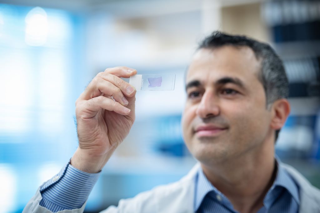
Dr Ali Bashashati observes an endometrial cancer sample on a microscope slide. Credit: University of British Columbia

A discovery by researchers at the University of British Columbia promises to improve care for patients with endometrial cancer, the most common gynaecologic malignancy.  Using artificial intelligence (AI) to spot patterns across thousands of cancer cell images, the researchers have pinpointed a distinct subset of more stubborn endometrial cancer that would otherwise go unrecognised by traditional pathology and molecular diagnostics.

The findings, published in Nature Communications, will help doctors identify patients with high-risk disease who could benefit from more comprehensive treatment.

“Endometrial cancer is a diverse disease, with some patients much more likely to see their cancer return than others,” said Dr Jessica McAlpine, professor at UBC. “It’s so important that patients with high-risk disease are identified so we can intervene and hopefully prevent recurrence. This AI-based approach will help ensure no patient misses an opportunity for potentially lifesaving interventions.”

AI-powered precision medicine

The discovery builds on work by Dr McAlpine and colleagues in the Gynaecologic Cancer Initiative, who in 2013 helped show that endometrial cancer can be classified into four subtypes based on the molecular characteristics of cancerous cells, with each posing a different level of risk to patients.

Dr McAlpine and team then went on to develop an innovative molecular diagnostic tool, called ProMiSE, that can accurately discern between the subtypes. The tool is now used across parts of Canada and internationally to guide treatment decisions.

Yet, challenges remain. The most prevalent molecular subtype, encompassing approximately 50% of all cases, is largely a catch-all category for endometrial cancers lacking discernible molecular features.

“There are patients in this very large category who have extremely good outcomes, and others whose cancer outcomes are highly unfavourable. But until now, we have lacked the tools to identify those at-risk so that we can offer them appropriate treatment,” said Dr McAlpine.

Dr McAlpine turned to long-time collaborator and machine learning expert Dr.Ali Bashashati, an assistant professor of biomedical engineering and pathology and laboratory medicine at UBC, to try and further segment the category using advanced AI methods.

Dr Bashashati and his team developed a deep learning AI model that analyses images of tissue samples collected from patients. The AI was trained to differentiate between different subtypes, and after analysing over 2300 cancer tissue images, pinpointed the new subgroup that exhibited markedly inferior survival rates.

“The power of AI is that it can objectively look at large sets of images and identify patterns that elude human pathologists,” said Dr Bashashati. “It’s finding the needle in the haystack. It tells us this group of cancers with these characteristics are the worst offenders and represent a higher risk for patients.”

Bringing the discovery to patients

The team is now exploring how the AI tool could be integrated into clinical practice alongside traditional molecular and pathology diagnostics.

“The two work hand-in-hand, with AI providing an additional layer on top of the testing we’re already doing,” said Dr McAlpine.

One benefit of the AI-based approach is that it’s cost-efficient and easy to deploy across geographies. The AI analyses images that are routinely gathered by pathologists and healthcare providers, even at smaller hospital sites in rural and remote communities, and shared when seeking second opinions on a diagnosis.

The combined use of molecular and AI-based analysis could allow many patients to remain in their home communities for less intensive surgery, while ensuring those who need treatment at a larger cancer centre can do so.  

“What is really compelling to us is the opportunity for greater equity and access,” said Dr Bashashati. “The AI doesn’t care if you’re in a large urban centre or rural community, it would just be available, so our hope is that this could really transform how we diagnose and treat endometrial cancer for patients everywhere.”

Source: University of British Columbia

Life Healthcare Concludes Agreement to Sub-License “RM2”

Photo by Khwanchai Phanthong on Pexels

Life Healthcare through its wholly owned subsidiary Life Molecular Imaging Limited (LMI), has entered into a contract with Lantheus Holdings Inc. (“Lantheus”), to sub-license one of LMI’s early-stage novel radiotherapeutic and radio diagnostic products (RM2).

“As part of Life Healthcare’s strategy to monetise LMI’s product development portfolio, we are delighted to have found a partner for our RM2 product”, said Pete Wharton-Hood, Life Healthcare, CEO.  “Through this agreement, LMI has secured a partnership for the development of this early-stage diagnostic and therapeutic product through to commercialisation. This exciting opportunity unlocks some of the value in LMI’”, continued Wharton-Hood.

Lantheus will make an upfront payment of $35 million for the sub-licensing rights to RM2, as per the agreement. In addition, several payments will potentially be paid to LMI on the achievement of development and regulatory milestones as well as royalty payments when the product is sold commercially.

The sub-licensing agreement secures Lantheus’ rights to develop the product and complete the early development in collaboration with LMI. “LMI is uniquely positioned to assist in this area, says Wharton -Hood and we are pleased by this development as it showcases and harnesses the specialised, dedicated and focused talent within LMI”. “With Lantheus’ experience in developing and providing access to radiotheranostics in cancer, we are confident in our decision to hand them the reins for this promising theranostic pair and are honored to work with them toward improving the future of people with prostate and breast cancer,” said Ludger Dinkelborg, CEO, Life Molecular Imaging.

Lantheus Holdings, Inc. is listed on NASDAQ in the United States of America and is the leading radiopharmaceutical-focused company committed to delivering life-changing science to enable clinicians to Find, Fight and Follow disease to deliver better patient outcomes. Lantheus has been providing radiopharmaceutical solutions for more than 65 years and has identified value and commercial opportunity in continuing the development of RM2.

LMI is a wholly owned subsidiary in Life Healthcare and is registered in the United Kingdom. The company has a product Neuraceq® which has been approved in many countries and is used to detect amyloid plaque in the brain through a PET-CT Scan and has multiple products in early clinical development. LMI also provides clinical research services for pharmaceutical companies.

Life Healthcare has retained R1bn to provide for funding requirements of LMI as part of the Alliance Medical Group disposal which was concluded earlier this year “This transaction will reduce the quantum required and Life Healthcare will consider distributing a portion of the surplus to shareholders as part of the full year dividend,” stated Wharton-Hood.

About RM2

RM2 is a 9 amino acid peptide that binds to Gastrin Releasing Peptide receptor (GRPr); and can be used to treat multiple malignant tumors like prostate, breast, lung, glioma, and ovarian tumors.

About Life Molecular Imaging

LMI is a wholly owned subsidiary in Life Healthcare and is registered in the United Kingdom. The company has one globally approved product Neuraceq ® that is used to detect amyloid plaque in the brain through a PET-CT scan and has multiple products in early clinical development as well as providing clinical research services for pharmaceutical companies.