Rates of multiple myeloma (MM), the second most common blood cancer in the United States, are increasing and are twice as high in men than in women. A new study published by Wiley online in CANCER, a peer-reviewed journal of the American Cancer Society, provides insights that may help to explain this disparity.
To investigate the sex difference in MM, researchers analyzed data on 850 patients with newly diagnosed MM enrolled in the Integrative Molecular And Genetic Epidemiology (IMAGE) study at the University of Alabama at Birmingham.
Compared with female patients, male patients were more likely to have advanced (International Staging System stage III) disease at the time of diagnosis. Males were also more likely to have high myeloma load—serum monoclonal protein (an abnormal protein produced by cancerous blood cells), more organ failure (especially kidney failure), and bone damage. Men were less likely than women to have low bone mineral density, and myeloma-defining features tended to differ between the two sexes. These differences were apparent even after taking numerous factors into account – including race, age, body mass index, education, income, smoking, and alcohol use.
Analyses suggested that certain chromosomal abnormalities that lead to initiation of myeloma occurring more often in younger males may help to explain some of the differences seen in this study.
“This research suggests that sex-specific mechanisms promote multiple myeloma pathogenesis, which may account for the excess risk seen in men,” said lead author Krystle L. Ong, PhD, of the O’Neal Comprehensive Cancer Center at the University of Alabama at Birmingham. “These findings may be used to improve risk stratification, diagnosis, and tailored treatments for both men and women with newly diagnosed multiple myeloma or related early precursor conditions.”
Cases of cardiovascular, leg/foot, kidney complications, and eye disease all higher in men Sex differences in complication rates persist regardless of disease duration
Photo by Photomix Company on Pexels
Men are at greater risk than women of the major health effects of diabetes (types 1 and 2), suggests a long term study published online in the Journal of Epidemiology & Community Health.
Rates of cardiovascular disease, leg, foot, and kidney complications, and the sight-threatening eye disease diabetic retinopathy are all higher in men, regardless of whether they had diabetes for more or less than 10 years, the findings show.
The global prevalence of diabetes is similar in men and women, and is projected to rise to 783 million by 2045, note the researchers.
But while cardiovascular disease is more common in men, overall, it’s not clear if this sex difference is apparent in the incidence of the complications associated with diabetes, say the researchers. Nor is it clear whether the length of time lived with diabetes might be influential, they add.
To explore this further, the researchers drew on survey responses from the 45 and Up Study, Australia, a large prospective study of 267 357 people over the age of 45 living in New South Wales (NSW).
These responses were linked to medical records for a total of 25 713 people, all of whom had either type 1 or type 2 diabetes, to monitor the development of any of the major health issues associated with diabetes
These include cardiovascular disease – ischaemic heart disease, mini stroke or TIA, stroke, heart failure, diabetic cardiomyopathy; eye problems – cataract, diabetic retinopathy; leg/foot problems – peripheral neuropathy (nerve damage), ulcers, cellulitis, osteomyelitis (bone inflammation), peripheral vascular disease (poor circulation), and minor or major amputation; and kidney problems – acute kidney failure, chronic kidney disease, chronic kidney failure, dialysis, and kidney transplant.
Almost half of the group were aged 60 to 74, and over half (57%; 14,697) were men, a higher proportion of whom were overweight (39% vs 29% of women) and had a history of heart disease.
Although a similar proportion of men and women were current smokers, a higher proportion of men were ex-smokers: 51% vs 29% of the women.
Of the 19 277 (75%) people with diabetes whose age was recorded at their diagnosis, 58% had been living with the disease for less than a decade and 42% had lived with it for 10 or more years.
Men had higher rates, and were at greater risk, of the complications associated with diabetes.
Over an average monitoring period of 10 years, and after factoring in age, 44% of the men experienced a cardiovascular disease complication while 57% had eye complications. Similarly, 25% of the men had leg/foot complications, and 35% kidney complications. The equivalent figures for women were, respectively, 31%, 61%, 18% and 25%.
Overall, men were 51% more likely to develop cardiovascular disease than women, 47% more likely to have leg and foot complications, and 55% more likely to have kidney complications.
Although there was little difference in the overall risk of eye complications between the sexes, men were at slightly higher risk (14%) of diabetic retinopathy.
While complication rates rose in tandem with the number of years lived with diabetes for both men and women, the sex difference in complication rates persisted.
By way of an explanation, the researchers point out that the men in the study were more likely to have well known risk factors. Men may also be less likely to make lifestyle changes, take preventive meds, or get health checks to lower their risks, they suggest.
This is an observational study, and as such, no firm conclusions can be drawn about causal factors, added to which people with a history of complications were excluded from the study. And information on potentially influential factors, such as diabetes medications, and glucose, blood fat, and blood pressure control wasn’t available.
But based on their findings, the researchers suggest: “For every 1000 people with diabetes, our findings suggest that an average of 37, 52, 21, and 32 people will develop cardiovascular disease, eye, lower limb, and kidney complications every year.”
While the risks of complications are lower in women with diabetes, they are still high, emphasise the researchers.
And they conclude: “Although men with diabetes are at greater risk of developing complications, in particular [cardiovascular disease], kidney and lower-limb complications, the rates of complications are high in both sexes.
“The similar sex difference for those with shorter compared with longer diabetes duration highlights the need for targeted complication screening and prevention strategies from the time of diabetes diagnosis.
“Further investigation into the underlying mechanisms for the observed sex differences in diabetes complications are needed to inform targeted interventions.”
Fitness amongst young adults varies widely from one country to another, and is strongly associated with both socioeconomic development and gender equality, a new study from Karolinska Institutet published in the Journal of Sport and Health Science reports. The results indicate that levels of development and gender equality in a society can affect differences in physical capacity and therefore public health in general.
Cardiorespiratory fitness (CRF) is an important factor of health and life-expectancy. For this present study, researchers systematically reviewed data from 95 studies in 24 countries involving a total of over 119 000 adult participants.
CRF is measured by what is known as the VO2peak, which is the highest oxygen uptake a body achieves during physical exertion.
The group, which included researchers from KI and Shanghai University of Sport, studied correlations between CRF, the Human Development Index (HDI) and the Gender Inequality Index (GII).
HDI is a measure of societal parameters like education, income and life-expectancy, while GII reflects differences between women and men in terms of health, education and labour.
Clear correlation in women
The results show that people in countries with a higher HDI were, on average, fitter, a correlation that was particularly salient amongst women, where young women in countries with a medium HDI had a higher VO2peak than women in countries with a low HDI (31.2 versus 28.5mL/kg/min). However, a further HDI increase from medium to high gave only small improvements.
“Our results suggest that societal structures impact greatly on people’s access to exercise and thus their fitness levels,” says the study’s lead author Nicolas Pillon, researcher at the Department of Physiology and Pharmacology, KI.
The study also shows that higher gender equality (a lower GII) correlates with a higher level of fitness in both women and men. Again, the difference was the most notable amongst young women, who in countries with high gender equality had on average a 6.5mL/kg/min higher VO2peak than their peers in countries with low gender equality.
“Our results underpin the importance of societal interventions and guidelines that reduce social and gender-related hindrances to physical exercise but point out that more research is needed from countries with a lower HDI, and on the obstacles facing different ethnic and socioeconomic communities,” says Barbara Ainsworth, researcher at Shanghai University of Sport and head of the study.
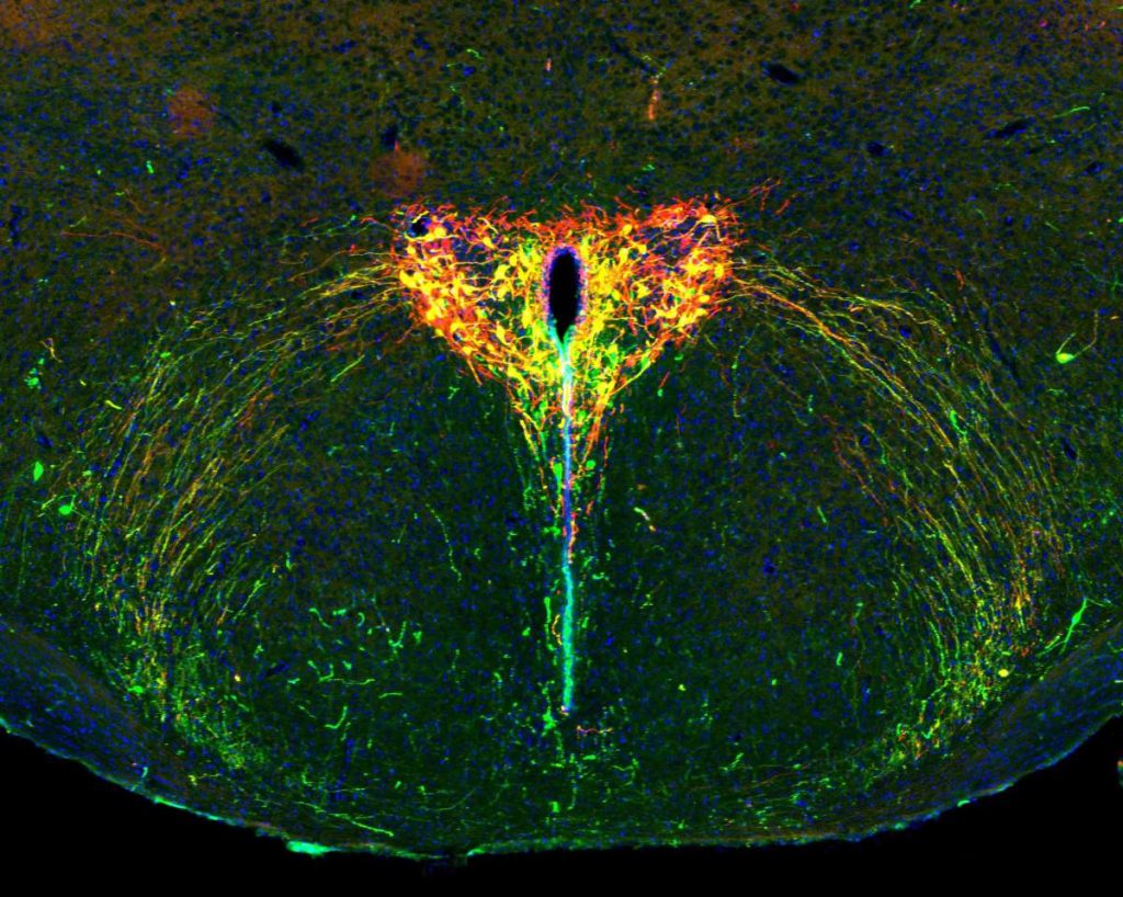
The developing brain of a two-week-old mouse pup under the microscope. The oxytocin system appears in green, the light-sensitive protein in red and cells that carry both show up in yellow. Cell nuclei are in blue. Credit: Weizmann Institute of Science
According to attachment theory, the attachment between an infant and a primary caregiver shapes the baby’s future social ties. Yet little is known about the biological mechanisms underlying childhood attachment, mainly because it is so difficult to study the young brain in natural conditions.
Now, scientists in Prof Ofer Yizhar’s laboratory at the Weizmann Institute of Science have developed a new, noninvasive research method that makes it possible to silence selected nerve cells deep within the brains of mouse pups without disrupting their natural behaviour. Using this method, the researchers investigated the role of oxytocin, a short protein released from nerve cells in the brain. While most oxytocin research has focused on adults, the new findings, published in Science, show that oxytocin also shapes the social behaviour of pups and may underlie emotional differences between males and females that emerge early in life.
Oxytocin, sometimes referred to as the “love hormone,” was once thought to simply promote sociability in adults. Over time, however, it became clear that its role is far more complex: In some circumstances, it intensifies behaviors and emotions far removed from love, such as anxiety or aggression. Recent research has also shown that young mammalian brains – including those of human children – are especially sensitive to oxytocin. In brain regions responsible for sensory processing, emotional regulation and social behavior, the number of oxytocin receptors peaks during early childhood: around ages two to three in humans, and two to three weeks in mice. Some studies have even linked oxytocin deficiency to childhood autism. Still, without sufficiently precise tools to examine neural activity deep within the developing brain, many aspects of the role of oxytocin in early life have remained a mystery.
“The findings may offer a clue as to why males and females diverge in their social behaviors and emotional worlds long before puberty”
To shed light on the subject, a team led by Dr Daniel Zelmanoff, a physician-scientist in Yizhar’s lab, developed a noninvasive technique to probe specific nerve cells in the young brain. The group, pioneers in the field of optogenetics – a technology that uses light to switch individual cells on or off – devised a method in which the targeted brain cells of mouse pups are infected with an engineered virus. This otherwise harmless virus introduces a foreign gene of mosquito origin that encodes a light-sensitive protein; when exposed to light, the protein “turns off” the nerve cell. In fact, the protein is so light-sensitive that the researchers could silence selected nerve cells deep inside the brain simply by shining red light on the pups’ heads.
“This new method allows us to peek inside the brain without disturbing the pups’ everyday lives, making it a powerful tool for studying nervous system development,” Yizhar explains. “It is especially useful for studying oxytocin because this hormone’s effects depend on social context – and our method lets us switch off the oxytocin system on demand, only during the exact situation we want to study.”
The researchers focused on oxytocin’s role during the temporary separation of a mouse pup from its mother and their reunion a few hours later – a situation familiar to every parent of a young child. The scientists observed increased oxytocin activity in the pup’s brain during separation, which returned to normal after reunion with the mother. Pups with an active oxytocin system during the separation gradually adapted to being alone in an unfamiliar environment, producing fewer ultrasonic vocalizations – the mouse equivalent of a baby’s cry. In contrast, pups whose oxytocin system was silenced did not adapt; they continued emitting distress calls at the same rate until reunited with their mothers. These findings show that the so-called “love hormone” also plays a critical role in coping with loneliness.
Attachment theory holds that children who are securely attached to their parents show distress when separated from them but are able to calm down over time, feeling free to explore their surroundings. “We discovered that mouse pups need an active oxytocin system in order to adapt to separation from their mothers,” says Yizhar. “This suggests that the oxytocin system plays a role not only in the brain of the parent, which was already known, but also in that of the infant. In addition, since oxytocin receptors are present in the sensory processing centers of the young brain, we hypothesize that this hormone also helps sharpen a pup’s senses when it is alone.”
Children do not quickly forget the experience of being separated from their parents, and this separation shapes how they behave when reunited. For example, a securely attached child separated from a parent for a few hours will seek contact upon reunion, and is quickly calmed. The researchers found that activation of the oxytocin system in mouse pups during separation not only strengthened them in the moment but also determined how they behaved when their mothers returned. These pups emitted more ultrasonic calls than usual, and the frequency of the calls grew as they got closer to their mothers. Using artificial intelligence, the team identified a distinct vocal pattern: Before attaching to the mother’s nipple, the pups made high-pitched, frequent calls; afterwards, their calls dropped in pitch and slowed in tempo.
“Activating the oxytocin system during separation increases the pup’s motivation to regain closeness to the mother when reunited,” Yizhar explains. “This is reflected in the heightened rate and unique pattern of their calls. We now understand that these ultrasonic vocalizations are much more than just crying: The high-pitched, rapid calls appear to signal a request for closeness, while the lower-pitched, slower-paced calls likely express a quick return to calm and a wish to remain attached. Of course, more research is needed to pin down the exact meaning of each vocalization type.”
In the next stage, the researchers explored whether oxytocin’s role in pups differs between the sexes, as it does in older animals. They found that female pups with an active oxytocin system emitted many more ultrasonic calls when reunited with their mothers than females with silenced oxytocin systems, whereas the calls of male pups were unaffected by the status of their oxytocin systems. “This is the first sex difference observed in oxytocin system activity at such an early stage of development,” Yizhar notes. “It may offer a clue as to why males and females diverge in their social behaviours and emotional worlds long before puberty.”
“Most known functions of oxytocin are shared by all mammals,” Yizhar concludes. “Still, future studies must check whether the hormone affects the development of social behaviour, emotional maturity and maternal attachment in the brains of children. If so, this could help us better understand what can go wrong in emotional and social development – as in autism spectrum disorder, for example – and how to intervene at an early stage.”
Could a keto diet affect males differently from females? A study from The University of Texas Health Science at San Antonio (UT Health San Antonio) suggests so, and oestrogen could promote different protections against adverse effects of the diet like the accumulation of cells expressing markers of age, or senescence.
The study, published Aug. 26 in the journal Cell Reports, found that male, but not female, mice on a ketogenic diet showed the accumulation of cells in organs expressing markers of cellular senescence. A keto diet is a popular low-carbohydrate, high-fat regimen that can help some Type 2 diabetes patients control blood sugar and those with epilepsy manage seizures. Cells expressing senescence markers can contribute to age-related declines in overall bodily function.
“These results suggest sex specificity alters the effects of a ketogenic diet, with important clinical implications,” said David Gius, MD, PhD, assistant dean of research and professor with the Department of Radiation Oncology at UT Health San Antonio, associate cancer director for translational research at the institution’s Mays Cancer Center and investigator for its Barshop Institute for Longevity and Aging Studies.
He is lead author of the study, titled, “Divergent sex-specific effects on a ketogenic diet: Male, but not female, mice exhibit oxidative stress and cellular senescence.”
Ketogenic diets induce ketogenesis, the generation of ketone bodies or water-soluble molecules from fat for use as fuel in place of glucose. They have shown benefits in controlling refractory epilepsy and are being investigated as potential therapies for other health conditions.
In the past decades, keto diets also have become popular in North America and Europe for weight loss.
While the diets can improve certain health parameters, evidence from mice and clinical studies suggest the effects may be dependent on multiple variables, including adherence, metabolism and, importantly, sex, suggesting that hormone status may impact response.
Gius says the role of gender in the response to keto diets has been understudied. One reason is that male mice have been used extensively for in vivo basic and translational research because it was assumed that females would give less consistent results due to variability from the oestrous cycle. Recent studies, however, suggest that largely is unfounded.
In the new study, Gius’ team observed a keto-diet-induced increase in cellular senescence only in male mice, except when they were given the female hormone oestrogen. Male mice on a keto diet also exhibited an increase in markers of oxidative stress, which is known to contribute to senescence in cells.
Notably, the researchers found, estrogen or estradiol treatment prevented increases in cell senescence and oxidated stress in male mice on a keto diet, as did several established antioxidants.
They also observed that when females were administered tamoxifen, a “selective oestrogen receptor inhibitor” that blocks the effects of oestrogen, they then exhibited an increase in oxidative stress and cells expressing senescence markers, the same as male mice. “These results strongly suggest that oestrogen is an important variable in the response to a ketogenic diet,” Gius said.
The researchers also found that a high-fat diet – comprising more carbohydrates than a keto diet – also induces cellular senescence in male, but not female, mice.
Genomic and hormone study of white Europeans finds 22 additional disease-related variants
Ball and stick 3D model of testosterone. Source: Wikimedia CC0
Researchers identified almost two dozen previously unknown genetic variants linked to type 2 diabetes by including participants’ hormone levels in their analysis. Yan V. Sun of Emory University, USA, and colleagues reports these findings in the open-access journal PLOS Genetics.
Type 2 diabetes affects an increasing number of people worldwide, and more often affects men than women. The disease is caused by a mix of genetic and lifestyle factors, but little is known about how someone’s environment – both inside and outside the body – interacts with their genes to impact a person’s risk of developing the disease.
In the new study, researchers performed genome-wide interaction studies to investigate whether a person’s hormone levels interact with their genetic variants to affect their risk of developing type 2 diabetes. They grouped males and females independently and considered measurements of three types of sex hormones – total testosterone, bioavailable testosterone and sex-hormone binding globulin. The information came from white European participants in the UK Biobank, which contains biological samples and health data from half a million people.
The researchers used statistical analyses to identify relevant variants in the genomes of individuals with and without type 2 diabetes. By taking into account hormone levels, the analysis was able to identify 22 spots on the genome that increased a person’s risk for type 2 diabetes. These variants had not been reported previously in the most recent genomic study for type 2 diabetes.
The new study suggests that a person’s hormone levels may be interacting with their genes to increase their odds of having type 2 diabetes. For future studies, the researchers recommended that additional hormone measurements for each participant and more diverse cohorts should be included. They conclude that this approach, which includes environmental factors in genomic studies, may help us to identify additional disease-related genes and gain a better understanding of the mechanisms behind complex diseases.
The authors add, “We found that sex hormone levels contribute to differences in genetic risk factors for type 2 diabetes in men and women. By analyzing data for men and women separately, we identified new genetic associations with type 2 diabetes.”
The lead analyst, Amonae Dabbs-Brown notes, “I actually used to work at the CDC developing methods to measure some of these sex hormones. It’s really exciting to see what happens downstream. Maybe one day I’ll even get to see how these analyses are used in the clinic!”
Provided by PLOS
In your coverage, please use this URL to provide access to the freely available paper in PLOS Genetics: https://plos.io/3ViXDKH
Citation: Dabbs-Brown A, Liu C, Hui Q, Wilson PW, Zhou JJ, Gwinn M, et al. (2025) Identification of gene-sex hormone interactions associated with type 2 diabetes among men and women. PLoS Genet 21(9): e1011470. https://doi.org/10.1371/journal.pgen.1011470
Author countries: United States
Funding: This work is supported in part by funding from the National Institutes of Health (HL154996 to YVS, DK139632 to YVS, and HL156991 to YVS). The funders had no role in study design, data collection and analysis, decision to publish, or preparation of the manuscript. YVS received salary support from the National Institutes of Health.
Competing interests: The authors have declared that no competing interests exist.
Schizophrenia and bipolar disorder are serious mental illnesses that affect both males and females, but research in Acta Psychiatrica Scandinavica indicates that sex may influence the characteristics and course of these conditions.
The research included 1516 individuals from the multicentre PsyCourse Study: 543 with bipolar disorder, 517 with schizophrenia, and 456 healthy controls.
Several differences between groups and sexes were identified in age at diagnosis, age at treatment, illness duration, illicit drug use, and smoking. For example, females in the schizophrenia group were older than males at first outpatient treatment compared with females in the bipolar disorder group. Moreover, those who were older at first outpatient treatment presented a longer duration of illness. Regarding substance use, the highest rates were observed in males with schizophrenia. People with bipolar disorder showed better functioning and neurocognitive performance than those with schizophrenia. Among individuals with bipolar disorder, females reported better performance in verbal memory and psychomotor speed than males. Both females and males with serious mental illnesses showed higher rates of thyroid alterations than healthy controls.
“Our findings reveal a clear message: sex-sensitive treatment is essential for improving clinical outcomes, promoting healthy habits, and managing comorbidities,” said corresponding author Anabel Martinez-Arán, PhD, of the Hospital Clinic of Barcelona.
Cardiologists have long known that up to half of all heart attacks and strokes occur among apparently healthy individuals who do not smoke and do not have high blood pressure, high cholesterol, or diabetes, the “standard modifiable risk factors” which doctors often call “SMuRFs.” How to identify risk among the “SMuRF-Less” has been an elusive goal in preventive cardiology, particularly in women who are often under-diagnosed and under-treated.
A new study by Mass General Brigham researchers that leverages data from the Women’s Health Study has found hsCRP, a marker of inflammation, can help identify women who are at risk but are missed by current screening algorithms. Results are presented at a late-breaking clinical science session at the European Society of Cardiology Congress (ESC) and simultaneously published in The European Heart Journal.
“Women who suffer from heart attacks and strokes yet have no standard modifiable risk factors are not identified by the risk equations doctors use in daily practice,” said Paul Ridker, MD, MPH, a preventive cardiologist at Mass General Brigham’s Heart and Vascular Institute. “Yet our data clearly show that apparently healthy women who are inflamed are at substantial lifetime risk. We should be identifying these women in their 40s, at a time when they can initiate preventive care, not wait for the disease to establish itself in their 70s when it is often too late to make a real difference.”
As part of the federally funded study, researchers studied 12 530 initially healthy women with no standard modifiable risk factors who had the inflammatory biomarker hsCRP measured at study entry and who were then followed over 30 years. Despite the lack of traditional risks, women who were inflamed as defined by hsCRP levels > 3 mg/L had a 77% increased lifetime risk of coronary heart disease, a 39% increased lifetime risk of stroke, and a 52% increased lifetime risk of any major cardiovascular event.
Additionally, researchers released a new analysis of randomised trial data showing that “SMuRF-Less but Inflamed” patients can reduce their risk of heart attack and stroke by 38% using statin therapy.
“While those with inflammation should aggressively initiate lifestyle and behavioural preventive efforts, statin therapy could also play an important role in helping reduce risk among these individuals,” said Ridker.
Analysis of lipid blood levels in women with Alzheimer’s disease has shown noticeable loss of unsaturated fats, such as those that contain omega fatty acids, compared to healthy women.
In men with Alzheimer’s, no significant difference was found in the same lipid molecule composition disease compared to healthy men, which suggests that those lipids have a different role in the disease according to sex. Fats perform important roles in maintaining a healthy brain, so this study could indicate why more women are diagnosed with the disease.
The study, published today in Alzheimer’s & Dementia: The Journal of the Alzheimer’s Association by scientists from King’s College London and Queen Mary University London, is the first to reveal the important role lipids could have in the risk for Alzheimer’s between the sexes.
Women are disproportionately impacted by Alzheimer’s Disease and are more often diagnosed with the disease than men after the age of 80. One of the most surprising things we saw when looking at the different sexes was that there was no difference in these lipids in healthy and cognitively impaired men, but for women this picture was completely different. The study reveals that Alzheimer’s lipid biology is different between the sexes, opening new avenues for research.
Dr Cristina Legido-Quigley, Reader in Systems Medicine
The scientists took plasma samples from 841 participants who had Alzheimer’s Disease, mild cognitive impairment and cognitively health controls and and were measured for brain inflammation and damage.
They used mass spectrometry to analyse the 700 individual lipids in the blood. Lipids are a group of many molecules. Saturated lipids are generally considered as ‘unhealthy’ or ‘bad’ lipids, while unsaturated lipid, which sometime contains omega fatty acids, are generally considered ‘healthy’.
Scientists saw a steep increase in lipids with saturation – the ‘unhealthy lipids’ – in women with Alzheimer’s compared to the healthy group. The lipids with attached omega fatty acids were the most decreased in the Alzheimer’s group.
Now, the scientists say there is a statistical indication that there is a causal link between Alzheimer’s Disease and fatty acids. But a clinical trial is necessary to confirm the link.
Dr Legido-Quigley added: “Our study suggests that women should make sure they are getting omega fatty acids in their diet – through fatty fish or via supplements. However, we need clinical trials to determine if shifting the lipid composition can influence the biological trajectory of Alzheimer’s Disease.”
Dr Asger Wretlind, first author of the study from the School of Cancer & Pharmaceutical Sciences, said: “Scientists have known for some time that more women than men are diagnosed with Alzheimer’s disease.
Although this still warrants further research, we were able to detect biological differences in lipids between the sexes in a large cohort, and show the importance of lipids containing omegas in the blood, which has not been done before. The results are very striking and now we are looking at how early in life this change occurs in women.
Dr Asger Wretlind, School of Cancer & Pharmaceutical Sciences
Many diseases affect men and women differently. Asthma tends to strike men earlier in life, yet more women develop asthma as they get older. Parkinson’s is more common in men, but Alzheimer’s is more common in women.
The differences are even more stark when it comes to autoimmune disease. Women are around two and a half times more likely than men to develop multiple sclerosis and nine times more likely to develop lupus.
Why would some diseases strike one sex more than another? And why do some tissues, such as the lungs and brain, seem especially vulnerable to these sex-based differences?
To answer these questions, scientists at La Jolla Institute for Immunology (LJI) are leading new research into how our immune cells defend specific parts of the body.
“In just the last two years, LJI scientists have uncovered a whole new body of information about how the immune systems of men and women are very different,” says Saphire. “We’re looking at what is genetically encoded in our XX or XY chromosomes, and how hormones like oestrogen and testosterone affect what is genetically programmed into our immune cells.”
In the paper, the researchers define biological sex (in an immunology context) as the presence of XX chromosomes in females and XY chromosomes in males. “Every cell in your body is either XX or XY,” says Saphire. “That X chromosome has many, many immune-related genes. Women have two copies of each. That gives them, in a sense, twice the palette of colours to paint from in formulating an immune response. It can also give them a stronger immune response for those genes that are doubly active – active in both copies simultaneously.
Sex hormones are important for much more than reproductive function. Immune cells can also sense hormones such as oestrogen and testosterone and use them to determine which genes to turn on or off and which ones to turn on more brightly or dim. This means similar immune cells can do different things, depending on whether that cell is from a male or a female.
Further, female cells vary in which of their two copies of X is “turned on.” As a result, women have organs with a collage, or mosaic, of immune cells that work differently in different tissues. This innate “variety” of immune cells appears to be an effective way to ward off infectious disease (women are better than men at fighting off pathogens such as SARS-CoV-2).
But scientists have also found that having more genes from X chromosomes may predispose women to autoimmune disease. This increased X chromosome “dosage” is closely linked to a higher risk for autoimmune diseases such as Sjögren’s syndrome and scleroderma.
New research into sex-based immune system differences is also critical for developing new cancer immunotherapies, Sharma explains.
“We’re increasingly understanding how sex-based differences affect disease outcomes. When it comes to medicine, one size doesn’t fit everybody,” says Sharma, who directs LJI’s Center for Sex-Based Differences in the Immune System. “This is leading to new research, particularly in the cancer field, toward precision medicine. We’re asking how a person’s individual immune system is contributing to controlling that cancer through immunotherapy.”
Saphire and Sharma also highlight environmental factors, such as nutrition and chemical exposures, that may add to the complex interplay of chromosomes and sex hormones. Men and women also appear to have some signature differences in their skin and gut microbiomes.
The researchers hope these foundational discoveries can lead to medical advances for all, and they’re working with collaborators across the country to move this research forward. “It takes a team to translate these findings,” says Sharma.