A Korean population-based cohort study investigated the risk of Alzheimer’s disease (AD) among breast cancer survivors compared to age-matched controls without cancer. The study, published in JAMA Network Open, found that breast cancer survivors had an 8% lower risk of AD than controls, with a significant association in survivors over 65 years old – though the effect did not persist past five years. Radiotherapy was associated with a lower risk of AD among breast cancer survivors – but not other treatments.
Breast cancer survivors may experience long-term health consequences, including cognitive function and risk of dementia. The risk of AD among breast cancer survivors is still unclear and may vary depending on age at diagnosis, treatment received, and time since treatment.
Previous studies reported mixed results on the risk of AD among breast cancer survivors, with some finding no increase in risk and others finding a 35% increased risk for those diagnosed at age 65 or older. These studies have been hampered by a number of methodological issues, including not accounting for risk factors.
Cytotoxic chemotherapy can cause cognitive decline termed ‘chemobrain’. Other chemotherapy drugs such as anthracycline may reduced AD risk by reducing the formation of amyloid deposits. Endocrine therapy may increase the risk of dementia by lowering oestrogen, but studies suggest that the use of tamoxifen and aromatase inhibitors is associated with a lower risk of AD. An increase in dementia is seen in radiotherapy for head and neck cancers.
To investigate the risk of AD among breast cancer survivors, researchers used the Korean National Health Insurance Service (K-NHIS) database, exploring whether there is an association with cancer treatment and various confounding factors.
Among 70 701 breast cancer survivors (mean age, 53.1 years), 1229 cases of AD were detected, with an incidence rate of 2.45 per 1000 person-years. Survivors exhibited a slightly lower risk of AD compared with cancer-free controls, especially among individuals 65 years or older (SHR, 0.92; 95% CI, 0.85-0.99). But landmark analyses found that this lower risk did not persist beyond five years of survival. Radiotherapy was associated with reduced risk of AD among survivors, while chemotherapy and endocrine therapy had no significant impact. Anthracycline use, however, did show a non-significant decrease in risk.
Differences in doses and timing of radiotherapy may influence the effects. The incident exposure to the brain is estimated to be 0.2Gy from a breast cancer radiotherapy dose of 50Gy. A pilot study found that patients with AD who received low-dose whole-brain radiotherapy at 3Gy showed a temporary improvement in cognitive function. This improvement is believed to be due to a neuroprotective effect on microglia. Other studies have noted a transient risk reduction for AD in breast cancer radiotherapy; however, patients receiving radiotherapy usually do so in conjunction with breast-conserving surgery – those opting for this procedure are younger, with fewer comorbidities and smaller tumours.
The study suggests that cancer treatment may have benefits against AD development, but the risk of AD may differ depending on the duration of survival.
The findings indicate that breast cancer treatment may not directly lead to AD, and that managing modifiable risk factors for AD, such as smoking and diabetes, is a feasible option to lower AD risk among breast cancer survivors.
A diabetes medication that lowers brain fluid pressure has cut monthly migraine days by more than half, according to a new study presented at the European Academy of Neurology (EAN) Congress 2025
A diabetes medication that lowers brain fluid pressure has cut monthly migraine days by more than half, according to a new study presented at the European Academy of Neurology (EAN) Congress 2025.1
Researchers at the Headache Centre of the University of Naples “Federico II” gave the glucagon-like peptide-1 (GLP-1) receptor agonist liraglutide to 26 adults with obesity and chronic migraine (defined as ≥ 15 headache days per month). Patients reported an average of 11 fewer headache days per month, while disability scores on the Migraine Disability Assessment Test dropped by 35 points, indicating a clinically meaningful improvement in work, study, and social functioning.
GLP-1 agonists have gained recent widespread attention, reshaping treatment approaches for several diseases, including diabetes and cardiovascular disease.2 In the treatment of type 2 diabetes, liraglutide helps lower blood sugar levels and reduce body weight by suppressing appetite and reducing energy intake.3,4,5
Importantly, while participants’ body-mass index declined slightly (from 34.01 to 33.65), this change was not statistically significant. An analysis of covariance confirmed that BMI reduction had no effect on headache frequency, strengthening the hypothesis that pressure modulation, not weight loss, drives the benefit.
“Most patients felt better within the first two weeks and reported quality of life improved significantly”, said lead researcher Dr Simone Braca. “The benefit lasted for the full three-month observation period, even though weight loss was modest and statistically non-significant.”
Patients were screened to exclude papilledema (optic disc swelling resulting from increased intracranial pressure) and sixth nerve palsy, ruling out idiopathic intracranial hypertension (IIH) as a confounding factor. Growing evidence closely links subtle increases in intracranial pressure to migraine attacks.6 GLP-1-receptor agonists such as liraglutide reduce cerebrospinal fluid secretion and have already proved effective in treating IIH.7 Therefore, building on these observations, Dr Braca and colleagues hypothesised that exploiting the same mechanism of action might ultimately dampen cortical and trigeminal sensitisation that underlie migraine.
“We think that, by modulating cerebrospinal fluid pressure and reducing intracranial venous sinuses compression, these drugs produce a decrease in the release of calcitonin gene-related peptide (CGRP), a key migraine-promoting peptide”, Dr Braca explained. “That would pose intracranial pressure control as a brand-new, pharmacologically targetable pathway.”
Mild gastrointestinal side effects (mainly nausea and constipation) occurred in 38% of participants but did not lead to treatment discontinuation.
Following this exploratory 12-week pilot study, a randomised, double-blind trial with direct or indirect intracranial pressure measurement is now being planned by the same research team in Naples, led by professor Roberto De Simone. “We also want to determine whether other GLP-1 drugs can deliver the same relief, possibly with even fewer gastrointestinal side effects”, Dr Braca noted.
If confirmed, GLP-1-receptor agonists could offer a new treatment option for the estimated one in seven people worldwide who live with migraine,8 particularly those who do not respond to current preventives. Given liraglutide’s established use in type 2 diabetes and obesity, it may represent a promising case of drug repurposing in neurology.
References
Braca S., Russo C. et al.GLP-1R Agonists for the Treatment of Migraine: A Pilot Prospective Observational Study. Abstract A-25-13975. Presented at the 11th EAN Congress (Helsinki, Finland).
Zheng, Z., Zong, Y., Ma, Y. et al. Glucagon-like peptide-1 receptor: mechanisms and advances in therapy. Sig Transduct Target Ther9, 234 (2024).
Lin, C. H. et al. An evaluation of liraglutide including its efficacy and safety for the treatment of obesity. Expert Opin. Pharmacother.21, 275–285 (2020).
Moon, S. et al. Efficacy and safety of the new appetite suppressant, liraglutide: A meta-analysis of randomized controlled trials. Endocrinol. Metab. (Seoul.)36, 647–660 (2021).
Jacobsen, L. V., Flint, A., Olsen, A. K. & Ingwersen, S. H. Liraglutide in type 2 diabetes mellitus: clinical pharmacokinetics and pharmacodynamics. Clin. Pharmacokinet.55, 657–672 (2016).
De Simone R, Sansone M, Russo C, Miele A, Stornaiuolo A, Braca S. The putative role of trigemino-vascular system in brain perfusion homeostasis and the significance of the migraine attack. Neurol Sci. 2022 Sep;43(9):5665-5672. doi: 10.1007/s10072-022-06200-x. Epub 2022 Jul 8. PMID: 35802218; PMCID: PMC9385793.
Mitchell J.L., Lyons H.S., Walker J.K. et al. (2023). The effect of GLP-1RA exenatide on idiopathic intracranial hypertension: a randomised clinical trial. Brain. 146(5):1821-1830.
Steiner T.J., Stovner L.J., Jensen, R. et al. (2020). Migraine remains second among the world’s causes of disability. The Journal of Headache and Pain. 21:137.
Prevention and treatment campaigns are not adequately targeting the particular needs of the 50+ years age group.
Photo by Sergey Mikheev on Unsplash
Indeed, between 2000 and 2016, the number of adults aged 50 years and older living with HIV in sub-Saharan Africa doubled. At present, their HIV prevalence is exceeding that of younger adults.
By 2040, one-quarter of people living with HIV in Africa will be aged 50 years and older; tailored awareness and treatment campaigns are pressing.
Dr Luicer Olubayo, a researcher at the Sydney Brenner Institute for Molecular Bioscience (SBIMB) at Wits University and the first author of a study published in The Lancet Healthy Longevityjournal, which investigated HIV in older people in Kenya and South Africa, noted that perceptions on who acquires HIV are limited. “We often think of HIV as a disease of younger people. It doesn’t help that intervention campaigns are mainly targeted at the youth.”
Moreover, older adults are less likely to believe that they can get HIV. This misconception is pervasive and has consequences for reaching global targets to achieve UNAIDS’ 95-95-95 targets by 2030 (95% of people living with HIV know their status, 95% of people who know their status are on treatment, and 95% have a suppressed viral load).
“While HIV prevalence among individuals over 50 years of age is similar to or even exceeds that of younger adults, HIV surveys focus on younger individuals, leaving considerable gaps in understanding HIV prevalence, incidence and treatment outcomes in older populations,” says Associate Professor F. Xavier Gómez-Olivé, at the MRC/Wits-Agincourt Research Unit.
Stigma remains a barrier to treatment
The uptake of HIV testing among older adults is poor, which delays diagnosis and limits access to care. This is, indeed, one of the signifiers of the pervasiveness of stigma surrounding the disease.
“We know that there is significant social stigma related to HIV infection. This is why understanding HIV-related stigma in older adults remains crucial as a way to inform interventions to support older people’s mental health and overall well-being,” says Olubayo.
Interventions could focus on repeated testing, the use of pre-exposure prophylaxis (PrEP), and campaigns to increase awareness and reduce infections among the elderly.
“HIV can be managed alongside other chronic conditions, too, since HIV is managed as a long-term illness,” says Gómez-Olivé.
Non-communicable diseases, such as hypertension, diabetes, and obesity, have dramatically increased in sub-Saharan Africa, particularly among older people. HIV treatment and intervention can be included in the healthcare ecosystem of long-term illnesses.
Apart from stigma, a complex interplay of factors shapes HIV risk
The study shows that age, education, gender, and where people live all affect their risk of HIV. Even though more people now have access to HIV treatment, older adults—especially in rural areas—still face significant challenges in preventing HIV, such as low education levels and gender inequality.
Widowed women had the highest HIV rate (30.8%). This may be due to losing a partner to HIV, stigma, and a greater risk of unsafe behaviours like transactional sex and limited power to negotiate condom use. People without formal education and those with low income also had higher rates of HIV infection.
The benefit of longitudinal data to make decisions
An important added value of this study is the provision of longitudinal insights into the HIV epidemic among older adults in sub-Saharan Africa. “Our study is beneficial in that older populations are under-represented, and not much is known about them over time. What changes are occurring? We have to answer these kinds of questions. With longitudinal data, we can look at the effectiveness of antiretroviral therapy coverage in older people,” says Gómez-Olivé.
The study used data collected in urban Kenya and in urban and rural sites across South Africa during two data collection waves: 2013-2016 and 2019-2022.
Throughout a decade of research, the team has been gaining a deeper understanding of this ageing HIV epidemic. Numerous important insights about HIV in older populations have been achieved, and research gaps are being covered.
Data for the study were drawn from the Africa Wits-INDEPTH Partnership for Genomic Research (AWI-Gen) from adults aged 40 years and older. AWI-Gen is a multicentre, longitudinal cohort study conducted at six research centres in four sub-Saharan African countries (South Africa, Kenya, Burkina Faso, and Ghana) to investigate various health determinants.
Caption: Hearty chefs (from left) Heleen Meyer, Herman Lensing, Isabella Niehaus, Monché Muller, and Zola Nene blend DASH principles with bold flavours – proving heart-healthy meals can be both delicious and fun.
South African heart-health is in the global spotlight this month, with Pharma Dynamics celebrating two major honours at the 30th Gourmand World Cookbook Awards, that took place during the Cascais World Food Summit in Lisbon, Portugal from 18–22 June 2025.
Hearty – Pharma Dynamics’ recently launched digital heart-healthy recipe collection was awarded first place in the category: Best Free Recipe Resourcein the World and also secured second place in the Free Resources for Healthcare Professionals category.
As part of its milestone celebration, Gourmand International reviewed three decades of award-winning cookbooks to identify the “cream of the crop” from each country. Cooking from the Heart’s DASH Edition, which won the Gourmand Award in 2023 in the Professional Health and Nutrition category, was selected as one of 20 standout South African titles of the past 30 years, nominated for the Gourmand’s 30th Anniversary Showcase.
The Cascais World Food Summit and Gourmand Awards bring together more than 500 food professionals from over 80 countries, celebrating excellence in culinary publishing and innovation.
“It is an incredible honour to see both our current innovation, Hearty and our longstanding Cooking from the Heart project recognised on the global stage,” says Nicole Jennings, spokesperson for Pharma Dynamics.
“Hearty represents the next evolution of our mission – combining the creativity of South Africa’s top chefs with proven heart-healthy principles. At the same time, having Cooking from the Heart’s DASH Edition selected for the 30th Anniversary Showcase reaffirms the enduring value of our work in empowering South Africans to eat healthier.”
Hearty showcases the culinary talents of five South African chefs and food writers who’ve each won the Gourmand’s “Best in the World” award:
Heleen Meyer – Food consultant, stylist and co-creator of Pharma Dynamics’ Cooking from the Heart series; multiple Gourmand honouree.
Herman Lensing – Author of seven cookbooks and award-winning magazine editor; Best in the World Celebrity Chef at Gourmand 2023.
Isabella Niehaus – Chef, stylist and author of several celebrated cookbooks; winner in the Entertainment and Vegan categories at Gourmand.
Monché Muller – Executive Chef of international wine label Oddo Vins et Domaines; winner of Best International Book at Gourmand 2023.
Zola Nene – Celebrity chef and TV personality; multiple Gourmand World Cookbook Awards winner.
Developed in collaboration with the Heart and Stroke Foundation South Africa, Hearty builds on the principles of the DASH (Dietary Approaches to Stop Hypertension) eating plan, but brings an indulgent, gourmet twist designed to challenge the notion that heart-healthy eating is bland or restrictive.
“Hearty is about celebrating food and flavour, while supporting cardiovascular wellness,” adds Jennings. “It’s a joy to collaborate with such an extraordinary line-up of chefs – each bringing their unique flair and creativity to inspire South Africans to embrace heart-healthy eating in a fresh and exciting way.”
The platform complements Pharma Dynamics’ Cooking from the Heart series – now in its eighth edition, which offers practical, dietitian-approved recipes designed to help South Africans manage and prevent cardiovascular disease. According to South African and international guidelines for hypertension, cholesterol and diabetes, lifestyle factors, such as healthy eating and maintaining a healthy weight are essential for effectively treating and controlling these conditions.
As South Africa’s leading supplier of cardiovascular medicines, Pharma Dynamics has provided patients and healthcare professionals with its Cooking from the Heart (CFTH) series since 2012. All eight titles are endorsed by the Heart and Stroke Foundation SA.
Pharma Dynamics continues to champion the shift from treatment to prevention through initiatives that promote sustainable lifestyle change.
“To be recognised at this level is a powerful reminder that our work – from the kitchen to the clinic – truly makes a difference,” emphasises Jennings. “It inspires us to keep finding new ways to help South Africans eat well, live well and thrive.”
A medical team at Erasmus University Medical Center in the Netherlands uses the new imaging probe with a Quest camera to get a better view of cancerous tumors during non-brain cancer surgery. Photo courtesy of Erasmus University Medical Center
In a significant leap forward for successful cancer surgery, researchers at the University of Missouri and collaborators have developed a new imaging probe to help surgeons more accurately identify and remove aggressive tumours during operations.
The tool is expected to be a critical advancement in the fight against glioblastoma, one of the most difficult-to-treat brain cancers. In the future, it is intended to be expanded for image-guided surgery of various other solid tumours.
Described in a new study in Nature Publishing Group Imaging, the innovation works by pairing a fluorescent dye with a fatty acid molecule that cancer cells readily absorb. When introduced into the body, the compound is taken up by tumour cells, causing them to glow under near-infrared light, revealing cancer that might otherwise remain hidden.
Glioblastoma is considered surgically incurable because the tumour doesn’t stay in one place – it spreads and invades healthy brain tissue in a diffuse, microscopic way. This makes it impossible to remove completely without risking serious damage to brain function.
“Surgery remains one of the primary treatments for many cancers,” Elena Goun, associate professor of chemistry in the College of Arts and Science and one of the lead authors of the study, said. “In breast or prostate cancer, surgeons can often remove the tumour along with surrounding tissue. In brain cancer, that’s simply not possible. You must preserve healthy brain tissue. But if even a few cancer cells are left behind, the disease will return.”
That dilemma is especially acute with glioblastoma, which doesn’t form a neatly contained mass. Instead, it sends out microscopic extensions — finger-like projections that blend into healthy brain tissue and are invisible to the naked eye.
Because of this, surgeons must walk a fine line: removing as much tumour as possible while avoiding harm to vital brain areas. The more thoroughly the tumour is removed, the more effective follow-up treatments like radiation and chemotherapy tend to be.
The new small-molecule probe, known as FA-ICG, is engineered to solve that problem. It links a natural long-chain fatty acid (FA) to indocyanine green (ICG), an FDA-approved near-infrared dye widely used in surgical imaging. This fatty acid-based approach means the probe is highly selective: glioblastoma cells, which thrive on fatty acids, absorb it more than normal brain cells. That makes the cancer stand out more clearly.
The result is a tool that takes advantage of cancer’s altered metabolism to highlight tumour cells from within.
“Surgeons would view a monitor during surgery showing where the probe is lighting up,” Goun explained. “If they still see fluorescent signals, it means cancer is still present and more tissue needs to be removed. When the light disappears, they would know they’ve cleared the area.”
In the operating room, surgeons already use a variety of tools to guide tumour removal – including microscopes, ultrasound and fluorescent dyes. Of those, fluorescent dyes are particularly useful because they make otherwise invisible tumour cells light up under special lighting.
Right now, the only approved imaging dye for glioblastoma surgery is 5-ALA, which fluoresces under blue light. But 5-ALA comes with major limitations: The operating room must be darkened in order to see it, tissue penetration is shallow and the fluorescent signal is often weak and non-specific.
It also comes with side effects, including photosensitivity, meaning patients must avoid bright light exposure after surgery due to the risk of skin and eye damage.
That’s where the FA-ICG probe shines – both literally and functionally.
Compared to 5-ALA, FA-ICG is brighter, works under normal surgical lighting, and offers real-time visualisation under the microscope – no need to turn the lights off mid-surgery. This saves time and makes procedures more efficient. The signal-to-background ratio is also higher, meaning it’s easier to distinguish tumour tissue from healthy brain.
The FA-ICG probe is not only easier to see, it’s also easier to use. Its longer half-life allows more flexibility in scheduling surgeries, and the logistics of administration are simpler than with current probes.
“The upside of fluorescence-guided surgery is that you can make little remnants much more visible using the light emitting properties of these tumour cells when you give them a dye,” said Rutger Balvers, a neurosurgeon at Erasmus University Medical Center in the Netherlands, who is expected to lead human clinical trials of the probe. “And we think that the upside of FA-ICG compared to what we have now is that it’s more select in targeting tumour cells. The visual properties of the probe are better than what we’ve used before.”
Michael Chicoine is a neurosurgeon at MU Health Care and chair of Mizzou’s School of Medicine’s Department of Neurosurgery. While he’s not directly involved in the research, Chicoine understands the potential benefits firsthand.
Currently, he said, MRIs are the gold standard for imaging tumours; however, they’re expensive and time-consuming, especially when required during an operation.
“This fluorescent metabolically linked tool gives you real-time imaging,” he said. “We could merge techniques, using the probe during surgery and saving the MRI for a sort of final exam. It’s definitely an exciting advancement.”
Researchers are also excited about other uses for the probe, including for other types of cancers and for use during follow-up treatments.
“After radiation or chemotherapy, it becomes very difficult to distinguish between scar tissue and active tumor,” Chicoine said. “This probe could give us a definitive answer – helping doctors know whether to continue treatment or adjust it, or consider another surgery. Eliminating the current uncertainty would be really helpful.”
Another promising use of the probe could be in photodynamic therapy either during or after surgery. Since the dye also has light-activated properties that can kill cancer cells, researchers are exploring whether it could double as a treatment tool, not just a diagnostic one.
Clinical trials for use in glioblastoma cases are expected to start in Europe, with strong interest already growing among neurosurgical teams.
The upcoming Phase 1 trial will focus on how patients tolerate the probe, whether there are any side effects at an effective dose and how its performance compares to existing tools. Ultimately, the goal is to make brain tumour surgery safer, helping surgeons remove all cancerous tissues while preserving as much healthy brain tissue as possible.
If results are positive, future studies could expand the use of FA-ICG beyond brain tumours to other cancers with high fatty acid metabolism, such as pancreatic cancer,according to fellow corresponding author Laura Mezzanotte from the Erasmus’ Department of Radiology and Nuclear Medicine.
Mitochondrial pathways help melanoma cells become aggressive, and some currently available drugs target these pathways.
3D structure of a melanoma cell derived by ion abrasion scanning electron microscopy. Credit: Sriram Subramaniam/ National Cancer Institute
Researchers have discovered that the most aggressive melanomas, the deadliest form of skin cancer, overactivate two key processes in mitochondria. Blocking these pathways with currently available drugs effectively killed melanoma cells. The findings are published by Wiley online in CANCER, a peer-reviewed journal of the American Cancer Society.
By mapping the proteins expressed in 151 tumour and normal skin samples, investigators found that the most aggressive melanomas hyper-activate the machinery that builds mitochondrial proteins and the mitochondrial system that turns nutrients into energy.
Remarkably, blocking these pathways effectively halted or killed melanoma cells cultured in lab dishes. Two types of drugs accomplished this: antibiotics, originally designed to block bacterial protein synthesis machinery, which closely resembles the machinery found in mitochondria, and specialised energy-production inhibitors. Importantly, non-cancerous skin cells remained mostly unaffected, highlighting the safety and specificity of these treatment approaches.
“This discovery identifies melanoma’s excessive reliance on mitochondrial energy as its Achilles’ heel, revealing a therapeutic vulnerability that we can exploit with existing drugs,” said senior author Jeovanis Gil, PhD, of Lund University in Sweden. “By pairing mitochondrial blockers with today’s standards of care, we may cut off a major escape route that cancers use to resist therapy and come back.”
Dr Gil added that the mitochondrial-protein signature his team discovered can be measured in routine biopsy material and could serve as a biomarker to identify patients most likely to benefit from mitochondrial-targeted add-on therapies. By enabling clinicians to match treatments to each patient’s tumour biology, these findings mark a step forward for precision medicine in melanoma. Moreover, because mitochondrial rewiring fuels resistance across many cancers, success in melanoma could open the door to similar personalised combination strategies in other hard-to-treat cancers.
A sample of Aspergillus flavus cultured in the Gao Lab. (Credit: Bella Ciervo)
University of Pennsylvania-led researchers have turned a deadly fungus into a potent cancer-fighting compound. After isolating a new class of molecules from Aspergillus flavus, a toxic crop fungus linked to deaths in excavating ancient tombs, the researchers modified the chemicals and tested them against leukaemia cells. The result was a promising cancer-killing compound that rivals FDA-approved drugs and opens up new frontiers in the discovery of more fungal medicines.
“Fungi gave us penicillin,” says Sherry Gao, Presidential Penn Compact Associate Professor in Chemical and Biomolecular Engineering (CBE) and in Bioengineering (BE) and senior author of a new paper in Nature Chemical Biology on the findings. “These results show that many more medicines derived from natural products remain to be found.”
From Curse to Cure
A. flavus, named for its yellow spores, has long been a microbial villain. After archaeologists opened King Tutankhamun’s tomb in the 1920s, a series of untimely deaths among the excavation team fuelled rumours of a pharaoh’s curse. Decades later, doctors theorised that fungal spores, dormant for millennia, could have played a role.
In the 1970s, a dozen scientists entered the tomb of Casimir IV in Poland. Within weeks, 10 of them died. Later investigations revealed the tomb contained A. flavus, whose toxins can lead to lung infections, especially in people with compromised immune systems.
Now, that same fungus is the unlikely source of a promising new cancer therapy.
A Rare Fungal Find
The therapy in question is a class of ribosomally synthesised and post-translationally modified peptides, or RiPPs, pronounced like the “rip” in a piece of fabric. The name refers to how the compound is produced – by the ribosome, a tiny cellular structure that makes proteins – and the fact that it is modified later, in this case, to enhance its cancer-killing properties.
“Purifying these chemicals is difficult,” says Qiuyue Nie, a postdoctoral fellow in CBE and the paper’s first author. While thousands of RiPPs have been identified in bacteria, only a handful have been found in fungi. In part, this is because past researchers misidentified fungal RiPPs as non-ribosomal peptides and had little understanding of how fungi created the molecules. “The synthesis of these compounds is complicated,” adds Nie. “But that’s also what gives them this remarkable bioactivity.”
Hunting for Chemicals
To find more fungal RiPPs, the researchers first scanned a dozen strains of Aspergillus, which previous research suggested might contain more of the chemicals.
By comparing chemicals produced by these strains with known RiPP building blocks, the researchers identified A. flavus as a promising candidate for further study.
Genetic analysis pointed to a particular protein in A. flavus as a source of fungal RiPPs. When the researchers turned the genes that create that protein off, the chemical markers indicating the presence of RiPPs also disappeared.
This novel approach – combining metabolic and genetic information – not only pinpointed the source of fungal RiPPs in A. flavus, but could be used to find more fungal RiPPs in the future.
A Potent New Medicine
After purifying four different RiPPs, the researchers found the molecules shared a unique structure of interlocking rings. The researchers named these molecules, which have never been previously described, after the fungus in which they were found: asperigimycins.
Even with no modification, when mixed with human cancer cells, asperigimycins demonstrated medical potential: two of the four variants had potent effects against leukaemia cells.
Another variant, to which the researchers added a lipid found in bees’ royal jelly, performed as well as cytarabine and daunorubicin, two FDA-approved drugs that have been used for decades to treat leukaemia.
Cracking the Code of Cell Entry
To understand why lipids enhanced asperigimycins’ potency, the researchers selectively turned genes on and off in the leukaemia cells. One gene, SLC46A3, proved critical in allowing asperigimycins to enter leukaemia cells in sufficient numbers.
That gene helps materials exit lysosomes, the tiny sacs that collect foreign materials entering human cells. “This gene acts like a gateway,” says Nie. “It doesn’t just help asperigimycins get into cells, it may also enable other ‘cyclic peptides’ to do the same.”
Like asperigimycins, those chemicals have medicinal properties – nearly two dozen cyclic peptides have received clinical approval since 2000 to treat diseases as varied as cancer and lupus – but many of them need modification to enter cells in sufficient quantities.
“Knowing that lipids can affect how this gene transports chemicals into cells gives us another tool for drug development,” says Nie.
Disrupting Cell Division
Through further experimentation, the researchers found that asperigimycins likely disrupt the process of cell division. “Cancer cells divide uncontrollably,” says Gao. “These compounds block the formation of microtubules, which are essential for cell division.”
Notably, the compounds had little to no effect on breast, liver or lung cancer cells – or a range of bacteria and fungi – suggesting that asperigimycins’ disruptive effects are specific to certain types of cells, a critical feature for any future medication.
Future Directions
In addition to demonstrating the medical potential of asperigimycins, the researchers identified similar clusters of genes in other fungi, suggesting that more fungal RiPPS remain to be discovered. “Even though only a few have been found, almost all of them have strong bioactivity,” says Nie. “This is an unexplored region with tremendous potential.”
The next step is to test asperigimycins in animal models, with the hope of one day moving to human clinical trials. “Nature has given us this incredible pharmacy,” says Gao. “It’s up to us to uncover its secrets. As engineers, we’re excited to keep exploring, learning from nature and using that knowledge to design better solutions.”
Acute myocarditis, or sudden inflammation of the heart, causes mild symptoms in most cases, but about 10% of acute myocarditis cases can be sudden and severe, leading to cardiac arrhythmias, heart pump failure, or even death. Current therapies for the condition are built on limited data and may not effectively target the underlying disease mechanisms. Patients may even require mechanical circulatory support for life support when the heart is failing.
Now a team at UC San Francisco is using a new class of drugs that target inflammation to treat acute fulminant myocarditis patients.
In an article in Circulation, the UCSF group reports on the successful treatment of a patient with sudden and severe (acute fulminant) myocarditis using an immune modulating medication known to inhibit the activity of enzymes that can trigger inflammation in the body.
“Our group has developed several animal models of myocarditis and by studying these, we became interested in a group of enzymes called Janus kinases, or JAKs, that seem to serve as communication nodes between immune cells,” said Javid Moslehi, MD, William Grossman Distinguished Professor in Cardiology and UCSF Section Chief of Cardio-Oncology and Immunology. “JAKs become hyperactive during acute heart inflammation, exacerbating the already activated immune system.”
Moslehi and his team therefore reasoned that targeting these enzymes using a novel class of therapies called JAK inhibitors would be a possible treatment modality for acute fulminant myocarditis. In recent years, the team has examined the effects of JAK inhibitor treatment on the immune cell populations for both RNA and proteins, showing acute benefit in various laboratory models of myocarditis.
Heart function improved dramatically
In the current case, the group treated a 20-year-old woman who came to UCSF with acute fulminant myocarditis.
“This patient’s heart was effectively falling apart and there was no time to lose,” said Connor O’Brien, MD, cardiologist, critical care specialist and UCSF assistant professor of Medicine. “We had put the patient on extracorporeal membrane oxygenation (ECMO) to maintain blood flow through vital organs and started the process of listing her for heart transplantation.”
O’Brien was on clinical service at the time, caring for the patient. The medical team had tried using corticosteroids as a treatment but without success. O’Brien then coordinated with Moslehi, given his expertise in myocarditis as well as some of his laboratory results with JAK inhibitors.
O’Brien added ruxolitinib, a JAK inhibitor, to the treatment strategy. The patient’s cardiac arrhythmias slowed down and her cardiac enzyme (a measure of heart damage) decreased. Over the next few days, her heart function improved dramatically allowing her to wean off ECMO support. She was successfully discharged from the hospital one week later.
Since that time, the UCSF Health cardiology team has gone on to successfully treat other patients with JAK inhibitors for acute myocarditis, but Moslehi adds a note of caution.
“The gold standard of any new treatment is a clinical trial, and it is important to note that we have not yet done this,” said Moslehi. “But the team is hopeful that these early results can lead to an eventual trial for patients.”
Moslehi and his team established the UCSF Myocarditis Center in March 2023 as part of the newly formed section of cardio-oncology and immunology. This multi-disciplinary group is focused on diagnosis and treatment for myocarditis, bringing laboratory scientists together with clinicians for more comprehensive care of patients.
“The Myocarditis Center really takes advantage of the incredible scientific environment at UCSF and specifically our cardiology team working closely with the cardiovascular research institute (CVRI) to help our patients,” Moslehi adds.
In addition to cadmium, molybdenum and zinc found to have particularly high risk increases
Right side heart failure. Credit: Scientific Animations CC4.0
A new multi-cohort study at Columbia University Mailman School of Public Health, has found that exposure to certain metals, detected in urine, is associated with a higher risk of heart failure (HF). Published in the Journal of the American College of Cardiology, it is the largest investigation of its kind to date, reinforcing the importance of reducing environmental metal exposure to reduce heart failure risk. While environmental metals are recognised as cardiovascular disease risk factors, until now the role of metal exposure in heart failure risk had remained understudied.
“Most previous studies have assessed individual metals in isolation. By examining metals as a mixture, our analysis more closely reflects real-world exposure patterns,” said Irene Martinez-Morata, MD, PhD, postdoctoral research scientist in Environmental Health Sciences at Columbia Mailman School, and lead author. “In our analysis of over 10 000 adults across diverse geographic, racial, and ethnic backgrounds, we observed consistent associations between elevated urinary metal levels and increased HF risk over long-term follow-up after accounting for other established traditional risk factors for the disease such as diabetes and obesity.”
The study pooled data from three large cohorts with more than 20 years of follow-up:
MESA (Multi-Ethnic Study of Atherosclerosis), U.S. adults aged 18–85 from six urban-suburban areas in Maryland, Illinois, North Carolina, California, Minnesota and New York.
SHS (Strong Heart Study), American Indian adults aged 18–65 in the U.S. from Oklahoma, Arizona, North Dakota and South Dakota.
Hortega Study, a general population cohort in Spain
Among the 10 861 participants, a thousand people developed heart failure. In a subset, researchers assessed left ventricular function, which measures how effectively the heart pumps blood.
Metals were measured in urine samples, which can indicate how much metal is in the body and how much is being eliminated from it. Health and lifestyle data – including medication use, cholesterol levels, blood pressure, glucose, BMI, and more – were collected via questionnaires, lab tests, and physical exams. The team used advanced machine learning models to evaluate the combined effects of five urinary metals as a mixture.
Key findings included:
Higher levels for the mixture of five metals in urine: arsenic, cadmium, molybdenum, selenium, and zinc, was associated with a 55% higher risk of heart failure in rural American Indian adults (SHS), a 38% higher risk in urban and suburban diverse populations (MESA) and a 8% increased risk in adults in Spain (Hortega).
In the analysis of metals individually, a doubling in the levels of urine cadmium, a toxic metal found in tobacco products, foods and industrial waste, was associated with 15% higher risk of heart failure.
Similarly, a doubling in the levels of molybdenum and zinc was associated with 13% and 22% higher risk of heart failure across the three cohorts. These metals have an essential function in the body, but high levels can be toxic.
“The strongest association between the 5-metal mixture and HF risk was seen in the SHS cohort,” said Martinez-Morata. “This population faces a historically high burden of contaminant metal exposure and cardiovascular disease and public health action is urgently needed.”
The sources of exposure to these metals can vary from urban and rural environments. Toxic metals such as arsenic, cadmium, and tungsten can occur as a result of mining and industrial activity leading to contamination of drinking water, foods that grow in contaminated soils, and air pollution. Many of these metals are also present in smoking devices, consumer products, and certain foods, observes Martinez and her co-authors. “Essential metals such as zinc and selenium are needed for biological functions, but high levels can be toxic.”
“We consistently found higher urinary levels of cadmium, molybdenum and zinc linked to increased heart failure risk,” noted Ana Navas-Acien, MD, PhD, Columbia Mailman School professor and chair of the Department of Environmental Health Sciences. “Even after adjusting for diabetes – a known HF risk factor – the zinc association remained significant.”
These results support the relevance of metal exposures as contributors to heart failure risk. “In ongoing research, we aim to clarify biological mechanisms and to explore the role of environmental interventions in cardiovascular disease prevention,” said Navas-Acien, who also is senior author.
“This study’s strengths include its large, diverse sample size, high-quality data, and robust, long-term follow-up,” said Martinez-Morata. “Our findings underscore the importance of continuing efforts to monitor and reduce environmental metal exposures, particularly in communities with historically high exposure levels as an innovative approach to improve cardiovascular health.”
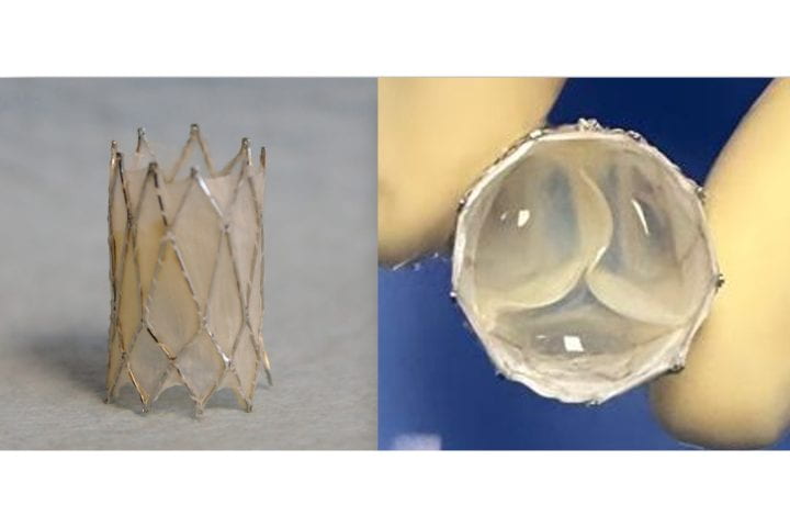
The Iris Valve, a transcatheter, growth-accommodating pulmonary valve designed for very young children, was developed at UC Irvine and is currently progressing toward FDA clinical approval. Arash Kheradvar
Researchers at the University of California, Irvine have successfully performed preclinical laboratory testing of a replacement heart valve intended for toddlers and young children with congenital cardiac defects, a key step toward obtaining approval for human use. The results of their study were published recently in the Journal of the American Heart Association.
The management of patients with congenital heart disease who require surgical pulmonary valve replacement typically occurs between the ages of 2 and 10. To be eligible for a minimally invasive transcatheter pulmonary valve procedure, patients currently must weigh at least 20.4kg. For children to receive minimally invasive treatment, they must be large enough so that their veins can accommodate the size of a crimped replacement valve. The Iris Valve designed and developed by the UC Irvine team can be implanted in children weighing as little as 7.7 to 10kg and gradually expanded to an adult diameter as they grow.
Research and development of the Iris Valve has been supported by the Eunice Kennedy Shriver National Institute of Child Health and Human Development; the National Heart, Lung, and Blood Institute; and the National Science Foundation.
This funding has enabled benchtop fracture testing, which demonstrated the valve’s ability to be crimped down to a 3mm diameter for transcatheter delivery and subsequently enlarged to 20mm without damage, as well as six-month animal studies that confirmed successful device integration within the pulmonary valve annulus, showing valve integrity and a favourable tissue response.
“We are pleased to see the Iris Valve performing as we expected in laboratory bench tests and as implants in Yucatan mini pigs, a crucial measure of the device’s feasibility,” said lead author Arash Kheradvar, UC Irvine professor of biomedical engineering. “This work represents the result of longstanding collaboration between our team at UC Irvine and Dr Michael Recto at Children’s Hospital of Orange County built over several years of joint research and development.”
Congenital heart defects affect about 1% of children born in the United States and Europe, with over 1 million cases in the US alone. These conditions often necessitate surgical interventions early in life, with additional procedures required to address a leaky pulmonary valve and prevent right ventricular failure as children grow.
The Iris Valve can be implanted via a minimally invasive catheter through the patient’s femoral vein. The Kheradvar group employed origami folding techniques to compress the device into a 12-French transcatheter system, reducing its diameter to no more than 3mm. Over time, the valve can be balloon-expanded up to its full 20mm diameter.
This implantation method, along with the ability to begin treatment earlier in very young patients, helps mitigate the risk of complications from delayed care and reduces the need for multiple surgeries in this vulnerable population.
“Once the Iris Valve comes to fruition, it will save hundreds of children at least one operation – if not two – throughout the course of their lives,” said Recto, an interventional paediatric cardiologist at CHOC who’s also a clinical professor of paediatrics at UC Irvine. “It will save them from having to undergo surgical pulmonary valve placement, as the Iris Valve is delivered via a small catheter in the vein and can be serially dilated to an adult diameter and also facilitate the future placement of larger transcatheter pulmonary valves – with sizes greater than 20 millimetres, like the Melody, Harmony and Sapien devices – if needed.”