New study highlights the importance of preserving fossilised soft tissues
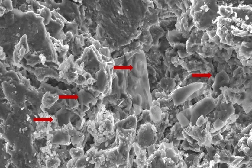
An image of fossilised erythrocyte-like structures. Credit: Anglia Ruskin University
New techniques used to analyse soft tissue in dinosaur fossils may hold the key to new cancer discoveries, according to a new study published in the journal Biology.
Researchers from Anglia Ruskin University (ARU) and Imperial College London analysed dinosaur fossils using advanced paleoproteomic techniques, a method that holds promise for uncovering molecular data from ancient specimens.
The researchers discovered red blood cell-like structures in a fossil while studying a Telmatosaurus transsylvanicus, a duck-billed, plant eating “marsh lizard” that lived between 66-70 million years ago in the Hateg Basin in present-day Romania.
The new study used Scanning Electron Microscopy (SEM) techniques to identify low-density structures resembling erythrocytes, or red blood cells, in the fossilised bone.
The findings raise the possibility that soft tissue and cellular components are more commonly preserved in ancient remains than previously thought.
By identifying preserved proteins and biomarkers, scientists believe they can gain insights into the diseases that affected prehistoric creatures, including cancer, potentially influencing future treatments for humans.
The authors of the new study highlight the necessity of prioritising the collection and preservation of fossilised soft tissue, rather than just dinosaur skeletons, as future advancements in molecular techniques will enable deeper insights into disease evolution.
A separate study had previously identified evidence of cancer in Telmatosaurus transsylvanicus, indicating its deep evolutionary roots.
More precise treatment options with robotic technology
Friday, 20 June 2025:Prostate cancer is a major risk to men’s health, with South African men facing a one in eight chance of developing this most common of male cancers.
Urologists Dr Hannes Brummer and Dr Johan Coetzee, who practise at Netcare Greenacres Hospital, are encouraging men to prioritise prostate cancer screening this Men’s Health Month.
“Usually, men do not feel any symptoms until prostate cancer has progressed significantly, which is why they need to be proactive about booking their routine prostate cancer screenings,” explains Dr Coetzee.
“With the advanced prostate specific antigen [PSA] screening blood test available from GPs these days, there is so much more opportunity for prostate cancer to be detected earlier when it is still at a highly treatable stage.”
“For men who are diagnosed with prostate cancer following a needle biopsy, the treatment options available have improved to such an extent that there is more hope than ever before. Even where surgery is needed, prostate cancer does not necessarily pose a significant disruption to your life,” Dr Brummer adds.
“A prostate cancer diagnosis can be daunting. We have walked this path with so many men, and robotic assisted surgery offers some important advantages for the removal of cancerous tissue in the prostate gland, in particular the precision of this minimally invasive option.”
Over 1 000 robotic assisted procedures have been performed at Netcare Greenacres Hospital since the introduction of this technology in August 2017.
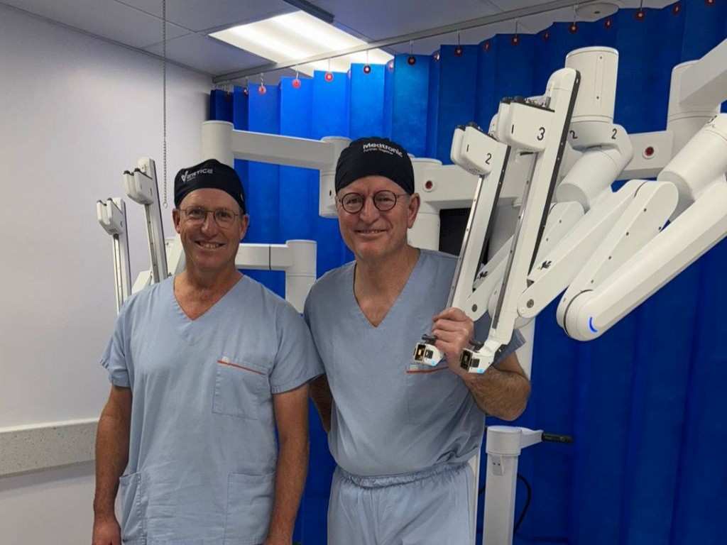
At Netcare Greenacres Hospital, Dr Brummer and Dr Coetzee use the da Vinci X robotic assisted surgical system to operate through tiny punctures in the skin using slender instruments more dexterous than the human hand.
Dr Brummer and Dr Coetzee emphasise that the surgeon remains in control of the robotic system at all times. With magnified 3D imaging capabilities, including a large fixed-focus area at the highest resolution, the nerves, blood vessels and tumour are visible with great clarity for the intricate procedure.
“This robotic system is especially useful for operating on the prostate, as we can more clearly distinguish the nerves controlling erectile function and urinary continence. In most cases, there is less need for blood transfusion and reduced risk of complications,” Dr Brummer explains.
“Another of the advantages of this robotic technology for prostate tumours is that there is much less tissue damage in this sensitive area. Compared with traditional surgery, this means men usually experience much less downtime with less discomfort after the procedure. This translates into shorter hospital stays and faster recovery with robotic assisted prostatectomies overall.”
General manager of Netcare Greenacres Hospital, Reon van Rensburg, joined the urologists in reinforcing the importance of prostate cancer awareness. “Let’s talk to our brothers, fathers, sons and grandsons about health issues, and get to know your family risk for both prostate cancer and breast cancer.”
Van Rensburg thanked Dr Brummer and Dr Coetzee for their continued dedication to making the world-class minimally-invasive robotic assisted surgical option available for patients local to Gqeberha and from as far afield as Knysna, George and East London, inland regions of the Eastern Cape, and parts of the southern Free State, the Northern Cape and the north-eastern region of the Western Cape.
“This Men’s Health Month, let’s pledge to be decisive about booking those routine health checks. Making the time now and every year could help to save your life in future,” Dr Brummer and Dr Coetzee concluded.
Knee braces, water therapy, and exercise are the most beneficial non-drug therapies, per meta-analysis of more than 100 clinical trials involving nearly 10 000 people
Knee braces, water therapy and exercise are the most promising non-drug therapies for treating knee osteoarthritis, according to a new meta-analysis publishing June 18, 2025 in the open-access journal PLOS Oneby Yuan Luo of the First People’s Hospital of Neijiang, China.
Knee osteoarthritis (KOA) is a common and often debilitating condition that affects millions of older adults, causing pain and stiffening of the knee joint. Treatment often includes anti-inflammatory drugs, which are linked to gastrointestinal and cardiovascular adverse events.
In the new study, researchers examined the current evidence on non-drug therapies for treating KOA. They looked at data from 139 clinical trials involving nearly 10 000 people to compare 12 different non-drug treatments. These included laser therapy, electrical stimulation, braces, insoles, kinesiology tape, water-based therapy, exercise, and ultrasound. By combining results from all these studies into a powerful network meta-analysis, the team could rank the therapies based on how well they worked.
Knee braces came out on top across most categories, including reducing pain, improving function, and relieving stiffness. Hydrotherapy—exercises or treatments performed in warm water—was particularly effective at easing pain and general exercise was also consistently effective, improving both pain and physical function. High-intensity laser therapy and shock wave therapy showed some benefits, while ultrasound consistently scored the lowest in effectiveness.
The authors caution that differences in study design, small sample sizes, and variability in treatment duration between the 139 included studies may limit the precision of the rankings. However, they conclude that physical therapy has promising effects on KOA, offering potential treatments without the risks of anti-inflammatory drugs. Future studies should examine the clinical efficacy of combined therapies, as well as their cost-effectiveness.
The authors add: “Knee braces, hydrotherapy, and exercise are the most effective non-drug therapies for knee osteoarthritis. They reduce pain and improve mobility without the gastrointestinal or cardiovascular risks linked to common pain medications. Patients and clinicians should prioritize these evidence-based options.”
“Our analysis of nearly 10 000 patients reveals that simple, accessible therapies like knee bracing and water-based exercise outperform high-tech options like ultrasound. This could reshape clinical guidelines to focus on safer, lower-cost interventions.”
Health workers have long relied on Body Mass Index as a way to measure whether people are within a healthy weight range. Now, a collection of top researchers have made the case for a new way to understand and diagnose obesity. In part two of this special Spotlight series, we take a look at what this new framing might mean for South Africa.
If we are going to tackle the global rise in obesity, our understanding of the condition needs to change. That is according to a Lancet Commission convened by a global group of 58 experts from different medical specialties. While we have historically thought of obesity as a risk factor for other diseases like diabetes, the commission’s recent report published in the journal Lancet Diabetes and Endocrinology concludes that obesity is sometimes better thought of as a disease itself – one that can directly cause severe health symptoms (see part one of this series for a detailed discussion of this argument).
By categorising obesity as a disease, public health systems and medical aid schemes around the world would be more likely to cover people for weight-loss drugs or weight-loss surgery, according to the report. At present, these services are often only financed if a patient’s obesity has already led to other diseases. This is given that obesity is not viewed as a stand-alone chronic illness.
But if we’re going to redefine obesity as a disease, or at least some forms of it, then we need good clinical definitions and ways to measure it. For a long time, this has posed challenges, according to the Lancet report.
The perils of BMI
At present, health workers often rely on Body Mass Index (BMI) to gauge whether a patient is within a healthy weight range. BMI is measured by taking a person’s weight in kilograms and dividing it by their height in meters squared.
A healthy weight is typically considered to be between 18.5 and 25. A person whose BMI is between 25 and 30 is considered to be overweight, while someone with a BMI of over 30 is considered to have obesity. But according to the Lancet report, this is a crude measure, and one which provides very little information about whether a person is actually ill.
One basic issue is that a person can have a high BMI even if they don’t have a lot of excess fat. Instead, they may simply have a lot of muscle or bone. Indeed, the report notes that some athletes are in the obese BMI range.
Even when a high BMI does indicate that a person has obesity, it still doesn’t tell us where a person’s fat is stored and this is vital medical information. If excess fat is stored in the stomach and chest, then it poses more severe health risks than when it is stored in the limbs or thighs. This is because excess fat will do more harm if it surrounds vital organs.
The lead author of the Lancet report, Professor Frances Rubino, says that the pitfalls of BMI have long been understood, but practitioners have continued to use it.
“BMI is still by and large the most used approach everywhere, even though medical organisations have [raised issues] for quite some time,” he tells Spotlight.
“The problem is that even when we as individuals or organisations say BMI is no good, we haven’t provided an alternative. And so, inevitably, the ease of calculating BMI and the uncertainties about alternatives makes you default back to BMI.”
To deal with this problem, the report advocates for several alternative techniques for measuring obesity which offer more precision.
The first option is to use tools that directly measure body composition like a DEXA scanner. This is a sophisticated x-ray machine which can be used to distinguish between fat, bone and muscle. It can also be used to determine where fat is concentrated. It’s thus a very precise measurement tool, but the machines are expensive and the scans can be time-consuming.
Alternatively, the report recommends using BMI in combination with another measure like waist-to-hip ratio, waist-to-height ratio or simply waist circumference. If two of these alternative measures are used, then BMI can be removed from the picture.
These additional metrics are clinically useful because they provide information about where fat is stored. For instance, a larger waistline inevitably indicates a larger stomach. Indeed, studies have found that above a certain level, a larger waist circumference is linked to a higher chance of dying early, even when looking at people with the same BMI.
The report thus offers a more accurate way to measure obesity in the clinical setting. But its authors argue that this is only the first step when making a diagnosis. The second is to look at whether a patient’s obesity has actually caused health problems as this isn’t automatically the case. They acknowledge for instance that there are some people with obesity who “appear to be able to live a relatively healthy life for many years, or even a lifetime”.
The report refers to these cases as “preclinical obesity”. Such patients don’t have a disease as such, according to the report, but still have an increased risk of facing health issues in the future. As such, the report’s authors argue that they should be monitored and sometimes even treated, depending on factors like family history.
By contrast, cases of obesity which have directly caused health problems are referred to as “clinical obesity”. These cases, according to the report, should be treated immediately just like any other serious disease. It lists a series of medical symptoms associated with clinical obesity that would allow health workers to make an appropriate diagnosis.
The recommendation is thus for health workers to determine whether a person has obesity through the metrics listed above, and then to determine whether it is clinical or preclinical by evaluating a patient’s symptoms. This will inevitably guide the treatment plan.
How does this relate to SA?
Professor Francois Venter, who runs the Ezintsha research centre at WITS university, says the Lancet report offers a good starting point for South Africa, but it has to be adapted for our own needs and context.
“It’s a big step forward from BMI which grossly underdiagnoses and overdiagnoses obesity,” says Venter, who adds that additional metrics like waist circumference are a “welcome addition”.
The view that clinical obesity is a disease that needs to be immediately treated is also correct, according to Venter. Though he adds that the public health system in South Africa is not in a financial position to start handing out weight-loss medicine to everyone who needs it.
“The drugs are hugely expensive,” says Venter, “and they have side effects, so you need a lot of resources to support people taking them.” But while it may not yet be feasible to treat all cases of clinical obesity in South Africa, Venter believes we should use the diagnostic model offered by the Lancet Commission to begin identifying at least some people with clinical obesity so that they can begin treatment.
“You have to start somewhere, and for that you need a good staging system,” he says. “Let’s use the Lancet Commission and start to see if we can identify a few priority people and screen them and start to work on the drug delivery system.”
Yet while Venter believes that the commission makes important contributions, he also cautions that we need more data on obesity in Africa before we can apply all of its conclusions to our own context.
“If you go to the supplement of the Lancet Commission, there’s not a single African study there. It all comes from Europe, North America and Asia. It’s not the commission’s fault but [there is a lack of data on Africa].”
This is important as findings that apply to European or Asian populations may not necessarily hold for others. Consider the following case.
As noted, the commission states that BMI is not sufficient to determine whether someone is overweight and must therefore be complemented with other measures. But it states that if someone’s BMI is above 40 (way above the current threshold for obesity), then this can “pragmatically be assumed” without the need for further measures.
But this may not hold in Africa, says Venter.
“The commission says that if your BMI is over 40, which is very big, you can infer that this person has got obesity and they are sick and need to lose weight. I don’t know if we can say that in Africa, where we often have patients who are huge, and yet they are very active, and when you [look at] their blood pressure and all their metabolics, they’re actually pretty healthy,” he notes. “So, I think they’re sometimes jumping to conclusions about African populations that we don’t have data on,” adds Venter.
Is South Africa ready to move past BMI?
Another concern is that while the Lancet Commission may offer useful recommendations for advanced economies, its starting assumptions may not be as relevant for countries like South Africa.
For instance, while specialists agree that BMI is a crude measure of obesity, direct measures like DEXA scans are “out of our reach economically”, according to Professor Susan Goldstein, who leads PRICELESS-SA, a health economics unit at the South African Medical Research Council.
And while supplementing BMI with the other metrics like waist circumference may be doable, health experts told Spotlight that at present healthcare workers in South Africa aren’t even measuring BMI alone.
Dr Yogan Pillay, a former deputy director-general at the national health department who now runs TB and HIV delivery at the Gates Foundation, told Spotlight: “I can’t tell you how few people in the public sector have their BMI monitored at all. Community health workers are supposed to be going out and measuring BMI, but even that’s not happening”.
Goldstein also suggests that the monitoring of BMI in South Africa is limited. “If you go into the clinic for your blood pressure, do they say: ‘How’s your BMI?’ No, I doubt that,” says Goldstein. “It’s just not one of the measures that [gets done].”
She adds that South Africa could introduce the combination of metrics proposed by the commission, like waist circumference combined with BMI, but says it would simply require “a lot of re-education of health workers”.
Prevention vs treatment
For Goldstein, the commission is correct to regard clinical obesity as a disease which needs to be treated, but we also shouldn’t view medication as the only way forward.
“We have to remember that prevention is very important,” says Goldstein. “We have to focus on food control, we have to look at ultra-processed foods, and unless we do that as well [in addition to medication] we are going to lose this battle.”
The National Health Department already has a strategy document for preventing obesity, but some of its recommendations have been critiqued for focusing on the wrong problems. For instance, to prevent childhood obesity, the strategy document recommends reforming the Life Orientation curriculum and educating tuck shop vendors so that both students and food sellers have more information about healthy eating. But as Spotlight previously reported, there are no recommendations to subsidise healthy foods or to increase their availability in poor areas, which several experts believe is more important than educational initiatives.
Venter also highlights the importance of obesity prevention, though he emphasises that this shouldn’t be in conflict with a treatment approach – instead, we need to push for both.
“The [prevention] we need to do is fix the food supply… and the only way you do that is to decrease the cost of unprocessed food.” But while this may help prevent future cases of obesity, it doesn’t help people who are already suffering from obesity, says Venter. And since such people comprise such a large share of the population, we can’t simply ignore them, he says.
“Even if you fix the entire food industry tomorrow, those [people who are already obese] are going to remain where they are because simply changing your diet isn’t going to do diddly squat [when you already have obesity],” he adds. (Part 1 discusses this in more detail).
Goldstein adds that increasing access to treatment would also inevitably reduce the costs of “hypertension, diabetes, osteoarthritis, and a whole range of other illnesses if it’s properly managed”.
One way to advance access to medication would be for the government to negotiate reduced prices of GLP-1 drugs, she says. (Spotlight previously reported on the prices and availability of these medicines in South Africa here.)
Funding
A final concern that has been raised about the Lancet commission is about its source of funding.
“I don’t know how one gets around this,” says Goldstein, “but there were 58 experts on the commission, 47 declared conflicts of interest.”
Indeed, the section of the commission that lists conflicts of interest spans over 2 000 words (roughly the size of this article). This includes research grants and consulting fees from companies like Novo Nordisk and Eli Lilly, which produce anti-obesity drugs.
In response, Rubino told Spotlight that “people who work in the medical profession obviously work and consult, and the more expertise they have, the more likely they are to be asked by somebody to advise. So sometimes people have contracts to consult a company – but that doesn’t mean that they necessarily make revenue if the company has better sales. You get paid fees for your services as a consultant”.
Rubino says this still has to be declared as it may result in some bias, even if it is unconscious, but “if you wanted to have experts who had zero relationship [to companies] of any sort then you might have to wonder if there is expertise available there… the nature of any medical professional is that the more expertise they have, the more likely that they have engaged in work with multiple stakeholders”.
For Venter, there is some truth to this. “It’s very difficult to find people in the obesity field that aren’t sponsored by a drug company,” he says. “Governments don’t fund research… and everyone else doesn’t fund research. Researchers go where the research is funded.”
This doesn’t actually solve the problem, says Venter, as financing from drug companies can always influence the conclusions of researchers. It simply suggests that the problem is bigger than the commission. Ultimately, he argues that the authors should at least be applauded for providing such granular details about conflicts of interest.
Rubino adds that while researchers on the commission may have historically received money from drug companies for separate research studies or consulting activities, none of them received money for their work on the commission itself.
“This commission has been working for more than four years since conception… An estimate of how many meetings we had is north of 700, and none of us have received a single penny [for doing this],” he says.
Disclosure: The Gates Foundation is mentioned in this article. Spotlight receives funding from the Gates Foundation but is editorially independent – an independence that the editors guard jealously. Spotlight is a member of the South African Press Council.
By delivering an HIV vaccine candidate along with two adjuvants, researchers showed they could generate many more HIV-targeting B cells in mice.
Anne Trafton | MIT News
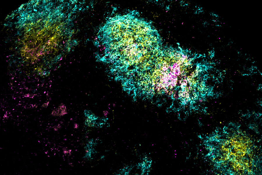
Image shows the vaccine antigen (pink) being concentrated in a germinal center (yellow) within B cell follicles (cyan), triggered by the researchers’ combination adjuvant vaccine.
Credits: Image: Courtesy of the researchers
Researchers at MIT and the Scripps Research Institute have shown that they can generate a strong immune response to HIV with just one vaccine dose, by adding two powerful adjuvants — materials that help stimulate the immune system.
In a study of mice, the researchers showed that this approach produced a much wider diversity of antibodies against an HIV antigen, compared to the vaccine given on its own or with just one of the adjuvants. The dual-adjuvant vaccine accumulated in the lymph nodes and remained there for up to a month, allowing the immune system to build up a much greater number of antibodies against the HIV protein.
This strategy could lead to the development of vaccines that only need to be given once, for infectious diseases including HIV or SARS-CoV-2, the researchers say.
“This approach is compatible with many protein-based vaccines, so it offers the opportunity to engineer new formulations for these types of vaccines across a wide range of different diseases, such as influenza, SARS-CoV-2, or other pandemic outbreaks,” says J. Christopher Love, the Raymond A. and Helen E. St. Laurent Professor of Chemical Engineering at MIT, and a member of the Koch Institute for Integrative Cancer Research and the Ragon Institute of MGH, MIT, and Harvard.
Love and Darrell Irvine, a professor of immunology and microbiology at the Scripps Research Institute, are the senior authors of the study, which appears today in Science Translational Medicine. Kristen Rodrigues PhD ’23 and Yiming Zhang PhD ’25 are the lead authors of the paper.
More powerful vaccines
Most vaccines are delivered along with adjuvants, which help to stimulate a stronger immune response to the antigen. One adjuvant commonly used with protein-based vaccines, including those for hepatitis A and B, is aluminum hydroxide, also known as alum. This adjuvant works by activating the innate immune response, helping the body to form a stronger memory of the vaccine antigen.
Several years ago, Irvine developed another adjuvant based on saponin, an FDA-approved adjuvant derived from the bark of the Chilean soapbark tree. His work showed that nanoparticles containing both saponin and a molecule called MPLA, which promotes inflammation, worked better than saponin on its own. That nanoparticle, known as SMNP, is now being used as an adjuvant for an HIV vaccine that is currently in clinical trials.
Irvine and Love then tried combining alum and SMNP and showed that vaccines containing both of those adjuvants could generate even more powerful immune responses against either HIV or SARS-CoV-2.
In the new paper, the researchers wanted to explore why these two adjuvants work so well together to boost the immune response, specifically the B cell response. B cells produce antibodies that can circulate in the bloodstream and recognise a pathogen if the body is exposed to it again.
For this study, the researchers used an HIV protein called MD39 as their vaccine antigen, and anchored dozens of these proteins to each alum particle, along with SMNP.
After vaccinating mice with these particles, the researchers found that the vaccine accumulated in the lymph nodes — structures where B cells encounter antigens and undergo rapid mutations that generate antibodies with high affinity for a particular antigen. This process takes place within clusters of cells known as germinal centers.
The researchers showed that SMNP and alum helped the HIV antigen to penetrate through the protective layer of cells surrounding the lymph nodes without being broken down into fragments. The adjuvants also helped the antigens to remain intact in the lymph nodes for up to 28 days.
“As a result, the B cells that are cycling in the lymph nodes are constantly being exposed to the antigen over that time period, and they get the chance to refine their solution to the antigen,” Love says.
This approach may mimic what occurs during a natural infection, when antigens can remain in the lymph nodes for weeks, giving the body time to build up an immune response.
Antibody diversity
Single-cell RNA sequencing of B cells from the vaccinated mice revealed that the vaccine containing both adjuvants generated a much more diverse repertoire of B cells and antibodies. Mice that received the dual-adjuvant vaccine produced two to three times more unique B cells than mice that received just one of the adjuvants.
That increase in B cell number and diversity boosts the chances that the vaccine could generate broadly neutralizing antibodies — antibodies that can recognize a variety of strains of a given virus, such as HIV.
“When you think about the immune system sampling all of the possible solutions, the more chances we give it to identify an effective solution, the better,” Love says. “Generating broadly neutralizing antibodies is something that likely requires both the kind of approach that we showed here, to get that strong and diversified response, as well as antigen design to get the right part of the immunogen shown.”
Using these two adjuvants together could also contribute to the development of more potent vaccines against other infectious diseases, with just a single dose.
“What’s potentially powerful about this approach is that you can achieve long-term exposures based on a combination of adjuvants that are already reasonably well-understood, so it doesn’t require a different technology. It’s just combining features of these adjuvants to enable low-dose or potentially even single-dose treatments,” Love says.
The research was funded by the National Institutes of Health; the Koch Institute Support (core) Grant from the National Cancer Institute; the Ragon Institute of MGH, MIT, and Harvard; and the Howard Hughes Medical Institute.
This story is republished courtesy of MIT News (web.mit.edu/newsoffice/), a popular site that covers news about MIT research, innovation and teaching.
Brain networks responsible for sensing, understanding, and responding emotionally to pain develop at different rates in infants, with the conscious understanding of pain not fully developed until after birth, finds a new study led by UCL (University College London) researchers.
The authors of the study, published in the journal Pain, investigated how different types of pain processing develop very early on, by scanning the brains of infants born prematurely.
Lead author Professor Lorenzo Fabrizi (UCL Neuroscience, Physiology & Pharmacology) said: “Pain is a complex experience with physical, emotional, and cognitive elements. In adults, pain processing relies on a functional network of brain regions called the ‘pain connectome’, with different regions working together to help us experience pain, each part responsible for different aspects of it.
“In newborn babies, this network is underdeveloped, which could mean that pain experience in newborns is totally different from the way we, as adults, understand it.”
The scientists, based at UCL, UCLH and King’s College London, were looking at three different components of pain processing: sensory-discriminative (identifying and localising the intensity and quality of pain), affective-motivational (resulting in the emotional response to pain), and cognitive-evaluative (the appraisal and interpretation of pain).
Using advanced brain imaging data from two of the largest available databases of brain magnetic resonance imaging (MRI) in the world – the Developing Human Connectome Project and the Human Connectome Project – the researchers mapped how these networks grow in a group of 372 infants, mostly born preterm, from less than 32 weeks up to 42 weeks after conception. The infants were all less than two weeks old when the scans took place, to ensure that the findings reflected the intrinsic brain maturation, without being affected by different experiences post-birth.
The researchers compared these findings to brain data from adults, as the mature pain-processing networks have previously been mapped out in other studies. The researchers analysed how much the brain networks known to be responsible for processing pain were functionally connected in infants at different ages.
The scientists found that the first subnetwork to reach adult levels in strength and connectivity is the sensory-discriminative network, at around 34-36 weeks after conception, so that babies can sense pain but are not yet fully capable of responding emotionally or interpreting the pain. Before this point, infants may have difficulty identifying what part of their body is experiencing pain. At around 36-38 weeks, the affective-motivational subnetwork reaches maturity, so that infants can identify pain as unpleasant and threatening.
The cognitive-evaluative subnetwork does not reach maturity until more than 42 weeks after conception, meaning that babies born at full term have still not fully developed the brain networks required to understand pain.
The research team had previously found in a 2023 study that preterm babies do not habituate to repeated pain experiences in medically necessary procedures (that is, their reaction to repeated pain does not reduce over time). The new finding that preterm babies have not fully developed the brain connections responsible for appraising pain may help to explain this.
Professor Fabrizi said: “Our results suggest that preterm babies may be particularly vulnerable to painful medical procedures during critical stages of brain development. The findings therefore emphasise the importance of informed paediatric care, including the role of tailored pain management and carefully planned timing of medical interventions for newborns, particularly those born preterm.”
A new study in teenagers with type 1 diabetes shows promise in reducing chronic kidney disease and informing future precision care.
Photo by Nataliya Vaitkevich on Pexels
A clinical trial involving adolescents with type 1 diabetes (T1D) has found a combination therapy may reduce chronic kidney disease and improve health outcomes. The findings could help guide more precision care for young people with T1D.
Led by Dr Farid Mahmud, Associate Scientist in the Translational Medicine program and Staff Physician in the Division of Endocrinology at The Hospital for Sick Children (SickKids), and published in Nature Medicine, the study evaluated a therapy that combines standard insulin treatment with the investigational drug dapagliflozin. Results of this combination therapy showed improved blood sugar control and kidney function, and reduced weight gain in adolescents with T1D.
While most people with T1D are diagnosed as adults, the condition often starts in childhood and early adolescence. The lifelong insulin therapy needed can lead to side effects such as weight gain and chronic kidney disease. In the trial, participants who received dapagliflozin alongside insulin had fewer of these side effects and better overall health outcomes.
“Our findings showed that adolescents who received this combination therapy were able to improve many symptoms typically associated with insulin-managed type one diabetes,” says Mahmud. “This could inform a new early intervention strategy for the growing population of teenagers with type one diabetes.”
Patient partner key to trial success
While previous research has shown similar results in adults, Mahmud’s team focused on designing a clinical trial specifically for teenagers, a group often underrepresented in clinical trials. Hormonal changes, psychological development, and the shared responsibility between teens and their parents for managing treatment protocols can make trial participation more complex for this age group.
To address these challenges, the research team worked closely with patient partner Lynne McArthur. Together, they enrolled 98 participants between 12 and 18 years old in the study, known as the ATTEMPT study, across three sites.
McArthur’s involvement in research began when one of her twin sons was diagnosed with T1D following a trip to the SickKids emergency department at just 18 months old. A few years later, his twin was also diagnosed. That experience led McArthur to become more involved in research efforts to improve diagnosis and treatment options for families like hers.
“Deciding to participate in a clinical trial is an important decision, but my goal has always been disease prevention. I knew that our participation could help build a future where children don’t get T1D.”
Lynne McArthur
Now that her sons are older, McArthur continues to be involved as a patient advisor. She reviews recruitment materials and provides feedback on trial design, helping ensure the research stays connected to the lived experience of people managing T1D.
“Participating in research, whether in a trial or as an advisor, is hugely rewarding. With my experience as trial participant, I can see how the plans on paper would impact the real lives of people living with diabetes,” explains McArthur.
A new, highly efficient process for performing this conversion could make it easier to develop therapies for spinal cord injuries or diseases like ALS.
Anne Trafton | MIT News
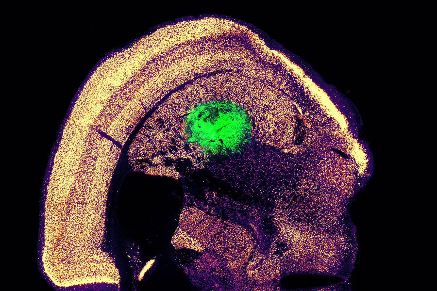
Researchers at MIT have devised a simplified process to convert a skin cell directly into a neuron. This image shows converted neurons (green) that have integrated with neurons in the brain’s striatum after implantation.
Credits :Image: Courtesy of the researchers
Converting one type of cell to another – for example, a skin cell to a neuron – can be done through a process that requires the skin cell to be induced into a “pluripotent” stem cell, then differentiated into a neuron. Researchers at MIT have now devised a simplified process that bypasses the stem cell stage, converting a skin cell directly into a neuron.
Working with mouse cells, the researchers developed a conversion method that is highly efficient and can produce more than 10 neurons from a single skin cell. If replicated in human cells, this approach could enable the generation of large quantities of motor neurons, which could potentially be used to treat patients with spinal cord injuries or diseases that impair mobility.
“We were able to get to yields where we could ask questions about whether these cells can be viable candidates for the cell replacement therapies, which we hope they could be. That’s where these types of reprogramming technologies can take us,” says Katie Galloway, the W. M. Keck Career Development Professor in Biomedical Engineering and Chemical Engineering.
As a first step toward developing these cells as a therapy, the researchers showed that they could generate motor neurons and engraft them into the brains of mice, where they integrated with host tissue.
Galloway is the senior author of two papers describing the new method, which appear today in Cell Systems. MIT graduate student Nathan Wang is the lead author of both papers.
From skin to neurons
Nearly 20 years ago, scientists in Japan showed that by delivering four transcription factors to skin cells, they could coax them to become induced pluripotent stem cells (iPSCs). Similar to embryonic stem cells, iPSCs can be differentiated into many other cell types. This technique works well, but it takes several weeks, and many of the cells don’t end up fully transitioning to mature cell types.
“Oftentimes, one of the challenges in reprogramming is that cells can get stuck in intermediate states,” Galloway says. “So, we’re using direct conversion, where instead of going through an iPSC intermediate, we’re going directly from a somatic cell to a motor neuron.”
Galloway’s research group and others have demonstrated this type of direct conversion before, but with very low yields – fewer than 1 percent. In Galloway’s previous work, she used a combination of six transcription factors plus two other proteins that stimulate cell proliferation. Each of those eight genes was delivered using a separate viral vector, making it difficult to ensure that each was expressed at the correct level in each cell.
In the first of the new Cell Systems papers, Galloway and her students reported a way to streamline the process so that skin cells can be converted to motor neurons using just three transcription factors, plus the two genes that drive cells into a highly proliferative state.
Using mouse cells, the researchers started with the original six transcription factors and experimented with dropping them out, one at a time, until they reached a combination of three – NGN2, ISL1, and LHX3 — that could successfully complete the conversion to neurons.
Once the number of genes was down to three, the researchers could use a single modified virus to deliver all three of them, allowing them to ensure that each cell expresses each gene at the correct levels.
Using a separate virus, the researchers also delivered genes encoding p53DD and a mutated version of HRAS. These genes drive the skin cells to divide many times before they start converting to neurons, allowing for a much higher yield of neurons, about 1100 percent.
“If you were to express the transcription factors at really high levels in nonproliferative cells, the reprogramming rates would be really low, but hyperproliferative cells are more receptive. It’s like they’ve been potentiated for conversion, and then they become much more receptive to the levels of the transcription factors,” Galloway says.
The researchers also developed a slightly different combination of transcription factors that allowed them to perform the same direct conversion using human cells, but with a lower efficiency rate – between 10 and 30 percent, the researchers estimate. This process takes about five weeks, which is slightly faster than converting the cells to iPSCs first and then turning them into neurons.
Implanting cells
Once the researchers identified the optimal combination of genes to deliver, they began working on the best ways to deliver them, which was the focus of the second Cell Systems paper.
They tried out three different delivery viruses and found that a retrovirus achieved the most efficient rate of conversion. Reducing the density of cells grown in the dish also helped to improve the overall yield of motor neurons. This optimised process, which takes about two weeks in mouse cells, achieved a yield of more than 1000 percent.
Working with colleagues at Boston University, the researchers then tested whether these motor neurons could be successfully engrafted into mice. They delivered the cells to a part of the brain known as the striatum, which is involved in motor control and other functions.
After two weeks, the researchers found that many of the neurons had survived and seemed to be forming connections with other brain cells. When grown in a dish, these cells showed measurable electrical activity and calcium signaling, suggesting the ability to communicate with other neurons. The researchers now hope to explore the possibility of implanting these neurons into the spinal cord.
The MIT team also hopes to increase the efficiency of this process for human cell conversion, which could allow for the generation of large quantities of neurons that could be used to treat spinal cord injuries or diseases that affect motor control, such as ALS. Clinical trials using neurons derived from iPSCs to treat ALS are now underway, but expanding the number of cells available for such treatments could make it easier to test and develop them for more widespread use in humans, Galloway says.
The research was funded by the National Institute of General Medical Sciences and the National Science Foundation Graduate Research Fellowship Program.
Eating a high-fat diet containing a large amount of oleic acid – a type of fatty acid commonly found in olive oil – could drive obesity more than other types of dietary fats, according to a study published in the journal Cell Reports.
The study found that oleic acid, a monounsaturated fat associated with obesity but also tentatively linked to cardiovascular benefits and often touted as a ‘healthy’ fatty acid, causes the body to make more lipid cells. By boosting a signalling protein called AKT2 and reducing the activity of a regulating protein called LXR, high levels of oleic acid resulted in faster growth of the precursor cells that form new lipid cells.
“We know that the types of fat that people eat have changed during the obesity epidemic. We wanted to know whether simply overeating a diet rich in fat causes obesity, or whether the composition of these fatty acids that make up the oils in the diet is important. Do specific fat molecules trigger responses in the cells?” said Michael Rudolph, PhD, assistant professor of biochemistry and physiology at the University of Oklahoma College of Medicine.
Rudolph and his team fed mice a variety of specialised diets enriched in specific individual fatty acids, including those found in coconut oil, peanut oil, milk, lard and soybean oil. Oleic acid was the only one that caused the precursor cells that give rise to fat cells to proliferate more than other fatty acids.
“You can think of the fat cells as an army,” Rudolph said. “When you give oleic acid, it initially increases the number of ‘fat cell soldiers’ in the army, which creates a larger capacity to store excess dietary nutrients. Over time, if the excess nutrients overtake the number of fat cells, obesity can occur, which can then lead to cardiovascular disease or diabetes if not controlled.”
Unfortunately, it’s not quite so easy to isolate different fatty acids in a human diet. People generally consume a complex mixture if they have cream in their coffee, a salad for lunch and meat and pasta for dinner. However, Rudolph said, there are increasing levels of oleic acid in the food supply, particularly when access to food variety is limited and fast food is an affordable option.
“I think the take-home message is moderation and to consume fats from a variety of different sources,” he said. “Relatively balanced levels of oleic acid seem to be beneficial, but higher and prolonged levels may be detrimental. If someone is at risk for heart disease, high levels of oleic acid may not be a good idea.”
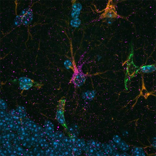
Using advanced single-nuclei RNA sequencing (snRNA-seq) and a widely used preclinical model for Alzheimer’s disease, researchers from Mass General Brigham and collaborators at SUNY Upstate Medical University have identified specific brain cell types that responded most to exercise. These findings, which were validated in samples from humans, shed light on the connection between exercise and brain health and point to future drug targets. Results are published in Nature Neuroscience.
“While we’ve long known that exercise helps protect the brain, we didn’t fully understand which cells were responsible or how it worked at a molecular level,” said senior author Christiane Wrann, DVM, PhD, a neuroscientist at Massachusetts General Hospital. “Now, we have a detailed map of how exercise impacts each major cell type in the memory centre of the brain in Alzheimer’s disease.”
Scientists identified a distinct subtype of brain support cells—astrocytes enriched in the protein cadherin-4 (CDH4), shown in magenta, that seem to protect nerve cells against cell death. In Alzheimer’s disease, these cells become less abundant, but exercise seems to strengthen them. (Image credit: Luis Moreira)
The study focused on a part of the hippocampus – a critical region for memory and learning that is damaged early in Alzheimer’s disease. The research team leveraged single-nuclei RNA sequencing, a relatively new technologies that allow researchers to look at activity at the molecular level in single cells for an in-depth understanding of diseases like Alzheimer’s.
The researchers exercised a common mouse model for Alzheimer’s disease using running wheels, which improved their memory compared to the sedentary counterparts. They then analysed gene activity across thousands of individual brain cells, finding that exercise changed activity both in microglia, a disease-associated population of brain cells, and in a specific type of neurovascular-associated astrocyte (NVA), newly discovered by the team, which are cells associated with blood vessels in the brain. Furthermore, the scientist identified the metabolic gene Atpif1 as an important regulator to create new neurons in the brain. “That we were able to modulate newborn neurons using our new target genes set underscores the promise our study,” said lead author Joana Da Rocha, PhD, a postdoctoral fellow working in Dr Wrann’s lab.
To ensure the findings were relevant to humans, the team validated their discoveries in a large dataset of human Alzheimer’s brain tissue, finding striking similarities.
“This work not only sheds light on how exercise benefits the brain but also uncovers potential cell-specific targets for future Alzheimer’s therapies,” said Nathan Tucker, a biostatistician at SUNY Upstate Medical University and co-senior of the study. “Our study offers a valuable resource for the scientific community investigating Alzheimer’s prevention and treatment.”