Combination Inhaler Cuts Asthma Attacks in Children by Nearly Half

Findings from a trial comparing the real-world effectiveness of asthma inhalers could reshape how children with asthma are treated.
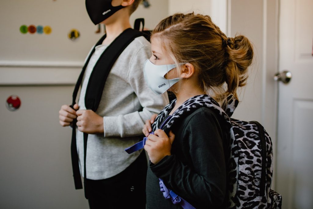
In the first randomised controlled trial to investigate the use of a 2-in-1 inhaler as the sole reliever therapy for children aged 5 to 15, an international team found the combined treatment to be more effective than salbutamol, the current standard for asthma symptom relief in children, with no additional safety concerns.
The results show that using a single 2-in-1 anti-inflammatory reliever inhaler – which combines the inhaled corticosteroid (ICS) budesonide and the fast-acting bronchodilator formoterol – reduced children’s asthma attacks by an average of 45%, compared to the widely-used salbutamol inhaler.
Asthma attacks in children may be life-threatening and reducing their frequency and severity is a public health priority.
The 2-in-1 budesonide-formoterol inhaler is widely recommended as the preferred reliever treatment for adults, but children are still usually prescribed salbutamol.
Researchers say the findings, published in The Lancet, provide the evidence needed to bring children’s global asthma guidelines into line with adults’, which could benefit millions of children around the world with mild-to-moderate asthma.
The CARE study (Children’s Anti-inflammatory REliever) was designed and led by the Medical Research Institute of New Zealand (MRINZ), in collaboration with Imperial College London, University of Otago Wellington, Starship Children’s Hospital, and the University of Auckland. It recruited 360 children across New Zealand who were then randomly assigned to receive either budesonide-formoterol or salbutamol for on-demand symptom relief.
The trial lasted a year and the budesonide-formoterol reliever resulted in a lower rate of asthma attacks than salbutamol reliever, with rates of 0.23 versus 0.41 per participant per year. This means that for every 100 children with mild asthma who are switched from salbutamol to a 2-in-1 budesonide-formoterol inhaler, there would be 18 fewer asthma attacks per year. Importantly, the study also confirmed the safety of the combined-inhaler approach, with no significant differences in children’s growth, lung function, or asthma control between the two groups.
Dr Lee Hatter, lead author of the study and Senior Clinical Research Fellow at the MRINZ, said: “This is a key step in addressing the evidence gap that exists between asthma management in adults and children. For the first time, we have demonstrated that the budesonide-formoterol 2-in-1 inhaler, used as needed for symptom relief, can significantly reduce asthma attacks in children with mild asthma. This evidence-based treatment could lead to improved asthma outcomes for children worldwide.”
Professor Richard Beasley, Director of MRINZ and senior author of the study, said: “Implementing these findings could be transformative for asthma management on a global scale. The evidence that budesonide-formoterol is more effective than salbutamol in preventing asthma attacks in children with mild asthma has the potential to redefine the global standard of asthma management.”
The burden of asthma in the estimated 113 million children and adolescents with asthma worldwide is substantial. The latest study builds on previous studies in adults led by MRINZ researchers which shaped international asthma treatment guidelines. These findings contributed to the recommended use of the 2-in-1 ICS–formoterol reliever inhaler as the preferred reliever treatment for adults with asthma around the world.
The incorporation of findings from the CARE study into global asthma treatment strategies could help reduce disparities in care and ensure that more children access effective, evidence-based treatments.
The researchers say that global health organisations have long advocated for child-targeted asthma interventions, and their findings provide crucial evidence to support those efforts.
However, the authors acknowledge some limitations of the clinical trial. It was undertaken during the COVID-19 pandemic, during which stringent public health measures and fewer circulating respiratory viruses contributed to the lower than predicted rate of severe asthma attacks. The authors also acknowledge the challenges with the identification of asthma attacks in children, and the potential bias with the lack of blinding of the randomised treatments. They say though that the study’s findings are generalisable to clinical practice due to its pragmatic, real-world design.
Professor Andrew Bush, from Imperial College London, senior respiratory paediatrician and co-author of the CARE study, said: “Having an asthma attack can be very scary for children and their parents. I’m so pleased that we’ve been able to prove that an inhaler that significantly reduces attacks – already a game-changer for adults – is safe for children with mild asthma too. We believe this will transform asthma care worldwide and are excited to be building on this work with the CARE UK study.”
Professor Helen Reddel, Chair of the Science Committee of the Global Initiative for Asthma (GINA), commented on the global significance of the study, saying that it fills a critically important gap for asthma management globally. Professor Reddel said: “Asthma attacks have a profound impact on children’s physical, social and emotional development and their prevention is a high priority for asthma care. It is in childhood, too, that lifelong habits are established, particularly reliance on traditional medications like salbutamol that only relieve symptoms and don’t prevent asthma attacks.”
Professor Bob Hancox, Medical Director of the New Zealand Asthma and Respiratory Foundation, said: “This is a very important study for children with mild asthma. We have known for some time that 2-in-1 budesonide/formoterol inhalers are better than the traditional reliever treatment in adults, but this had not been tested in children. This research shows that this 2-in-1 inhaler is effective and safe for children as young as 5. This information will help to reduce the burden of asthma for many children, and both they and their families will breathe easier because of it.”
Source: Imperial College London