Category: Lab Tests and Imaging

Student Designs a Prostate Checking Device to Replace the Digital Exam

Pro check, designed by Loughborough University student Devon Tyso.

A Loughborough University student has developed a new medical device that could transform how prostate health is assessed and monitored.

Devon Tyso, a Product Design and Technology student, has designed ‘PRO check’, an innovative tool designed to replace the traditional digital rectal examination (DRE), which involves a doctor manually assessing the prostate with a finger.

According to Devon, the current approach is heavily reliant on a clinician’s subjective judgement and experience, and many see the method as ‘intrusive’.

“As one in seven men will get prostate cancer, it’s vital to detect abnormalities early and track changes over time,” said Devon, “The current examination method involves a lot of guesswork.

“PRO check provides objective, measurable data and allows prostate health to be visualised – enabling more accurate diagnosis, and improved long-term monitoring.

“Having a device conduct the exam may also feel less invasive, which may encourage more men to get checked, potentially catching issues earlier.”

How the device works

Designed for use by GPs during routine prostate assessments, PRO check allows doctors to evaluate the size and texture of the prostate — two key indicators of potential health issues — in a more objective and consistent way than the traditional digital rectal examination.

The device is a handheld probe, and it is covered with a condom before being inserted into the body. Once in position, the condom inflates to different pressures, pressing against the surface of the prostate, causing it to compress. A laser grid is projected onto the inner surface of the condom so the shape of the underlying prostate can be captured.

Stereoscopic cameras capture images of the laser grid, tracking where the gridlines intersect and how these intersections shift as pressure changes. This information is then fed into mathematical equations to create 3D images — or ‘topographical representations’ — that reveal the prostate’s shape and surface structure under different pressures.

Studying the prostate’s surface details could help clinicians identify areas requiring further investigation. Healthy prostate tissue is typically soft and compressible, so regions that appear stiff or resist pressure could indicate potential abnormalities and warrant further investigation.

The device can also produce data on prostate volume – one of the measurements used to calculate prostate-specific antigen (PSA) density, which helps assess prostate cancer risk. Devon says currently volume estimates are often based on a clinician’s best judgement.

In addition, data from PRO check can be used to generate a compressibility-versus-pressure graph – a novel data type not currently available in clinical practice. This graph shows how the prostate compresses at different pressure levels, which Devon hopes could offer new insights into prostate health and complement existing diagnostic tools.

PRO check is designed to integrate with artificial intelligence, enabling automatic extraction of video data, real-time calculations, and the generation of 3D images for live display on a laptop or tablet during the examination.

The idea is that all examination data from PRO check would be stored on the patient’s records, helping to build a personalised prostate health profile that can be tracked and monitored over time.

Inspiration

Devon’s inspiration for PRO check came from a mix of personal experience – after his grandfather’s prostate cancer diagnosis – and unexpected technical research.

“It really hit home how common prostate issues are after my family member was found to have an enlarged prostate,” said the 22-year-old from Cardiff, “I realised nearly everyone I spoke to about it knew someone affected by it.

“When I started looking into prostate examinations, I kept thinking ‘how can a doctor remember what your prostate felt like four months ago?’ and how horrible it must be just be told whether you’re fine or not without seeing any data or anything visual.”

While researching non-invasive ways to assess tissue structure inside the body, Devon came across a technique used by NASA to map the surface of asteroids — projecting laser grids onto them, capturing images with satellite-mounted cameras, and analysing the gridline intersections to reveal the contours of the surface.

“I saw that NASA were mapping surface heights on a massive scale, and I thought – if they can do that in space, why can’t we use similar principles to examine something here on Earth?” said Devon, “I’ve basically used the exact same technique and scaled it down for PRO check.”

Prototypes

Devon designed PRO check as part of his final year project – which was exhibited at the School of Design and Creative Arts’ 2025 Degree Show – and has prototyped several of its key components.

He has built and tested two working prototypes. The first demonstrates how a laser grid and camera can be setup to map the surface of the prostate.

Devon designed a custom rig that enabled him to capture images of a laser grid projected onto different silicone prostate models — representing a healthy gland, a small tumour, a large tumour, and an enlarged prostate — from an optimal angle using a smartphone camera.
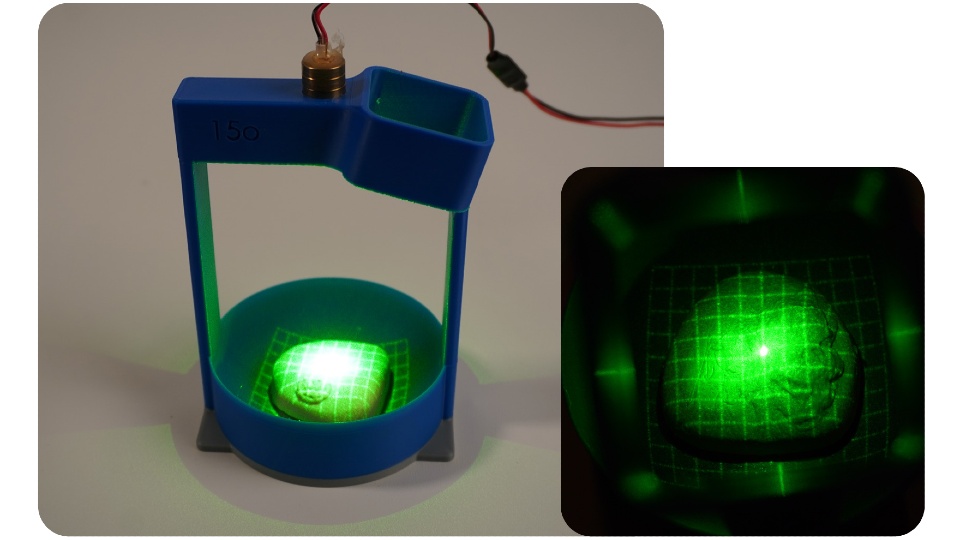
First prototype of pro checkPRO check prototype one demonstrated how laser gridlines and a camera can be used to image the surface of the prostate.

The second prototype features electronics that inflate a small balloon at controlled pressures, regulated by a pressure-sensing chip. Devon consulted three healthcare professionals to measure the pressure typically applied during prostate exams and replicated those levels in his design.

Devon tested the prototype using the silicone prostate models but encased them in a sponge disc to simulate surrounding tissue.

Devon manually extracted data on the gridline intersections from the camera footage and applied mathematical equations to generate 3D images of the prostate surfaces and surrounding tissue under different pressures.

Next steps

Devon hopes to collaborate with medical professionals and product developers to turn PRO check into a fully realised medical device.

When speaking about his ultimate goal, Devon said: “I’d love to see this used in GP surgeries across the UK one day.

“With early detection being so critical, anything that helps men get checked sooner and more comfortably – and provides reliable data and visualisations – has huge potential. I really believe this could make a difference.”

Further information on PRO check can be found on the Degree Show website.

Source: Loughborough University

Handheld TB Test for Low-cost Diagnosis in HIV Hotspots

A new handheld tuberculosis testing device by Tulane University is the size of a credit card, requires no electricity and significantly improves detection of the disease in those with HIV. (Vincent Postle/Tulane University)

Current tuberculosis infection tests struggle to detect the disease in those with HIV. A common co-infection, HIV can hide TB from traditional tests by eliminating the immune cells relied upon to sound the alarm.

While more than 90% of the 2 billion TB cases worldwide are latent – symptom-free and not contagious – the weakening of the immune system in those with HIV can allow latent TB to turn active, increasing the potential for new infections to spread and often resulting in fatal outcomes. Tuberculosis is the leading cause of death among those with HIV worldwide.

Now, Tulane University researchers have developed a new handheld TB test that significantly improves detection in people with HIV, according to a new study in Nature Biomedical Engineering. Powered by a beetle-inspired chemical reaction, the device requires no electricity and addresses a critical gap in TB infection detection that has long hobbled efforts to eliminate the world’s deadliest infectious disease. 

“The goal was to develop a TB test that could be taken anywhere and provide quicker, more accurate results for anybody.”

Tony Hu, PhD

Dubbed the ASTRA (Antigen-Specific T-cell Response Assay), the credit card-sized device requires only a drop of blood to provide same day diagnoses without need for a laboratory or trained staff. When tested against the traditional IGRA blood test (Interferon-Gamma Release Assay), the ASTRA detected TB in HIV-infected individuals with 87% specificity compared to IGRA’s 60%, while also outperforming in detection of TB without HIV co-infection. 

“The goal was to develop a TB test that could be taken anywhere and provide quicker, more accurate results for anybody,” said senior author Tony Hu, PhD, Chair in Biotechnology Innovation at Tulane University and director of the Tulane Center for Cellular & Molecular Diagnostics. “Current tests such as the IGRA are cost-prohibitive or require access to facilities that resource-limited communities don’t have. If we are going to eliminate TB, we have to diagnose and treat as many infection cases as possible.”

Added Bo Ning, lead author and assistant professor of biochemistry at Tulane University School of Medicine: “If your community has an immunocompromised population, someone may have latent TB.  This can help block the spread of TB and ensure that no one slips through the cracks.” 

To create a test that would not be stymied by HIV, the researchers identified two new biomarkers that could detect TB without relying on the immune cells susceptible to the virus. 

After adding a drop of blood to the device, it must incubate for 4 hours to allow a preloaded reagent to stimulate a response from the immune cells. The reagent acts as a “wanted poster” asking if they’ve seen tuberculosis bacteria before. 

To avoid the use of electricity, the researchers looked to an unlikely source for inspiration: the bombardier beetle. When threatened, these large insects combine two chemicals, and the resulting reaction produces a forceful spray. Similarly, two chemicals in the ASTRA are combined to propel the sample across a chip for final analysis and diagnosis. 

The new device delivers results in about 4 hours, compared to the IGRA, which takes 24 hours, and a common TB skin test, which can take between two and three days for a diagnosis. 

The ASTRA’s performance was validated using samples collected from a cohort in Eswatini, a country with high TB incidence and the highest reported HIV prevalence (27.3%) worldwide.

Increasing testing accuracy, access and speed is even more vital as TB resistance to drugs grows more robust, Hu said. 

“The sooner you have a diagnosis, the sooner you can begin the process of determining proper treatment,” Hu said. “TB is the No. 1 pathogen HIV patients worry about globally. If treatment is available, we should be working to kill these bacteria, latent or not.”

Source: Tulane University

Test Detects Cancers in the Bloodstream Three Years Prior to Diagnosis

Photo by Hush Naidoo on Unsplash

Genetic material shed by tumours can be detected in the bloodstream three years prior to cancer diagnosis, according to a study led by investigators at Johns Hopkins.

The study, partly funded by the National Institutes of Health, was published in Cancer Discovery.

Investigators were surprised they could detect cancer-derived mutations in the blood so much earlier, says lead study author Yuxuan Wang, MD, PhD, an assistant professor of oncology at the Johns Hopkins University School of Medicine. “Three years earlier provides time for intervention. The tumours are likely to be much less advanced and more likely to be curable.”

To determine how early cancers could be detected prior to clinical signs or symptoms, Wang and colleagues assessed plasma samples that were collected for the Atherosclerosis Risk in Communities (ARIC) study, a large National Institutes of Health-funded study to investigate risk factors for heart attack, stroke, heart failure and other cardiovascular diseases. They used highly accurate and sensitive sequencing techniques to analyse blood samples from 26 participants in the ARIC study who were diagnosed with cancer within six months after sample collection, and 26 from similar participants who were not diagnosed with cancer.

At the time of blood sample collection, eight of these 52 participants scored positively on a multicancer early detection (MCED) laboratory test. All eight were diagnosed within four months following blood collection. For six of the eight individuals, investigators also were able to assess additional blood samples collected 3.1–3.5 years prior to diagnosis, and in four of these cases, tumour-derived mutations could also be identified in samples taken at the earlier timepoint.

“This study shows the promise of MCED tests in detecting cancers very early, and sets the benchmark sensitivities required for their success,” says Bert Vogelstein, MD, Clayton Professor of Oncology, co-director of the Ludwig Center at Johns Hopkins and a senior author on the study.

“Detecting cancers years before their clinical diagnosis could help provide management with a more favourable outcome,” adds Nickolas Papadopoulos, PhD, professor of oncology, Ludwig Center investigator and senior author of the study. “Of course, we need to determine the appropriate clinical follow-up after a positive test for such cancers.”

Source: Johns Hopkins Medicine

Spotting Blood Clots Before They Strike

Thrombophilia. Credit: Scientific Animations CC4.0.

Researchers from the University of Tokyo have found a way to observe clotting activity in blood as it happens – without needing invasive procedures. Using a new type of microscope and artificial intelligence (AI), their study shows how platelet clumping can be tracked in patients with coronary artery disease (CAD), opening the door to safer, more personalised treatment.

“Platelets play a crucial role in heart disease, especially in CAD, because they are directly involved in forming blood clots,” explained Dr Kazutoshi Hirose, an assistant professor at the University of Tokyo Hospital and lead author of the study in Nature Communications. “To prevent dangerous clots, patients with CAD are often treated with antiplatelet drugs. However, it’s still challenging to accurately evaluate how well these drugs are working in each individual, which makes monitoring platelet activity an important goal for both doctors and researchers.”

That challenge pushed Hirose and his collaborators to develop a new system for monitoring platelets in motion, using a high-speed optical device and artificial intelligence.

“We used an advanced device called a frequency-division multiplexed (FDM) microscope, which works like a super high-speed camera that takes sharp pictures of blood cells in flow,” said co-author Yuqi Zhou, an assistant professor of chemistry at the University of Tokyo . “Just like traffic cameras capture every car on the road, our microscope captures thousands of images of blood cells in motion every second. We then use artificial intelligence to analyse those images. The AI can tell whether it’s looking at a single platelet (like one car), a clump of platelets (like a traffic jam), or even a white blood cell tagging along (like a police car caught in the jam).”

The research team applied this technique to blood samples from over 200 patients. Their images revealed that patients with acute coronary syndrome had more platelet aggregates than those with chronic symptoms – supporting the idea that this technology can track clotting risk in real time.

“Part of my scientific curiosity comes from the recent advances in high-speed imaging and artificial intelligence, which have opened up new ways to observe and analyse blood cells in motion,” said Keisuke Goda, a professor of chemistry at the University of Tokyo who led the research team. “AI can ‘see’ patterns beyond what the human eye can detect.”

One of the most important findings was that a simple blood drawn from the arm – rather than from the heart’s arteries – provided nearly the same information.

“Typically, if doctors want to understand what’s happening in the arteries, especially the coronary arteries, they need to do invasive procedures, like inserting a catheter through the wrist or groin to collect blood,” said Hirose. “What we found is that just taking a regular blood sample from a vein in the arm can still provide meaningful information about platelet activity in the arteries. That’s exciting because it makes the process much easier, safer and more convenient.”

The long-term hope is that this technology will help doctors better personalise heart disease treatment.

“Just like some people need more or less of a painkiller depending on their body, we found that people respond differently to antiplatelet drugs. In fact, some patients are affected by recurrent thrombosis and others are suffering from recurrences of bleeding events even on the same antiplatelet medications,” said Hirose. “Our technology can help doctors see how each individual’s platelets are behaving in real time. That means treatments could be adjusted to better match each person’s needs.”

“Our study shows that even something as small as a blood cell can tell a big story about your health,” Zhou added.

Source: University of Tokyo

Protein Clues in the Hunt for the Cause of Frontotemporal Dementia

Photo by Bill Oxford on Unsplash

Dementia usually affects older people, so when it occurs in middle age, it can be hard to recognise. The most common form is frontotemporal dementia (FTD), which is often mistaken for depression, schizophrenia, or Parkinson’s disease before the correct diagnosis is reached.

Now, as part of an NIH-funded study, researchers at UC San Francisco have found some clues about how FTD develops that could lead to new diagnostics and get more patients into clinical trials. The findings appear in Nature Aging on May 16.

The team measured more than 4000 proteins found in spinal tap fluid from 116 FTD patients and compared them to those from 39 of their healthy relatives. All 116 patients had inherited forms of FTD, enabling researchers to study the disease in living people with confirmed diagnosis, something that isn’t possible in non-inherited FTD cases, which can only be confirmed after death.

The composition of the proteins that changed in FTD suggest that these patients have problems with RNA regulation along with defects that affect connections in their brains. These proteins, researchers think, could be the first specific markers for FTD that emerge as the disease develops in middle age.

FTD is the most common form of dementia for people under 60. Because it occurs in younger people, it is often misdiagnosed as depression, early-onset Alzheimer’s, Parkinson’s, or a psychiatric condition. It takes an average of 3.6 years for patients to get an accurate diagnosis. There is no cure, and there are no treatments to slow or stop disease progression. It is distinct from Alzheimer’s in 3 major ways:

  • FTD features a gradual decline in behavior, language, or movement, but memory is intact.
  • It usually strikes younger people, between 45 and 65 years of age, and is less likely to strike after 65.
  • It’s less common, affecting 60 000 people in the U.S. (Alzheimer’s affects 6.7 million Americans).

“FTD affects people in the prime of their lives, stripping them of their independence,” said Rowan Saloner, PhD, professor in the UCSF Memory and Aging Center and corresponding author of the paper. “But there’s no definitive way to diagnose it in living patients, unlike other dementias like Alzheimer’s disease.”

“If we’re able to identify FTD early on, perhaps using some of the proteins we’ve identified, we can direct patients to the right resources, get them into the right therapeutic trials, and, ultimately, we hope, provide them with precision treatments.”

Source: University of California – San Francisco

Medical Imaging Experts can Outsmart Optical Illusions

Photo by Anna Shvets

Medical imaging experts are adept at solving common optical illusions, according to research from four UK universities, including the University of East Anglia. The correct analysis of medical images from scans, such as MRI, is critical for diagnosing cancer and many other conditions.

A new study published in Scientific Reports shows that people who do this professionally are also more accurate at judging the size of objects in common optical illusions.

In other words, medical imaging experts also literally see better in everyday life!

The research is also the first to show that people can be trained to do better at solving visual illusions, which was previously thought to be near-impossible.

Senior researcher Dr Martin Doherty, from UEA’s School of Psychology, said: “Optical illusions are designed to fool the brain. They can be a bit of fun, but they also help researchers shed light on how our brains works.

“We wanted to better understand whether people who are very experienced and skilled in visual recognition, do better at solving optical illusions.”

Participants were shown a series of visual illusions that made it hard to correctly judge the size of two similar objects – and asked to identify the larger one.

The object size differences varied, giving the research team an estimate of how much participants’ judgements were affected by the illusions.

They tested 44 radiographers and radiologists and compared their scores with a control group of 107 non-experts.

First author Dr Radoslaw Wincza, from the School of Medicine and Dentistry at University of Central Lancashire, said: “Many illusions are effective even if you know it is an illusion, and until now it was generally believed you could not train yourself to avoid the illusory effects.

“But this research suggests that training aimed at accurately perceiving objects in medical images has the effect of making experts less susceptible to visual illusions.

“This is the first time that it’s been shown that people can be trained to do better at solving visual illusions. And they could perhaps even be used for training medical image analysts in future.

“This is particularly important, given that 60 to 80 per cent of diagnostic errors are perceptual in nature,” he added.

Source: University of East Anglia

New Biomarker for Parkinson’s Disease Discovered in CSF

A misfolded protein facilitates reliable diagnosis even in the early stages of Parkinson’s disease in body fluids.

Source: CC0

Parkinson’s disease is a neurodegenerative disorder that is usually diagnosed in its late stage on the basis of clinical symptoms, mainly motor disorders. By this point, however, the brain is already severely and irreparably damaged. Moreover, diagnosis is difficult and often incorrect because the disease takes many forms and symptoms overlap with other disorders.

Researchers from the PRODI Center for Protein Diagnostics at Ruhr University Bochum, Germany, and the biotech company betaSENSE have now discovered a biomarker in cerebrospinal fluid (CSF) that facilitates a reliable diagnosis at an early stage and can shed light on the progression of the disease and the effect of a therapy. They report their findings in the journal EMBO Molecular Medicine.

Parkinson’s disease – an unstoppable condition

Parkinson’s disease is characterised by the loss of dopaminergic nerve cells in the brain, typically leading to increasing motor impairments as the symptoms progress. Dopamine supplements can compensate for the loss and temporarily alleviate the symptoms. The misfolding of the key protein alpha-synuclein (αSyn) from α-helical structures to β-sheet-rich structures plays a crucial role in the development of Parkinson’s disease. “These misfoldings make the protein sticky, leading to the formation of larger complexes, so-called oligomers. The oligomers then produce long fibrillar filaments and cause the aggregation of these filaments into macroscopically large Lewy bodies in the brain,” explains Professor Klaus Gerwert, founding and managing director at PRODI and CEO of betaSENSE.

Advanced platform technology

In two independent clinical cohorts with a total of 134 participants, the Bochum-based researchers showed that, with a sensitivity and specificity of well over 90%, this misfolding of αSyn in body fluids is a viable biomarker for the diagnosis of Parkinson’s disease. The research was conducted using cerebrospinal fluid samples from patients at the Parkinson’s centres in Bochum (St. Josef Hospital, Professor Lars Tönges, Professor Ralf Gold) and Kassel (Paracelsus-Elena-Klinik, Dr. Sandrina Weber, Professor Brit Mollenhauer). The measurements were carried out using the patented iRS (immuno-infrared sensor) technology from betaSENSE GmbH.

betaSENSE has already successfully implemented the iRS technology for diagnosing Alzheimer’s disease. In this case, it was shown that the misfolding of the biomarker Aβ can indicate the risk of Alzheimer’s dementia at a later stage with high accuracy up to 17 years before clinical diagnosis. “We have now transferred this approach to Parkinson’s for the misfolding of αSyn,” stresses Klaus Gerwert.

Development of Parkinson’s drugs

In addition to diagnostic applications, the technology can also help to develop new active substances and prove their efficacy in clinical trials.

Source: Ruhr-University Bochum

Preventing Unnecessary Pancreatic Cancer Surgery

Pancreatic cancer. Credit: Scientific Animations CC BY-SA 4.0

Pancreatic cysts are fluid-filled sacs that can form in the pancreas. Some remain benign, while others have the potential to develop into pancreatic cancer. A recent study, which followed 257 patients in Japan for an average of five years, showed that the presence or absence of invasive nodules in pancreatic cysts is key to assessing whether these cysts are benign or cancerous.

The findings, published in the journal Annals of Surgery, may help patients diagnosed with a high risk of pancreatic cancer to avoid unnecessary surgery.

Pancreatic cancer is one of the most life-threatening and rapidly growing cancers. Pancreatic cysts, known as pancreatic intraductal papillary mucinous neoplasms (IPMNs), are gaining attention as one of the precursors of the cancer that can be identified by radiological imaging. In this context, patients diagnosed with pancreatic cysts are referred for further evaluation, and if they meet the criteria for being at particularly high risk of developing cancer, called high-risk stigmata, they are often recommended for surgery.

However, it was not clear whether all patients who met the criteria would need to undergo surgery. “In fact, among patients who underwent surgery, there were a number of cases where pathological examination results showed that their IPMNs were still benign and had not progressed to cancer,” explained Ryohei Kumano from Nagoya University, the first author of the study. “Pancreatic surgery is a significant burden for patients, so we wanted to find a more accurate way to diagnose whether their IPMNs are benign or cancerous in order to avoid unnecessary surgery.”

A research group consisting of Professor Hiroki Kawashima and Dr Kumano from Nagoya University Graduate School of Medicine, Professor Eizaburo Ohno from Fujita Health University, and their colleagues focused on the presence or absence of invasive nodules in 257 IPMN patients with high-risk stigmata. The researchers evaluated the prognosis of the patients with and without these nodules.

Invasive nodules, solid growths within cysts that have begun to invade surrounding tissues, are difficult to detect with a conventional method that uses a CAT scan. Therefore, the researchers instead used contrast-enhanced endoscopic ultrasound, which is thought to detect invasive nodules more accurately.

To track the prognosis of patients with and without invasive nodules between surgical and non-surgical groups, the researchers followed them for an average of about five years (ranging from 6 months to 24 years, depending on the patient).

The results showed that the presence or absence of invasive nodules had a significant impact on their survival. For patients with invasive nodules, undergoing surgery had a positive effect on improving their survival. On the other hand, most patients without invasive nodules had a favorable outcome even without surgery. 

Endoscopic ultrasound (EUS) enables differentiation between non-invasive and invasive nodules within IPMN, providing crucial information for surgical decision making. (Credit: Ryohei Kumano) 

In this study, a total of 21 patients who did not have invasive nodules opted for clinical monitoring instead of surgery. Notably, their five-year survival rates were 84.7% for overall survival and 100% for disease-specific survival.

In addition, in patients at higher risk for surgery, such as the elderly, there was little difference in survival rates between patients who underwent surgery and those who did not, if they had no invasive nodules. “Avoiding surgery, especially in such patients, seems to be a reasonable treatment strategy, given the fact that pancreatic surgery is highly invasive, carries a high risk of complications, and requires a long recovery period,” Kumano said.  

“We expect that our findings will contribute to future clinical guidelines for IPMNs, leading to more accurate cancer diagnosis and optimised treatment selection.”

Source: Nagoya University

Simpler Blood Glucose Monitoring to Delay or Prevent Onset of Diabetes

Photo by Photomix Company on Pexels

The highs and lows of blood glucose aren’t just an energy rollercoaster; they could be a key to detecting diabetes risk early and spare a needle prick or two.

Researchers at the University of Tokyo have identified a simple, noninvasive method for assessing blood glucose regulation – an essential factor in diabetes risk. Their approach, based on continuous glucose monitoring (CGM) data, could improve early detection and risk assessment for diabetes without relying on blood samples and expensive or complex procedures.

The study is published in Communications Medicine.

Often called a “silent epidemic,” diabetes is an increasingly prevalent global health concern, with significant health and economic consequences. Early detection of impaired glucose regulation – an intermediate stage between normal blood glucose levels and diabetes – is essential for preventing or delaying the onset of Type 2 diabetes. Conventional diagnostic tools, however, often miss early signs because they rely on periodic blood samples rather than continuous monitoring.

“Traditional diabetes tests, while useful, do not capture the dynamic nature of glucose regulation under physiological conditions,” said Shinya Kuroda, a professor at the University of Tokyo’s Graduate School of Science and co-author of the current study.

To find a more practical alternative, the team turned to CGM, a wearable technology that continuously tracks glucose levels in real time, providing a clearer picture of blood glucose fluctuations in everyday life. Their goal was to identify a CGM-based method for estimating glucose handling capacity, which maintains stable levels, without invasive procedures.

The team analyzed 64 individuals without a prior diabetes diagnosis, using a CGM device, oral glucose tolerance tests (OGTT), and clamp tests that are used to assess insulin sensitivity and glucose metabolism. They then validated their findings with an independent dataset and mathematical simulations.

Their analysis showed that AC_Var, a measure of glucose-level fluctuations, strongly correlates with the disposition index, a well-established predictor of future diabetes risk. Moreover, the researchers’ model, which combines AC_Var with glucose standard deviation, outperformed traditional diabetes markers – such as fasting blood glucose, HbA1c and OGTT results – in predicting the disposition index.

“By analysing CGM data with our new algorithm, we identified individuals with impaired glycaemic control – even when standard diagnostic tests classified them as ‘normal,’” said Kuroda. “This means we can potentially detect issues much earlier, creating an opportunity for preventive interventions before diabetes is diagnosed.”

The team also showed that the method was more accurate than conventional diagnostic indicators in predicting diabetes complications such as coronary artery disease. To facilitate broader access to this approach, the research team has developed a web application that allows individuals and health care providers to easily calculate these CGM-based indices.

“Our ultimate goal is to provide a practical, accessible tool for widespread diabetes screening,” Kuroda said. “By enabling early detection of glucose regulation abnormalities, we hope to prevent or delay disease onset and reduce long-term complications.”

Source: University of Tokyo

Cancer Risk from CT Scans up to Four Times Higher than Previous Estimates

Photo by Mart Production on Pexels

Computed tomography (CT) scans may account for 5% of all cancers annually, according to a new study out of UC San Francisco that cautions against overusing and overdosing CTs. For children, the greatest risk comes from scans of the head.

The danger is greatest for infants, followed by children and adolescents. But adults are also at risk, since they are the most likely to get scans. In the U.S., nearly 103 000 cancers are predicted to result from the 93 million CT scans that were performed in 2023 alone. This is 3 to 4 times more than previous assessments, the authors said.

“CT can save lives, but its potential harms are often overlooked,” said first author Rebecca Smith-Bindman, MD, a UCSF radiologist and professor of epidemiology and biostatistics and obstetrics, gynaecology and reproductive sciences.

“Given the large volume of CT use in the United States, many cancers could occur in the future if current practices don’t change,” said Smith-Bindman.

“Our estimates put CT on par with other significant risk factors, such as alcohol consumption and excess body weight,” she said. “Reducing the number of scans and reducing doses per scan would save lives.”

Benefits and potential dangers

CT is both indispensable and widely used to detect tumours and diagnose many illnesses. Since 2007, the number of annual CT exams has surged by 30% in the U.S. But the ionising radiation dose from CT is a known cancer risk.

To assess the public health impact of current CT use, the study estimates the total number of lifetime cancers associated with radiation exposure in relation to the number and type of CT scans performed in 2023.

“Our approach used more accurate and individualised CT dose and utilisation data than prior studies, allowing us to produce more precise estimates of the number of radiation-induced cancers,” said co-author Diana Miglioretti, PhD, a breast cancer researcher and division chief of biostatistics at UC Davis. “These updated estimates suggest the excess risks – particularly among the youngest children – are higher than previously recognised.”

Researchers analysed 93 million exams from 61.5 million patients in the U.S. The number of scans increased with age, peaking in adults between 60 to 69 years old. Children accounted for 4.2% of the scans. The researchers excluded testing in the last year of a patient’s life because it was unlikely to lead to cancer.

Future cancers from radiation exposure

Adults 50 to 59 had the highest number of projected cancers: 10 400 cases for women, 9300 for men. The most common adult cancers were lung, colon, leukaemia, bladder and breast. The most frequently projected cancers in children were thyroid, lung and breast.

The largest number of cancers in adults would come from CTs of the abdomen and pelvis, while in children they came from CTs of the head. Projected cancer risks were highest among those who underwent CT when they were under 1 year old. They were 10 times more likely to get cancer compared to others in the study.

The researchers said some CT scans are unlikely to help patients, and are overused, such as those for upper respiratory infections or for headaches without concerning signs or symptoms. They said patients could lower their risk by getting fewer of these scans or by getting lower dose scans.

“There is currently unacceptable variation in the doses used for CT, with some patients receiving excessive doses,” Smith-Bindman said.

Co-author Malini Mahendra, MD, a UCSF assistant professor of Pediatric Critical Care, said it was important that families understand the risk of developing cancer from paediatric scans.

“Few patients and their families are counselled about the risk associated with CT examinations,” she said. “We hope our study’s findings will help clinicians better quantify and communicate these cancer risks, allowing for more informed conversations when weighing the benefits and risks of CT exams.”

Source: University of California – San Francisco