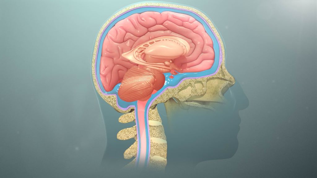
Cerebrospinal fluid (CSF) protects the central nervous system (CNS). Credit: Scientific Animations Wiki CC-BY 4.0
Deep inside your brain, a clear liquid is constantly on the move. Cerebrospinal fluid (CSF) cushions the brain, delivers nutrients, removes waste and keeps pressure stable. Think of it as an internal tide, circulating through cavities in the brain and around the spinal cord to keep this delicate organ in balance. When that flow is disrupted, the consequences can be serious.
For decades, doctors have relied mainly on static brain scans to guide treatment. But structure tells only part of the story. Researchers at the University of Pretoria (UP) are now focusing on something more dynamic: how fluid actually moves.
Two of the most common neurosurgical conditions worldwide – brain tumours and hydrocephalus (a dangerous build-up of fluid in the brain) – are closely tied to disturbed CSF circulation. Tumours can block or distort the pathways through which fluid moves. Hydrocephalus represents a more obvious breakdown, where fluid accumulates and pressure rises. In both cases, symptoms such as headaches, problems with vision and neurological decline are not simply caused by the presence of disease, but by changes in pressure and pulsating flow inside the skull.
Professor Llewellyn Padayachy, Head of the Department of Neurosurgery at BTC@UP explains: “The optic nerve, which connects the eye to the brain, is surrounded by the same protective layers as the brain itself. CSF flows along this nerve, meaning changes in brain pressure can subtly affect structures at the back of the eye. By using advanced, non-invasive eye imaging, researchers can detect signs of altered fluid flow and pressure without inserting monitors into the brain.”
This matters enormously for children with hydrocephalus and patients with brain tumours who require long-term monitoring. It offers a safer, repeatable way to track disease progression and treatment response. In low- and middle-income countries, where hydrocephalus is common but access to advanced imaging and neurosurgical infrastructure may be limited, such non-invasive tools could reduce reliance on costly technology while still delivering meaningful clinical insight.
The research also helps refine innovation. Modern shunts and endoscopic procedures increasingly aim to restore more natural fluid circulation rather than simply drain excess fluid. Objective eye-based markers provide measurable ways to evaluate whether these technologies truly improve flow.
While the link between the eye and brain pressure has long been recognised, what is new is the integration of advanced imaging, physiological modelling and continuous monitoring. This approach treats CSF flow as a living system, and shifts care from reacting to late damage towards detecting subtle change earlier.
Why this research matters
This work reframes brain disease through a simple but powerful idea: health depends on flow. By learning to read the movements of brain fluid, even through the eye, researchers are paving the way for safer monitoring, smarter surgery and more equitable neurological care worldwide.
Fast fact
The most common surgical treatment for hydrocephalus is the surgical placement of a shunt, which has one of the highest failure rates of any medical device on the market.
Abdominal contractions are tightly linked to gentle brain movements that help circulate CSF
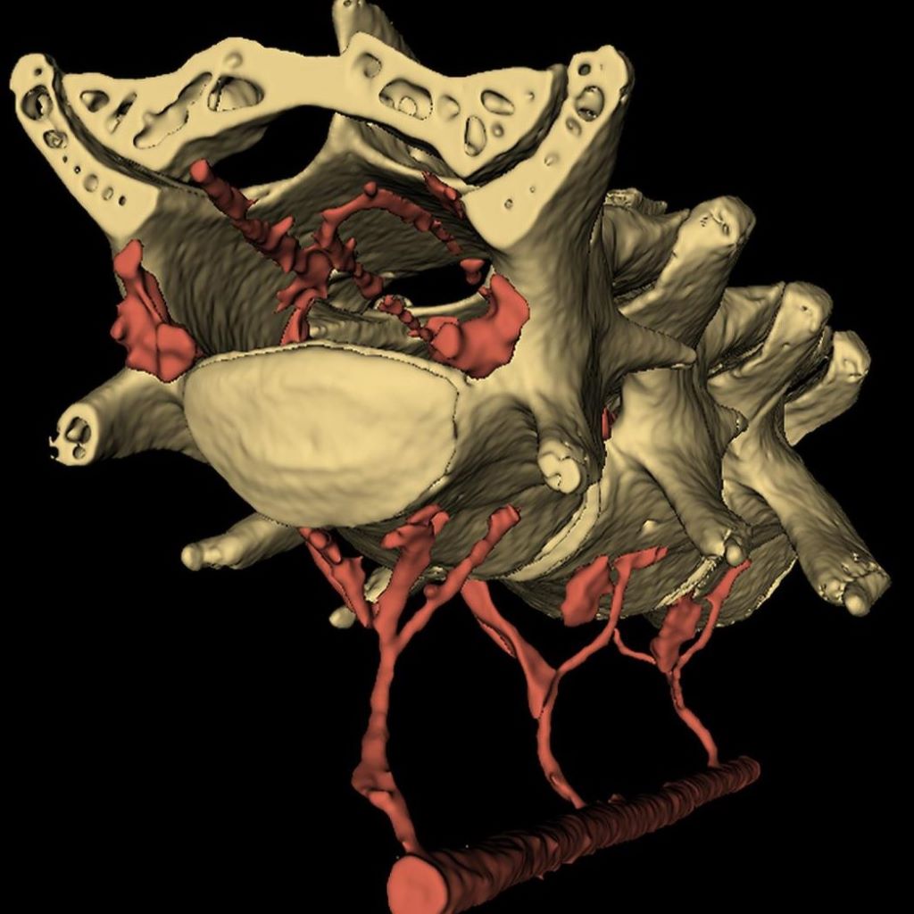
Using microCT scanning, which allows for high-resolution imaging of an organism’s internal structures, and other imaging techniques, researchers found that a network of veins serve as a mechanical connection between the abdominal cavity and the brain. Here, the veins in red run through the interior of a vertebrae and around the spine. Credit: Provided by Patrick Drew and team/Penn State. All Rights Reserved.
The brain is more mechanically connected to the body than previously appreciated, scientists reported in Nature Neuroscience. Through a study using mice and simulations, the team found a potential biological mechanism underlying why exercise is thought to benefit brain health: abdominal contractions compress blood vessels connected to the spinal cord and the brain, enabling the organ to gently move within the skull. This swaying facilitates the surrounding cerebrospinal fluid to flow over the brain, potentially washing away neural waste that could cause problems for brain function.
According to Patrick Drew, professor of engineering science and mechanics, of neurosurgery, of biology and of biomedical engineering at Penn State, the work builds on previous studies detailing how sleep and neuron loss can influence how and when cerebrospinal fluid flushes through the brain.
“Our research explains how just moving around might serve as an important physiological mechanism promoting brain health,” said Drew, corresponding author on the paper. “In this study, we found that when the abdominal muscles contract, they push blood from the abdomen into the spinal cord, just like in a hydraulic system, applying pressure to the brain and making it move. Simulations show that this gentle brain movement will drive fluid flow in and around the brain. It is thought the movement of fluid in the brain is important for removing waste and preventing neurodegenerative disorders. Our research shows that a little bit of motion is good, and it could be another reason why exercise is good for our brain health.”
Drew, who also holds the title of associate director of the Huck Institutes of the Life Sciences, explained how in a hydraulic system, a pump creates pressure that drives fluid flow. In this case, the pump is the abdominal contraction – which can be as light as the tensing prior to sitting up or taking a step. The contraction puts pressure on the vertebral venous plexus, a network of veins that connect the abdominal cavity to the spinal cavity, causing the brain to move.
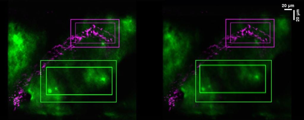
The researchers used two-photon microscopy — which allows for high-definition imaging of living tissue — to observe the brain shifting in the moments before the mouse moved, but right after the tightening of the abdominal muscles needed to spur the body into further movement. On the left, the brain, in green, sits during a stationary moment, while the image on the right shows the brain during movement. Credit: Provided by Patrick Drew and team/Penn State. All Rights Reserved.
The researchers visualised the process in moving mice with two advanced imaging technologies: two-photon microscopy, which allows for high-definition imaging of living tissue, and microcomputed tomography, which enables high-resolution 3D examination of whole organs. They observed the brain shifting in the moments before the mouse moved, but right after the tightening of the abdominal muscles needed to spur the body into further movement.
To confirm that it was abdominal contractions rather than other movement that acted as the pump, the researchers applied gentle and controlled pressure to the abdomens of lightly anaesthetised mice. With no other movement other than a localised mechanical pressure less than a human would experience with a blood pressure cuff, the mice’s brains shifted.
“Importantly, the brain began moving back to its baseline position immediately upon relief of the abdominal pressure,” Drew said. “This suggests that abdominal pressure can rapidly and significantly alter the position of the brain within the skull.”
With the abdominal contraction-brain movement link confirmed, Drew said the next step was to understand the fluid’s movement in the brain and if the brain’s movement could induce fluid flow. However, there previously were no existing imaging techniques to visualize the rapid, nuanced dynamics of such fluid flows.
“Luckily, our interdisciplinary team at Penn State was able to develop these techniques, including conducting the imaging experiments of living mice and creating computer simulations of fluid motion,” Drew said. “That combination of expertise is so important for understanding these types of complicated systems and how they impact health.”
Francesco Costanzo, professor of engineering science and mechanics, of biomedical engineering, of mechanical engineering and of mathematics, led the computational modelling.
“Modelling fluid flow in and around the brain offers unique challenges because there are simultaneous, independent movements, as well as time-dependent, coupled movements. Accounting for all of them requires accounting for the special physics that happens every time a fluid particle crosses one of the many membranes in the brain,” Costanzo said. “So, we simplified it. The brain has a structure similar to a sponge, in the sense that you have a soft skeleton and fluid can move through it.”
By simplifying the geometry of the brain to that of a sponge, Costanzo explained that the team could model how fluid flows through a structure with varied spaces, like wrinkles in the brain, or pores in the sponge.
“Keeping with the idea of the brain as a sponge, we also thought of it as a dirty sponge – how do you clean a dirty sponge?” Costanzo asked. “You run it under a tap and squeeze it out. In our simulations, we were able to get a sense of how the brain moving from an abdominal contraction can help induce fluid flow over the brain to help clear waste products.”
Drew emphasised that while more work is needed to understand the full implications in humans, this study suggests that body movement may help to cycle cerebrospinal fluid around and in the brain, removing waste and helping to protect against neurodegenerative disorders associated with waste buildup.
“This kind of motion is so small. It’s what’s generated when you walk or just contract your abdominal muscles, which you do when you engage in any physical behaviour. It could make such a difference for your brain health,” Drew said.
Researchers have discovered new diagnostic and prognostic markers for multiple sclerosis.
This is a pseudo-colored image of high-resolution gradient-echo MRI scan of a fixed cerebral hemisphere from a person with multiple sclerosis.
Credit: Govind Bhagavatheeshwaran, Daniel Reich, National Institute of Neurological Disorders and Stroke, National Institutes of Health
Researchers from the Max Planck Institute of Biochemistry and the Technical University of Munich (TUM) have discovered new diagnostic markers for multiple sclerosis (MS), a disease which affects 3 million people worldwide.
Using mass spectrometry, about 1500 proteins were analysed simultaneously per sample in the cerebrospinal fluid (CSF) of 5000 patients. The study uncovered a set of marker proteins that improve differentiation of MS from other inflammatory brain diseases where classical MS markers are negative. Additionally, the study identified changes in the CSF proteome that may potentially predict disease progression. This approach could also open up new avenues for the diagnosis of other diseases. The study was published in Cell.
Unspecific neurological symptoms can lead to lengthy or even inaccurate diagnoses of diseases, which is why improved protein markers are needed for swift and clear diagnosis
Using a new mass spectrometry method, approximately 1500 proteins were analyzed per cerebrospinal fluid sample across 5000 patients, and up to 2000 proteins in a further improved method
A new set of disease markers enable improved differentiation of multiple sclerosis (MS) from other inflammatory brain diseases, in particular for patients lacking the classical markers
The proteome of the cerebrospinal fluid (CSF) at diagnosis is informative for various aspects of disease evolution in a patient, such as long-term disability, risk of conversion from relapsing to progressive disease course and time to conversion
The method also has the potential to discover other proteins that could be used as markers for the diagnosis of other neurological diseases
Biomarker needs for multiple sclerosis
Imagine living with unexplained neurological symptoms: numbness, visual disturbances, fatigue, but not receiving a clear diagnosis for months or years. Non-specific neurological symptoms can make diagnosis difficult because, despite modern imaging techniques, there are no reliable molecular biomarkers for many neurological diseases.
Professor Bernhard Hemmer, head of the Department of Neurology at TUM University Hospital, explains: “The diagnosis of neurological diseases such as multiple sclerosis is based on a combination of imaging techniques using magnetic resonance imaging (MRI) and cerebrospinal fluid (CSF) analysis. While MRI reveals inflammatory changes in the brain and spinal cord, CSF shows chronic immune activity in the nervous system. In most cases, this combination enables a reliable diagnosis. In individual cases, however, differentiation can be challenging. This can lead to lengthy and less reliable diagnoses and is associated with uncertain and delayed treatment decisions. For this reason, we need new biomarkers to better diagnose the various diseases. In addition to diagnostic challenges, predicting disease progression, particularly disability accumulation, to guide optimal treatment, remains a major unmet need in MS”.
Proteomic study of cerebrospinal fluid across neurological diseases
In order to find new biomarkers, neurologists Bernhard Hemmer and Christiane Gasperi, both experts in MS research at TUM, have joined forces with Professor Matthias Mann, a world-leading expert in proteomics research. Matthias Mann, director at the MPI of Biochemistry, explains: “We have been developing the technology for measuring proteins using mass spectrometry in our laboratory together with colleagues for decades. Now we can reliably and accurately measure proteins in body fluids. However, for a long time researchers could only measure tens to hundreds of samples and only those proteins with the highest concentration in a body fluid. These proteins often turned out not to be the best markers for diseases. To go one step further, we combined the latest advances in mass spectrometry hardware, software, and sample preparation and adapted the workflow to cerebrospinal fluid.”
In this study, CSF samples from more than 5000 people with a wide range of neurological diseases were analysed. Jakob Bader, first author of the study and postdoctoral researcher in the field of proteomics research, explains: “Proteomics is a scientific discipline that aims to characterise a biological system by measuring all proteins, or at least as many as possible. For our study, it is essential to cover as many proteins as possible in order to increase the likelihood of measuring and later finding real disease markers in our analyses. The great advantage of this proteomic approach is that the identity of the markers does not have to be known beforehand. This saves years of research work in which individual candidates are examined one after the other.”
To avoid misinterpreting random differences between people as disease markers, it is essential to have a sufficient number of patients. Similarly, it is only possible to determine whether a marker is specific to a particular disease by considering the many other relevant diseases in parallel. “The breakthrough was achieving both objectives simultaneously: Analysing thousands of proteins while studying thousands of patients across many neurological diseases.” Jakob Bader adds.
A systematic analysis of disease effects and possible confounders
The 5,000 CSF samples came from a wide range of neurological disorders, including stroke, brain cancer, infections, autoimmune diseases such as MS, and others. Additionally, patient samples were analysed from individuals who had provided CSF samples for the diagnosis of severe headache disorders but in whom no neurological disease was found. This allowed the researchers to use these samples as controls. Systematic comparison of these disorders revealed shared and specific protein deviations from the controls.
For diagnostic use, an elevated protein concentration rarely points unambiguously to a single disorder. The study further revealed that disease-unspecific other effects like a person’s age, sex, and in particular degradation of the barriers insulating the brain from the CSF have a very large impact on the composition of this fluid, which complicates the quest for disease markers.
Biomarkers for a hard-to-identify form of multiple sclerosis
To showcase the potential of proteomic analysis for biomarker discovery, the researchers focused on the search for diagnostic markers for MS, a challenging task but with a direct medical need. Physician Christiane Gasperi says: “In approximately 10% of MS patients, diagnosis of the disease is particularly difficult because they lack the typical MS marker of so-called oligoclonal bands of antibodies that are specific to the CSF and not found in the blood. This complicates and potentially delays the diagnosis.”
She continues: “However, for our patients, a quick and clear diagnosis of the disease is of enormous importance. While current therapies cannot cure MS, they can slow its progression and reduce the long-term disability. That makes it crucial to start treatment early. At the same time, these therapies can have significant side effects, so treatment decisions require a high level of diagnostic certainty. When this confidence is not reached yet, therapy is often delayed. Thus, MS patients really benefit from an early intervention that depends on a clear and early diagnosis.”
To find better markers, the researchers applied an enhanced version of the proteomic method to measure about 2000 proteins in samples of MS and other inflammatory diseases of the CNS, which can mimic MS, and thus pose the greatest diagnostic challenges. This let them identify a set of 22 proteins that distinguishes MS from these inflammatory diseases with better accuracy than other parameters in the CSF that are currently measured in clinical practice. Christiane Gasperi comments: “It is particularly encouraging that we have found a combination of marker proteins that help in the diagnosis of this particularly difficult-to-identify form of MS.”
Predicting disease progression at diagnosis
Beyond improving diagnosis, the study also addressed a second major challenge: Some patients remain relatively stable for many years, while others accumulate disability more rapidly or transition from the relapsing disease course that is typical early on to a progressive course where disability accumulates persistently. At the time of diagnosis, it is very difficult to predict which trajectory a patient will follow. This uncertainty complicates treatment decisions and can be deeply unsettling for those newly diagnosed.
By analyzing hundreds of MS patient samples, the researchers showed that the CSF proteome at the time of diagnosis was associated with the level of disability years later. In addition, these patterns reflected a higher risk for patients to convert from the relapsing to the progressive disease course, as well as shorter times until such conversion occurred. Bernhard Hemmer explains: “Our findings suggest that important aspects of future disability and disease course are reflected in the proteome from the very beginning. This demonstrates that the biological information required for a prognostic test is already present at diagnosis.”
He summaries the study: “For diagnosis, we were able to define and validate a focused protein panel that improves differentiation in difficult cases. Additionally, we found that the overall protein pattern in the CSF at the time of diagnosis is linked to how the disease develops years later. Together, these findings bring us closer to more precise diagnosis and a more individualized treatment strategy from the very beginning.
An avenue for efficient biomarker discovery in neurology
Matthias Mann sees broader potential: “Proteins control almost all biological processes in the body and have long been the most important group of diagnostic markers. Nevertheless, we are probably only at the beginning here. With the methodology established here, we can now analyse the proteome in the CSF of many patients with an unprecedented number of proteins. This technological progress changes how we should search for biomarkers. Comprehensive proteome analysis of large patient collectives promise to be the most efficient path to new and better biomarkers. Beyond MS, this approach opens up prospects for many other diseases of the central nervous system – from Alzheimer’s and Parkinson’s to brain tumours and other neurological disorders.”
A novel, multi-analyte test developed by researchers at Johns Hopkins Medicine can accurately identify brain cancers using small samples of cerebrospinal fluid (CSF), offering a promising new tool to guide clinical decision-making.
The findings, supported by funding from the National Institutes of Health, were published in Cancer Discovery and demonstrate that combining multiple biological markers, including tumour-derived DNA and immune cell signatures, is more effective for diagnosing central nervous system cancers than using any one marker alone.
“This study highlights how much more information we can gain when we evaluate several analytes together,” says senior study author Chetan Bettegowda, MD, PhD, Professor and Director of the Department of Neurosurgery at the Johns Hopkins University School of Medicine. “The ability to detect cancers with high specificity and also gain insight into the immune environment of the brain could be an important advance in the care of patients with brain tumours.”
To evaluate the potential of a multi-analyte approach, investigators analysed 206 CSF samples, including samples from patients with high-grade gliomas, medulloblastomas, metastases and central nervous system lymphomas. Their test, called CSF-BAM (cerebrospinal fluid–B/T cell receptor, aneuploidy and mutation), measured chromosomal abnormalities, tumour-specific mutations, and T and B cell receptor sequences. In combination, these markers identified brain cancers with more than 80% sensitivity (ability to detect cancer) and 100% specificity (correctly identified those who were cancer-free) in the validation cohort. The 100% specificity means no false positives were recorded among individuals with noncancerous conditions.
The study also showed that the assay could distinguish between the immune cell populations present in cancer and noncancer cases, offering additional biological context that could be helpful in more-challenging clinical scenarios. Investigators say this ability to categorize T and B cell populations in the CSF provides insights into both disease presence and immune response.
“Many patients with brain lesions face invasive diagnostic procedures to confirm a cancer diagnosis,” says Christopher Douville, MD, assistant professor of oncology and a senior study author. “A tool like this could help us make better-informed decisions about who really needs a biopsy and who doesn’t.”
Researchers say the test could be particularly useful for cases in which conventional imaging or cytology is inconclusive, or in situations when obtaining tissue for diagnosis is risky or not possible. The multi-analyte approach, they say, enables clinicians to better detect cancer and better understand the disease status, supporting a more tailored approach to patient care.
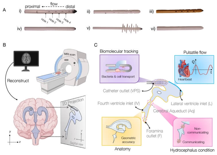
Schematic of approach to simulating brain shunt fluid dynamics. Credit: Harvard SEAS
Millions of people worldwide suffer from hydrocephalus, a condition which recently received greater attention when Billy Joel announced his diagnosis. Treatment usually involves surgical placement of shunts to divert cerebrospinal fluid away, but this procedure often leads to complications, infections, and multiple re-treatments.
Bioengineers in the Harvard John A. Paulson School of Engineering and Applied Sciences (SEAS) have now developed a new computational model to aid the creation of shunts tailored to individual patients’ anatomy and needs. The model combines brain anatomy, fluid flow, and biomolecular transport dynamics to simulate shunt performance with pinpoint accuracy.
The work was supported by federal funding from the National Science Foundation and published in Proceedings of the National Academy of Sciences. It was led by SEAS postdoctoral fellow Haritosh Patel, who works in the labs of Joanna Aizenberg, Professor of Materials Science at SEAS and Professor of Chemistry and Chemical Biology; and Venkatesh Murthy, Professor of Molecular and Cellular Biology and Director of the Center for Brain Science.
Repeat surgeries due to infection or obstruction
Tens of thousands of shunt procedures are performed annually in the U.S. — many of which are repeat surgeries due to the inserted devices becoming blocked or obstructed, or the patient suffering an infection.
“Some elderly patients told me they had had over 10 surgeries — one every two to three years,” Patel said. “We really wanted to understand why this was happening, and we realised that many of these obstructions and infections were tied to shunt designs that didn’t fully consider fluid dynamics as a fundamental part of their geometry. We noticed that the tubing geometry used in shunts closely resembles the kind of piping we rely on in household plumbing. While that simplicity has its advantages, we saw an opportunity to explore more creative, biomimetic solutions that better suit the complexity of the brain’s environment.”
Pursuing the problem from both a material and design perspective, the team quickly realized there was no universally accepted fluid flow model for the brain ventricle space to guide them. “Okay, well, we can’t test our devices in a model, so why don’t we first make a better model?” Patel said.
Computational tool simulates fluid flow in brain
The result is their computational tool, called BrainFlow, which combines detailed anatomical and physiological features of the brain to simulate the flow of cerebrospinal fluid flow in the presence of shunt implants.
The model incorporates patient-specific medical imaging data along with pulse-induced flow to mimic a patient’s cerebrospinal fluid dynamics, all to offer insight into optimal shunt design, placement, and even choice of materials.
“We believe that our model, combined with novel geometries and materials improvements such as anti-biofouling coatings developed in my lab, could lead to smoother integration of optimized, patient-specific medical devices into patients’ brains, with less likelihood of complications, and a better quality of life,” Aizenberg said.
The Harvard team is currently conducting studies that use the model to test different designs of shunts and calculate their efficacy.
A misfolded protein facilitates reliable diagnosis even in the early stages of Parkinson’s disease in body fluids.
Source: CC0
Parkinson’s disease is a neurodegenerative disorder that is usually diagnosed in its late stage on the basis of clinical symptoms, mainly motor disorders. By this point, however, the brain is already severely and irreparably damaged. Moreover, diagnosis is difficult and often incorrect because the disease takes many forms and symptoms overlap with other disorders.
Researchers from the PRODI Center for Protein Diagnostics at Ruhr University Bochum, Germany, and the biotech company betaSENSE have now discovered a biomarker in cerebrospinal fluid (CSF) that facilitates a reliable diagnosis at an early stage and can shed light on the progression of the disease and the effect of a therapy. They report their findings in the journal EMBO Molecular Medicine.
Parkinson’s disease – an unstoppable condition
Parkinson’s disease is characterised by the loss of dopaminergic nerve cells in the brain, typically leading to increasing motor impairments as the symptoms progress. Dopamine supplements can compensate for the loss and temporarily alleviate the symptoms. The misfolding of the key protein alpha-synuclein (αSyn) from α-helical structures to β-sheet-rich structures plays a crucial role in the development of Parkinson’s disease. “These misfoldings make the protein sticky, leading to the formation of larger complexes, so-called oligomers. The oligomers then produce long fibrillar filaments and cause the aggregation of these filaments into macroscopically large Lewy bodies in the brain,” explains Professor Klaus Gerwert, founding and managing director at PRODI and CEO of betaSENSE.
Advanced platform technology
In two independent clinical cohorts with a total of 134 participants, the Bochum-based researchers showed that, with a sensitivity and specificity of well over 90%, this misfolding of αSyn in body fluids is a viable biomarker for the diagnosis of Parkinson’s disease. The research was conducted using cerebrospinal fluid samples from patients at the Parkinson’s centres in Bochum (St. Josef Hospital, Professor Lars Tönges, Professor Ralf Gold) and Kassel (Paracelsus-Elena-Klinik, Dr. Sandrina Weber, Professor Brit Mollenhauer). The measurements were carried out using the patented iRS (immuno-infrared sensor) technology from betaSENSE GmbH.
betaSENSE has already successfully implemented the iRS technology for diagnosing Alzheimer’s disease. In this case, it was shown that the misfolding of the biomarker Aβ can indicate the risk of Alzheimer’s dementia at a later stage with high accuracy up to 17 years before clinical diagnosis. “We have now transferred this approach to Parkinson’s for the misfolding of αSyn,” stresses Klaus Gerwert.
Development of Parkinson’s drugs
In addition to diagnostic applications, the technology can also help to develop new active substances and prove their efficacy in clinical trials.
New research describes how a spreading wave of disruption and the flow of fluid in the brain triggers headaches, detailing the connection between the neurological symptoms associated with aura and the migraine that follows. The study, which appears in Science, also identifies new proteins that could be responsible for headaches and may serve as foundation for new migraine drugs.
“In this study, we describe the interaction between the central and peripheral nervous system brought about by increased concentrations of proteins released in the brain during an episode of spreading depolarization, a phenomenon responsible for the aura associated with migraines,” said lead author Maiken Nedergaard, MD, DMSc, co-director of the University of Rochester Center for Translational Neuromedicine. “These findings provide us with a host of new targets to suppress sensory nerve activation to prevent and treat migraines and strengthen existing therapies.”
It is estimated that one out of 10 people experience migraines and in about a quarter of these cases the headache is preceded by an aura, a sensory disturbance that can includes light flashes, blind spots, double vision, and tingling sensations or limb numbness. These symptoms typically appear five to 60 minutes prior to the headache.
The cause of the aura is a phenomenon called cortical spreading depression, a temporary depolarization of neurons and other cells caused by diffusion of glutamate and potassium that radiates like a wave across the brain, reducing oxygen levels and impairing blood flow. Most frequently, the depolarization event is located in the visual processing centre of the brain cortex, hence the visual symptoms that first herald a coming headache.
While migraines auras arise in the brain, the organ itself cannot sense pain. These signals must instead be transmitted from the central nervous system to the peripheral nervous system. The process of communication between the brain and peripheral sensory nerves in migraines has largely remained a mystery.
Fluid dynamics models shed light on migraine pain origins
Nedergaard and her colleagues at the University of Rochester and the University of Copenhagen are pioneers in understanding the flow of fluids in the brain. In 2012, her lab was the first to describe the glymphatic system, which uses cerebrospinal fluid (CSF) to wash away toxic proteins in the brain. In partnership with experts in fluid dynamics, the team has built detailed models of how the CSF moves in the brain and its role in transporting proteins, neurotransmitters, and other chemicals.
The most widely accepted theory is that nerve endings resting on the outer surface of the membranes that enclose the brain are responsible for the headaches that follow an aura. The new study, which was conducted in mice, describes a different route and identifies proteins, many of which are potential new drug targets, that may be responsible for activating the nerves and causing pain.
As the depolarization wave spreads, neurons release a host of inflammatory and other proteins into CSF. In a series of experiments in mice, the researchers showed how CSF transports these proteins to the trigeminal ganglion, a large bundle of nerves that rests at the base of the skull and supplies sensory information to the head and face.
It was assumed that the trigeminal ganglion, like the rest of the peripheral nervous system, rested outside the blood-brain-barrier, which tightly controls what molecules enter and leave the brain. However, the researchers identified a previously unknown gap in the barrier that allowed CSF to flow directly into the trigeminal ganglion, exposing sensory nerves to the cocktail of proteins released by the brain.
Migraine-associated proteins double during brain wave activity
Analysing the molecules, the researchers identified twelve proteins called ligands that bind with receptors on sensory nerves found in the trigeminal ganglion, potentially causing these cells to activate. The concentrations of several of these proteins found in CSF more than doubled following a cortical spreading depression. One of the proteins, calcitonin gene-related peptide (CGRP), is already the target of a new class of drugs to treat and prevent migraines called CGRP inhibitors. Other identified proteins are known to play a role in other pain conditions, such as neuropathic pain, and are likely important in migraine headaches as well.
“We have identified a new signaling pathway and several molecules that activate sensory nerves in the peripheral nervous system. Among the identified molecules are those already associated with migraines, but we didn’t know exactly how and where the migraine inducing action occurred,” said Martin Kaag Rasmussen, PhD, a postdoctoral fellow at the University of Copenhagen and first author of the study. “Defining the role of these newly identified ligand-receptor pairs may enable the discovery of new pharmacological targets, which could benefit the large portion of patients not responding to available therapies.”
The researchers also observed that the transport of proteins released in one side of the brain reaches mostly the nerves in the trigeminal ganglion on the same side, potentially explaining why pain occurs on one side of the head during most migraines.
A new study suggests that some patients diagnosed with behavioural-variant frontotemporal dementia (bvFTD) – a presently incurable, mentally debilitating condition – may instead have a cerebrospinal fluid leak, which is detectable on MRI scans and often treatable. The researchers say these findings, published in the peer-reviewed journal Alzheimer’s & Dementia: Translational Research and Clinical Interventions, could lead to a cure.
“Many of these patients experience cognitive, behavioural and personality changes so severe that they are arrested or placed in nursing homes,” said Wouter Schievink, MD, professor of Neurosurgery at Cedars-Sinai. “If they have behavioural-variant frontotemporal dementia with an unknown cause, then no treatment is available. But our study shows that patients with cerebrospinal fluid leaks can be cured if we can find the source of the leak.”
When cerebrospinal fluid (CSF) leaks into the body, the brain can sag, causing dementia symptoms. Schievink said many patients with brain sagging, detectable in MRI, go undiagnosed, and he advises clinicians to take a second look at patients with telltale symptoms.
“A knowledgeable radiologist, neurosurgeon or neurologist should check the patient’s MRI again to make sure there is no evidence for brain sagging,” Schievink said.
Clinicians can also ask about a history of severe headaches that improve when the patient lies down, significant sleepiness even after adequate night-time sleep, and whether the patient has ever been diagnosed with a Chiari brain malformation, a condition in which brain tissue extends into the spinal canal. Brain sagging, Schievink said, is often mistaken for a Chiari malformation.
Even when brain sagging is detected, the source of a CSF leak can be difficult to locate. When the fluid leaks through a tear or cyst in the surrounding membrane, it is visible on CT myelogram imaging with the aid of contrast medium.
Schievink and his team recently discovered an additional cause of CSF leak: the CSF-venous fistula. In these cases, the fluid leaks into a vein, making it difficult to see on a routine CT myelogram. To detect these leaks, technicians must use a specialized CT scan and observe the contrast medium in motion as it flows through the cerebrospinal fluid.
In this study, investigators used this imaging technique on 21 patients with brain sagging and symptoms of bvFTD, and they discovered CSF-venous fistulas in nine of those patients. All nine patients had their fistulas surgically closed, and their brain sagging and accompanying symptoms were completely reversed.
“This is a rapidly evolving field of study, and advances in imaging technology have greatly improved our ability to detect sources of CSF leak, especially CSF-venous fistula,” said Keith L. Black, MD, chair of the department of Neurosurgery at Cedars-Sinai. “This specialised imaging is not widely available, and this study suggests the need for further research to improve detection and cure rates for patients.”
The remaining 12 study participants, whose leaks could not be identified, were treated with nontargeted therapies designed to relieve brain sagging, such as implantable systems for infusing the patient with CSF. However, only three of these patients experienced relief from their symptoms.
“Great efforts need to be made to improve the detection rate of CSF leak in these patients,” Schievink said. “We have developed nontargeted treatments for patients where no leak can be detected, but as our study shows, these treatments are much less effective than targeted, surgical correction of the leak.”
Advances in neuro-imaging and molecular biology have unearthed a subtle, previously unknown layer in the brain. As described in the journal Science, the newly discovered layer forms a previously unknown component of brain anatomy that acts as both a protective barrier and platform from which immune cells monitor the brain for infection and inflammation.
“The discovery of a new anatomic structure that segregates and helps control the flow of cerebrospinal fluid (CSF) in and around the brain now provides us much greater appreciation of the sophisticated role that CSF plays not only in transporting and removing waste from the brain, but also in supporting its immune defenses,” said Maiken Nedergaard, co-director of the Center for Translational Neuromedicine at University of Rochester and the University of Copenhagen. Nedergaard and her colleagues have made significant findings in the field of neuroscience, including detailing the many critical functions of previously overlooked cells in the brain called glia and the brain’s unique process of waste removal, which the lab named the glymphatic system.
The study focuses on the series of membranes that encase the brain, creating a barrier from the rest of the body and keeping the brain bathed in CSF. The traditional understanding of what is collectively called the meningeal layer identifies the three individual layers as dura, arachnoid, and pia matter.
This new layer discovered by the international research team further divides the space between the arachnoid and pia layers, the subarachnoid space, into two compartments, separated by the newly described layer, which the researchers name SLYM (Subarachnoidal LYmphatic-like Membrane). While the paper mostly describes the function of SLYM in mice, it also reports its presence in the adult human brain as well.
SLYM is a type of membrane that lines other organs in the body, including the lungs and heart, called mesothelium. These membranes typically surround and protect organs, and harbour immune cells.
The new membrane is very thin and delicate, consisting of only a few cells in thickness. Yet SLYM is a tight barrier, allowing only very small molecules to transit and it also seems to separate “clean” and “dirty” CSF. This last observation hints at the likely role played by SLYM in the glymphatic system, which requires a controlled flow and exchange of CSF, allowing the influx of fresh CSF while flushing the toxic proteins associated with Alzheimer’s and other neurological diseases from the central nervous system. This discovery will help researchers more precisely understand the mechanics of the glymphatic system.
Central nervous system immune cells (indicated here expressing CD45) use SLYM as a platform close to the brain’s surface to monitor cerebrospinal fluid for signs of infection and inflammation.
The SLYM also appears important to the brain’s defences. The central nervous system has its own native population of immune cells, and the membrane’s integrity prevents outside immune cells from entering. In addition, the membrane appears to host its own population of central nervous system immune cells that use SLYM as an observation point close to the surface of the brain from which to scan passing CSF for signs of infection or inflammation.
Discovery of the SLYM opens the door for further study of its role in brain disease. For example, the researchers note that larger and more diverse concentrations of immune cells congregate on the membrane during inflammation and aging. Furthermore, when the membrane was ruptured during traumatic brain injury, the resulting disruption in the flow of CSF impaired the glymphatic system and allowed non-central nervous system immune cells to enter the brain.
These and similar observations suggest that diseases as diverse as multiple sclerosis, central nervous system infections, and Alzheimer’s might be triggered or worsened by abnormalities in SLYM function. They also suggest that the delivery of drugs and gene therapeutics to the brain may be impacted by SLYM, which will need to be considered as new generations of biologic therapies are being developed.
In a finding reminiscent of how vampires and zombies in fiction get sustenance from their victims, a team of researchers reported in the journal Nature that injecting cerebrospinal fluid (CSF) from young mice into old mice improves the memory and cognitive abilities of the older mice
Such an approach is nothing new, although the chief obstacle was safely harvesting such a tiny amount of CSF from the small animals. About two decades ago, studies had reported that transferring blood from younger mice to older ones notably improved the health of the older mice, giving them a ‘rejuvenating’ effect. It did not take long for people to take note of this discovery, with a startup company offering transfers of young people’s plasma for exorbitant amounts to wealthy older clients in the unproven hopes of reversing ageing. Fears of a dystopian future were averted when the US Food and Drug Administration released a statement stating such transfers had no clinical benefit, and the company folded. However, research continued.
Since ageing is too complex to measure in a clinical trial anyway, scientists have been focusing on tackling specific aspects of it, such as in neurodegenerative diseases like Alzheimer’s and research has continued in this direction. A few years ago, human umbilical cord plasma was shown to revitalise hippocampal function in aged mice, andprevious work led by Tony Wryss-Coray, PhD had found that young mouse blood improved age-related impairments in cognition. Studies of fear conditioning had shown that proliferation of oligodendrocyte precursor cells (OPCs) was necessary for fear formation, which raised the question of whether CSF might affect this.
Infusing CSF taken from 10 week old mice over seven days, researchers trained 18 month old mice to associate a flashing light with an electric shock to the foot. The CSF infusion was shown to improve recall of the fear stimulus in the older mice and induce greater OPC proliferation.
“The broad message here is that the aging process is malleable, which of course is not new because of this paper,” senior author Dr Wyss-Coray said in an interview with MedPage Today. “But it adds to the idea that aging is a potential therapeutic target, a process we can start to understand better and start to manipulate.”
“The other message – one that’s more brain-specific – is that if you improve the environment in which neurons live, you can actually have a substantial improvement in function,” he added. “That may be as important, or even more important sometimes, than targeting neuronal processes themselves.”
The researchers isolated fibroblast growth factor 17 (Fgf17) infusion as being necessary for OPC proliferation, and blocking it in young mice impaired cognition.
“This suggests that Fgf17 is not only able to recapitulate some of the useful effects of CSF from young mice, but it also seems to be necessary to make a young brain function at its full capacity,” Dr Wyss-Coray said.