While a hug or friendly pat can seem soothing, the question remains: can touch really help you feel better, and does it matter who it’s from or how they touch you? To explore these questions, researchers from the Social Brain Lab at the Netherlands Institute for Neuroscience and the University Hospital Essen conducted a large-scale analysis of studies exploring touch interventions. Their results, published in Nature Human Behaviour, reveal that the benefits of touch may not depend on who – or even what – does the touching.
Though individual studies provide conflicting results, this large-scale analysis shows that touch substantially improves both physical and mental wellbeing, for example via reduction of pain, anxiety, depression, and stress in adults. But in fact, those with physical or mental health problems (and therefore most in need of support) benefit even more from touch than healthy adults. “This is especially relevant considering how often touch interventions are overlooked,” notes first author Julian Packheiser.
“A key question of our study is to leverage the hundreds of individual studies out there to identify what type of touch works best,” adds professor Christian Keysers, director of the Social Brain Lab. “What if you don’t have a friend or partner close by to hug you? Would touch from a stranger or even a machine also help? And how often? The study clearly shows that touch can indeed be optimised, but the most important factors are not necessarily those we suspect.”
Interestingly, the person touching you, how they touch you, and the duration of their touch doesn’t make a difference in terms of impact. A long-lasting massage by a therapist could therefore be just as effective as a quick hug offered by a friend. That is, until the frequency of the intervention is considered. The more often a touch intervention is offered, the greater the impact. A quick hug could therefore be even more impactful than a massage if it is offered more frequently.
Human or non-human touch?
The next question was whether touch intervention needs to be human at all. As it turns out, object or robot interventions can be equally effective at improving physical wellbeing. “There are lots of people in need of wellbeing improvements, perhaps because they’re lonely but also because they may be inflicted by clinical conditions. These results indicate that a touch-robot, or even a simple weighted blanket has the potential to help those people,” last author Frédéric Michon explains. However, the benefits of robot and object interventions are less effective for mental wellbeing. Mental health disorders like anxiety or depression might therefore require human touch after all, “perhaps suggestive of the importance for an emotional component associated with the touch,” Michon points out.
While the researchers were equally curious about human-to-animal contact, studies exploring this question are still lacking. “It would be useful to see whether an animal’s or pet’s touch could improve wellbeing, and inversely if they also benefit from it, but unfortunately there simply aren’t enough studies, or properly controlled ones, for us to draw any general conclusions on these topics,” Michon clarifies.
Touch interventions across ages
When the team looked into the impact of touch on newborns, they found out that newborns also benefited significantly from touch. However, the person conducting the touch intervention was more important: the benefits of touch are higher when done by a parent instead of a healthcare worker. “This finding could be impactful,” Packheiser adds. “Death rates due to premature births are high in some countries and the knowledge that a baby benefits more from the touch of their own parent offers another easily implementable form of support for the baby’s health.”
Due to a lack of studies, it proved difficult to draw conclusions about children and teenagers. “Large scale studies like this help us draw more general conclusions but they also help us identify where research is lacking,” Michon explains. “We hope that our findings can steer future research to explore lesser-known questions. This includes animal touch, but also touch across ages, and in specific clinical settings like autistic patients, another category that has not been explored extensively.”
People with aphantasia – who cannot visualise an image in their mind’s eye – are less likely to remember the details of important past personal events or to recognise faces, according to a review of nearly ten years of research. People who cannot bring to mind visual imagery are also less likely to experience imagery of other kinds, like imagining music, according to new research by the academic who first discovered the phenomenon.
Professor Adam Zeman, of the University of Exeter, first coined the term aphantasia in 2015, to describe those who can’t visualise. Since then, tens of thousands of people worldwide have identified with the description. Many say they knew they processed information differently to others but were unable to describe how. Some of them expressed shock on discovering that other people can conjure up an image in their mind’s eye.
Now, Professor Zeman has conducted a review of around 50 recent studies, published in Trends in Cognitive Sciences, to summarise findings in a field that has emerged since his first publication. Research indicates that aphantasia is not a single entity but has subtypes. For example, not everyone with aphantasia has a poor autobiographical memory or difficulty in recognising faces, and in a minority of people, aphantasia appeared to be linked to autism. People who cannot visualise are more likely to have scientific occupations. Unexpectedly, although people with aphantasia can’t visualise at will, they often dream visually.
Professor Zeman’s review provides evidence that whether people have aphantasia or hyperphantasia – a particularly vivid visual imagination – is linked to variations in their physiology and neural connectivity in the brain, as well as in behaviour. For example, listening to scary stories alters skin conductance in those with imagery, meaning people sweat – but this does not occur in people with aphantasia.
Aphantasia is thought to affect around 1% of the population, while 3% are hyperphantasic. These figures rise to 5–10% with more generous criteria for inclusion. Both aphantasia and hyperphantasia often run in families, hinting at the possibility of a genetic basis.
Professor Zeman, who now holds honorary contracts at the universities of Exeter and Edinburgh, said: “Coining the term ‘aphantasia’ has unexpectedly opened a window on a neglected aspect of human experience. It is very gratifying that people who lack imagery have found the term helpful, while a substantial surge of research is shedding light on the implications of aphantasia.
“Despite the profound contrast in subjective experience between aphantasia and hyperphantasia, effects on everyday functioning are subtle – lack of imagery does not imply lack of imagination. Indeed, the consensus among researchers is that neither aphantasia nor hyperphantasia is a disorder. These are variations in human experience with roughly balanced advantages and disadvantages. Further work should help to spell these out in greater detail.”
“I struggle to fully immerse myself in role-play with my children”
Solicitor Mary Wathen’s frustration that she struggled to engage in role playing games with her two young children, when she found all other engagement with her children so fulfilling, was her sign that she had aphantasia, meaning she cannot visualise imagery.
The 43-year-old, from Newent near Cheltenham, said: “One of my friends said that he uses the images in his head to enhance role play. When I asked him to explain this in more detail it became clear that he – and everyone else in the room – could easily create an image in their head and use that as the backdrop for the role play. This was totally mind-blowing to me. I just cannot understand what they really mean – where is this image and what does it look like? To me, unless you can see something with your eyes, it’s not there.”
Mary’s shock intensified when she realised her husband, has such vivid visual imagery that he is probably hyperphantasic. “He thinks in moving pictures, like movies – sometimes to the point that he can mistake those thoughts for memories. To me, that’s unfathomable.”
Mary has come to realise that her lack of visual imagery may well account for her difficulties with memory. She said: “I can comprehend and retain concepts and principles really well but I’m unable to recall facts and figures. I can’t recreate something in my head or ‘re see’ something that is not actually there in that moment.
“I’ve found it quite saddening to learn that other people can call to mind an image of their children when they’re not there. I’d love to be able to do that, but I just can’t – but I’ve learned to compensate by taking plenty of photos, so that I can relive those memories through those images.
“Whilst I’m sure there are wonderful advantages to being able to think in pictures, I think it’s important to remind myself that there are advantages to having aphantasia too. I’m a really good written and verbal communicator – I think that’s because I’m not caught up with any pictures, so I just focus on the power of the word. I’m also a deeply emotional person and perhaps that’s my brain’s way of overcompensating; I feel things as a way of experiencing them, rather than seeing them.
“I think it’s really important to raise awareness that some people just don’t have this ability – particularly as using visual imagination is a key way that young children are taught to learn and engage. Primary teachers need to know that some children just won’t be able to visualise and that could be why they’re not engaging in those kinds of activities. We need to ensure we cater for everyone and encourage other ways of learning and engaging.”
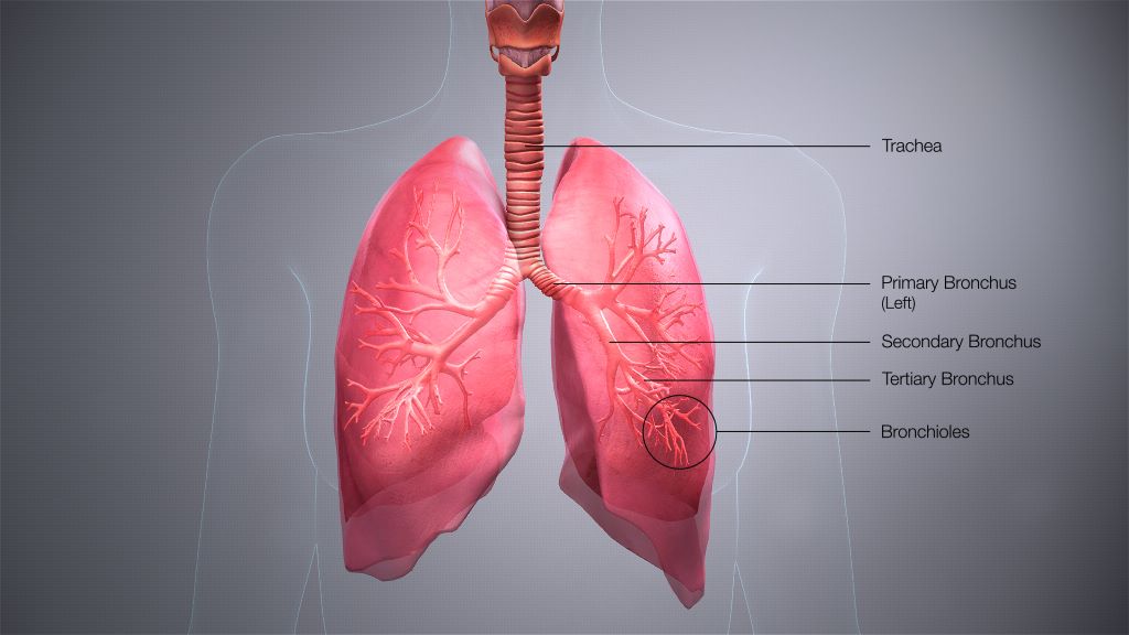
Scientists at King’s College London have discovered that the features of asthma attacks, a disease usually treated as being inflammatory, in fact stem from constriction of airways, making breathing difficult. The new study, published in Science, shows for the first time that many features of an asthma attack – inflammation, mucus secretion, and damage to the airway barrier that prevents infections – result from this mechanical constriction in a mouse model.
The findings suggest that blocking a process that normally causes epithelial cell death could prevent the damage, inflammation, and mucus that result from an asthma attack.
Professor Jody Rosenblatt from King’s College London said: “Our discovery is the culmination of more than ten years work. As cell biologists who watch processes, we could see that the physical constriction of an asthma attack causes widespread destruction of the airway barrier. Without this barrier, asthma sufferers are far more likely to get long-term inflammation, wound healing, and infections that cause more attacks. By understanding this fundamental mechanism, we are now in a better position to prevent all these events.”
Asthma symptoms include wheezing, coughing, feeling breathlessness and a tight chest. Triggers such as pollen or dust can make asthma symptoms worse and can lead to a life-threatening asthma attack.
Despite the disease commonality, the causes of asthma are still not understood. Current medications treat the consequences of an asthma attack by opening the airways, calming inflammation, and breaking up the sticky mucus which clogs the airway, which help control asthma, but do not prevent it.
The answer to stopping asthma symptoms may lie in cell extrusion, a process the researchers discovered that drives most epithelial cell death. Scientists used mouse lung models and human airway tissue to discover that when the airways contract, known as bronchoconstriction, the epithelial cells that line the airway get squeezed out to later die.
Because bronchoconstriction causes so many cell extrusions, it damages the airway barrier which causes inflammation and excess mucus.
In previous studies, the scientists found that the chemical compound gadolinium can block extrusion. In this study, they found it could work in mice to prevent the excess extrusion that causes damage and inflammation after an asthma attack. The authors note that gadolinium has not been tested in humans and has not been deemed to be safe or efficacious.
Professor Rosenblatt said: “This constriction and destruction of the airways causes the post-attack inflammation and excess mucus secretion that makes it difficult for people with asthma to breathe.
“Current therapies do not prevent this destruction – an inhaler such as Albuterol opens the airways, which is critical to breathing but, dishearteningly, we found it does not prevent the damage and the symptoms that follow an attack. Fortunately, we found that we can use an inexpensive compound, gadolinium which is frequently used for MRI imaging, to stop the airway damage in mice models as well as the ensuing inflammation and mucus secretion. Preventing this damage could then prevent the build-up of musculature that cause future attacks.”
Professor Chris Brightling from the University of Leicester and one of the co-authors of the study said: “In the last decade there has been tremendous progress in therapies for asthma particularly directed towards airway inflammation. However, there remains ongoing symptoms and attacks in many people with asthma. This study identifies a new process known as epithelial extrusion whereby damage to the lining of the airway occurs as a consequence of mechanical constriction and can drive many of the key features of asthma. Better understanding of this process is likely to lead to new therapies for asthma.”
The discovery of the mechanics behind cell extrusion could underlie other inflammatory diseases that also feature constriction such as cramping of the gut and inflammatory bowel disease.
In addition to shedding light on what people actually die of, autopsies can also play an important role in helping us to better understand disease. Tiyese Jeranji unpacks tuberculosis-related autopsy research in the Western Cape and delves into some of the fascinating complexities of this branch of TB research.
Figuring out how many people in South Africa die every year of tuberculosis (TB) is not straight-forward. On the one hand, Stats SA’s frequent mortality reports put the number at under 30 000, on the other hand, the World Health Organization (WHO) estimates that it is over 50 000.
While this may at first glance seem like a large discrepancy, there is a simple explanation. The Stats SA figures are based on what is written on death notifications, and these notifications very often do not tell the full story of what a person died of. The WHO estimate, is derived using mathematical modelling that triangulates estimates based on several data sources.
Looking at the numbers from studies that determine the cause of death (or what people actually died of) is one of the ways we know that relying on death notifications result in an undercount of TB deaths. Such autopsy studies have consistently found that many people had undiagnosed TB at the time of death and that the undiagnosed TB was often the actual cause of death.
One review study published in the journal AIDS concluded that “in resource-limited settings, TB accounts for approximately 40% of facility-based HIV/AIDS-related adult deaths” and that “almost half of this disease remains undiagnosed at the time of death”. According to WHO figures, of the estimated 280 000 people who fell ill with TB in South Africa in 2022, over 65 000 were not diagnosed.
Importance of autopsy research
Dr Muhammad Osman, Academic Portfolio Lead and Senior Lecturer: Public Health at the University of Greenwich, tells Spotlight that it is important to do TB autopsy studies because it enables us to identify TB that was not diagnosed during life – and this helps us understand the true burden of the disease.
Osman says identifying TB at autopsies has significant benefits. He says by overlaying health seeking behaviour (how people visit clinics), we can identify missed opportunities for TB screening and design interventions to improve screening for TB. “We could trace family contacts of the deceased and offer TB screening and prevention. This is not taking place at present,” he says.
Osman and his colleagues published a paper in the International Journal of Infectious diseases in 2021 looking at TB in people with sudden unexpected death (SUD) in Cape Town. They found that active TB was identified at post-mortems in 6.2% of the 770 cases they studied. More strikingly, in around 92% of those cases the TB had not been diagnosed while the person was alive.
Osman says that these days there is an increasing awareness of undiagnosed and untreated TB. He points out that new interventions to improve TB testing and diagnosis have been implemented such as targeted universal testing — an approach by which people who do not have any TB symptoms, but who are considered to be at high risk of TB, are routinely offered TB tests.
He says these days healthcare worker risk is considered more carefully and he stresses the importance of protecting forensic and pathology teams. (Forensics focuses on determining the cause and manner of death while pathology is the study and diagnosis of disease through examination of tissue, cells, autopsies, and so on.)
Closing the gaps
Osman says their study also identified a gap between the pathology services and access to routine health service records. “We thought that this is an essential gap to close – the forensic/pathology services need access to routine health service. For a limited number of these deaths we were able to match their records to the public health clinic and hospital records – and many of them had contact with the health services in the six months before death,” he says.
“If forensic pathologists are given full access to the health records, they would know the timing of previous TB and the treatment outcomes of those episodes. The lung changes seen with TB are different in the case of active TB and healed/recovered TB. There are well documented macroscopic (what’s is seen by the examination) and microscopic (seen through histology and microbiology) findings,” says Osman.
A complex disease
The study of TB is complicated by the fact that TB can occur at several stages on a continuum and can impact several different parts of the body.
Professor Threnesan Naidoo, research pathologist at the African Health Research Institute (AHRI), tells Spotlight that when people think of TB, they usually think of the person who’s been coughing for a few months, loss of weight, loss of appetite, having night sweats, and maybe coughing up some blood. “But there’s a journey to that point and then generally beyond that point, and clinically, there’s a continuum of the disease. We refer to it as latent disease, subclinical, active and then healed TB,” he says. It is an area in which things are changing fast – a paper published in the Lancet medical journal last week proposed dividing TB into five stages.
Naidoo says autopsies provide an opportunity to study TB at different stages (latent, subclinical, active, healed) especially when someone with TB dies of another cause. He says they can encounter people at any stage along the TB continuum because at any point someone could be shot, stabbed, or involved in a motor vehicle accident. “You (pathologist) have a unique opportunity to study the effect of TB on cells and tissue physically under a microscope and not through imaging (x-ray),” Naidoo says.
Autopsies also presents the opportunity to look at TB disease not only in the lung, but also the brain, thyroid gland, kidney or urinary system since TB has the capacity to spread everywhere, explains Naidoo.
“Autopsy gives you the opportunity to study TB everywhere,” he says. “Clinically (when someone is alive), you don’t go about investigating the entire body. Neither is it practical nor feasible or safe. But [with an] autopsy you’re examining the entire body anyway. We study TB in totality,” he says.
How it is done
The standard manner of doing an autopsy involves a thorough examination of the body. Naidoo explains that the process starts with an external examination to document injuries, marks, and other physical characteristics that are visible. The internal examination involves dissecting organs, tissues, and body cavities to identify any abnormalities or signs of disease. Samples may be taken for further analysis, such as toxicology tests, histological examination, or TB research.
Any findings from the samples, Naidoo notes, must be interpreted taking into account changes that occur in a dead body. “[In] the living, you know, it’s a living person and they’re able to do things and you’re able to see things on imaging (X-Ray), but in the dead you have to account for the fact that the person has now demised and certain changes occur after death.”
Autopsy study at UCT
An ongoing study at the University of Cape Town is exploring the role of lymph nodes in the spread or containment of TB disease by looking at tissue of the deceased.
Much TB research so far have been done on animals and not on humans, points out Dr Virginie Rozot, research officer at the South African TB Vaccine Initiative (SATVI) and co-principal investigator of the UCT study. “We have great non-human primate and great mice studies that try to underline the mechanism of the disease progression. However, animal models are not a true reflection of what happens in humans.
“For the longest time in these human studies, most studies have been done in the blood and what is happening in the blood has been taken to correlate with what is happening in the lung.”
In short, autopsies allow researchers to look directly at lung, brain and other tissue in a way that simply isn’t feasible in living people.
“So the only way you can actually access tissues is to do post mortem studies. Post mortem studies have been happening since the beginning of last century. And they were like fantastic studies, but the tools were not the same as we have today. I think that should come back to the front of the scene of research because then you can ask all the questions we’ve been trying to answer on what is happening in the tissue by looking into the blood,” she says. “Autopsy allows us to study the exact part we want to study not just the blood.”
Collecting samples
In collaboration with the Western Cape Forensic Pathology Service, UCT has created a postmortem sample collection platform to help with TB research. By leveraging the Inquest Act of 1959, which states that people that die of unnatural causes must undergo a medico-legal investigation to determine the cause of death, Rozot and her team come in to conduct a post-mortem to get their samples. They aim to do the post-mortem in less than 24 hours after death.
Since starting this study about eight months ago, they have done 125 autopsies , with a consent rate of 64%. “I think our consent rate is incredible. We are still putting together our findings to determine how many cases of TB we have found so far by looking at autopsies,” says Rozot.
Representative samples
Dr Laura Taylor, forensic pathologist at the Western Cape Forensic Pathology Services, says the bodies that they look at, in line with the Inquest Act relating to unnatural deaths, are representative of people in South Africa. “However, they are not exactly representative of the entire South African population because there are certain socio economic groups that are more likely to die of unnatural deaths due to increased prevalence of trauma and violence in their communities,” she says.
Because there is no central database, Taylor couldn’t say how many cases of TB they find among the deceased. “[T]here are autopsy records or reports which are written for each case, but there is no central database for TB specifically detected [through] autopsy,” she says.
Forensic autopsy and other diseases
Rozot and Naidoo share the view that, if done well, TB autopsy studies can help shed light on other diseases.
The value of this information is that people dying with or from TB will also have any of the other conditions such as hypertension, HIV, and diabetes, Naidoo says.
“You can work out all those variables… [people] don’t just come with diabetes, the diabetes changes the face of TB, HIV changes the face of TB and TB changes the face of those diseases as well. So, the complexity of it becomes something that we need to pay attention to, and look at all the common variables, like the association of TB and HIV is a big one. So studies might look at HIV infection and how it may affect TB and vice versa. Same with diabetes, hypertension, any of the other non-communicable diseases as well,” he concludes.
Frequent musculoskeletal pain is linked with an increased risk of exiting work and retiring earlier, according to a new study published this week in the open-access journal PLOS ONE by Nils Niederstrasser of the University of Portsmouth, UK, and colleagues.
Previous studies have shown higher rates of absenteeism, reduced working capacity and reduced income for people with chronic musculoskeletal pain. The prevalence of people living with musculoskeletal pain increases with age, but few studies have specifically focused on the effects of chronic pain on the employment status of older populations.
In the new study, Niederstrasser and colleagues used data on 1156 individuals aged 50+ living in England and taking part in the English Longitudinal Study of Ageing. Over the course of the 14-year data collection period, 1073 of the individuals retired.
The researchers found that people with more musculoskeletal pain complaints tended to retire earlier compared to pain-free participants (HR = 1.30, CI = 1.12–1.49). Participants suffering from musculoskeletal pain were also 1.25 times more likely to cease work sooner (CI = 1.10–1.43), whether or not they described themselves as retired. Other factors associated with earlier retirement age included higher work dissatisfaction and higher self-perceived social status. Frequent musculoskeletal pain remained a significant predictor of earlier retirement and risk of work cessation at earlier ages even when controlling for the influence of job satisfaction, depressive symptoms, self-perceived social status, sex, and working conditions.
The authors conclude that pain experiences can lead to poor work outcomes and point out that further research should establish the mechanisms and decision making involved in leaving the workforce for people with frequent musculoskeletal pain.
The authors add: “It is remarkable that pain predicts earlier retirement and work cessation to a similar extent or even more strongly than other variables, such as job satisfaction or specific job demands. It shows just how much impact pain can have on all aspects of people’s lives.”
Disturbed gut flora during the first years of life is associated with diagnoses such as autism and ADHD later in life. One explanation for this disturbance could be from antibiotic treatment. This is according to a study led by researchers at the University of Florida and Linköping University and published in the journal Cell.
The study is the first prospective study to examine gut flora composition and a large variety of other factors in infants, in relation to the development of the children’s nervous system. The researchers have found many biological markers that seem to be associated with future neurological development disorders, such as autism spectrum disorder, ADHD, communication disorder and intellectual disability.
“The remarkable aspect of the work is that these biomarkers are found at birth in cord blood or in the child’s stool at one year of age over a decade prior to the diagnosis,” says Eric W Triplett, professor at the Department of Microbiology and Cell Science at the University of Florida, USA, one of the study leaders.
Antibiotic treatment could be involved
The study is part of the ABIS (All Babies in Southeast Sweden) study led by Johnny Ludvigsson at Linköping University. More than 16 000 children born in 1997–1999, representing the general population, have been followed from birth into their twenties. Of these, 1197 children (7.3%), have been diagnosed with autism spectrum disorder, ADHD, communication disorder or intellectual disability. Many lifestyle and environmental factors have been identified through surveys conducted on several occasions during the children’s upbringing. For some of the children, the researchers have analysed substances in umbilical cord blood and bacteria in their stool at the age of one.
“We can see in the study that there are clear differences in the intestinal flora already during the first year of life between those who develop autism or ADHD and those who don’t. We’ve found associations with some factors that affect gut bacteria, such as antibiotic treatment during the child’s first year, which is linked to an increased risk of these diseases,” says Johnny Ludvigsson, senior professor at the Department of Biomedical and Clinical Sciences at Linköping University, who led the study together with Eric W. Triplett.
Children who had repeated ear infections before one year of age had a higher risk of a developmental neurological disorder diagnosis later in life. It is probably not the infection itself that is the culprit, but the researchers suspect a link to antibiotic treatment. They found that the presence of Citrobacter bacteria or the absence of Coprococcus bacteria increased the risk of future diagnosis. One possible explanation may be that antibiotic treatment has disturbed the composition of the gut flora in a way that contributes to neurodevelopmental disorders. The risk of antibiotic treatment damaging the gut flora and increasing the risk of diseases linked to the immune system, such as type 1 diabetes and childhood rheumatism, has been shown in previous studies.
“Coprococcus and Akkermansia muciniphila have potential protective effects. These bacteria were correlated with important substances in the stool, such as vitamin B and precursors to neurotransmitters which play vital roles orchestrating signalling in the brain. Overall, we saw deficits in these bacteria in children who later received a developmental neurological diagnosis,” says study first author Angelica Ahrens, Assistant Scientist in Eric Triplett’s research group at the University of Florida.
The present study also confirms that the risk of developmental neurological diagnosis in the child increases if the parents smoke. Conversely, breastfeeding has a protective effect, according to the study.
Differences at birth
In cord blood taken at the birth of children, the researchers measured substances such as fatty acids and amino acids, as well as exogenous ones such as nicotine and environmental toxins. They compared substances in the umbilical cord blood of 27 children diagnosed with autism with the same number of children without a diagnosis.
It turned out that children who were later diagnosed had low levels of several important fats in the umbilical cord blood. One of these was linolenic acid, which is needed for the formation of omega 3 fatty acids with anti-inflammatory properties and other effects in the brain. The same group also had higher levels than the control group of a PFAS substance, used as flame retardants and shown to negatively affect the immune system in several different ways. PFAS substances can enter the body via drinking water, food and the air we breathe.
Opens up new possibilities
As the relationships found in the Swedish children may not be generalisable to other populations, studies in other populations are needed. Another question is whether gut flora imbalance is a triggering factor or whether it has occurred as a result of underlying factors, such as diet or antibiotics. Yet even accounting for risk factors that might affect the gut flora, they found that the link between future diagnosis remained for many of the bacteria.
The research is at an early stage and more studies are needed, but the discovery that many biomarkers for future developmental neurological disorders can be observed at an early age opens up the possibility of developing screening protocols and preventive measures in the long term.
By HualinXMN – Own work, CC BY-SA 4.0, https://commons.wikimedia.org/w/index.php?curid=133759262
New research from Cedars-Sinai found that glucagon-like peptide-1 receptor agonists (GLP-1RAs) are associated with an increased risk of aspiration pneumonia following endoscopy. The large, population-based study is published in the leading peer-reviewed journal Gastroenterology.
One way the new obesity medications work is by slowing digestion, so people feel full longer, causing them to eat less.
This also means that food sits in the stomach longer. As a result, the stomach may not empty completely during the usual duration of fasting that is recommended ahead of a surgical procedure to decrease risk of aspiration, explained the study’s corresponding author, Ali Rezaie, MD, medical director of the GI Motility Program and director of bioinformatics at the MAST Program at Cedars-Sinai.
“Aspiration during or after endoscopy can be devastating,” Rezaie said.
“If significant, it can lead to respiratory failure, ICU admission and even death. Even mild cases may require close monitoring, respiratory support and medications including antibiotics. It is important we take all possible precautions to prevent aspiration from occurring.”
The study analysed data from nearly 1 million de-identified U.S. patients who underwent upper or lower endoscopy procedures between January 2018 and December 2020.
Patients who were prescribed GLP-1RA medications had a 33% higher chance of experiencing aspiration pneumonia than those who did not take these medications before the procedure.
This comparison also considered other variables that could influence the outcome to ensure a fair comparison between the two groups.
“When we apply this risk to the more than 20 million endoscopies that are performed in the U.S. each year, there may actually be a large number of cases where aspiration could be avoided if the patient safely stops their GLP-1RA medication in advance,” Rezaie said.
“The results of this study could change clinical practice,” said Yee Hui Yeo, MD, first author of the study and a clinical fellow in the Karsh Division of Gastroenterology and Hepatology at Cedars-Sinai. “Patients taking these medications who are scheduled to undergo a procedure should communicate with their healthcare team well in advance to avoid unnecessary and unwanted complications.”
Human colon cancer cells. Credit: National Cancer Institute
Researchers at Fred Hutchinson Cancer Center have found that a specific subtype of a microbe commonly found in the mouth is able to travel to the gut and grow within colorectal cancer tumours. This microbe is also a culprit for driving cancer progression and leads to poorer patient outcomes after cancer treatment.
The findings, published in Nature, could help improve therapeutic approaches and early screening methods for colorectal cancer, which is the second most common cause of cancer deaths in adults in the U.S. according to the American Cancer Society.
Examining colorectal cancer tumours removed from 200 patients, the Fred Hutch team measured levels of Fusobacterium nucleatum, a bacterium known to infect tumours. In about 50% of the cases, they found that only a specific subtype of the bacterium was elevated in the tumour tissue compared to healthy tissue.
The researchers also found this microbe in higher numbers within stool samples of colorectal cancer patients compared with stool samples from healthy people.
“We’ve consistently seen that patients with colorectal tumours containing Fusobacterium nucleatum have poor survival and poorer prognosis compared with patients without the microbe,” explained Susan Bullman, PhD, Fred Hutch cancer microbiome researcher and co-corresponding study author. “Now we’re finding that a specific subtype of this microbe is responsible for tumour growth. It suggests therapeutics and screening that target this subgroup within the microbiota would help people who are at a higher risk for more aggressive colorectal cancer.”
In the study, Bullman and co-corresponding author Christopher D. Johnston, PhD, Fred Hutch molecular microbiologist, along with the study’s first author Martha Zepeda-Rivera, PhD, a Washington Research Foundation Fellow and Staff Scientist in the Johnston Lab, wanted to discover how the microbe moves from its typical environment of the mouth to a distant site in the lower gut and how it contributes to cancer growth.
First they found a surprise that could be important for future treatments. The predominant group of Fusobacterium nucleatum in colorectal cancer tumours, thought to be a single subspecies, is actually composed of two distinct lineages known as “clades.”
“This discovery was similar to stumbling upon the Rosetta Stone in terms of genetics,” Johnston explained. “We have bacterial strains that are so phylogenetically close that we thought of them as the same thing, but now we see an enormous difference between their relative abundance in tumours versus the oral cavity.”
By separating out the genetic differences between these clades, the researchers found that the tumour-infiltrating Fna C2 type had acquired distinct genetic traits suggesting it could travel from the mouth through the stomach, withstand stomach acid and then grow in the lower gastrointestinal tract. The analysis revealed 195 genetic differences between the clades.
Then, comparing tumour tissue with healthy tissue from patients with colorectal cancer, the researchers found that only the subtype Fna C2 is significantly enriched in colorectal tumour tissue and is responsible for colorectal cancer growth.
Further molecular analyses of two patient cohorts, including over 200 colorectal tumours, revealed the presence of this Fna C2 lineage in approximately 50% of cases.
The researchers also found in hundreds of stool samples from people with and without colorectal cancer that Fna C2 levels were consistently higher in colorectal cancer.
“We have pinpointed the exact bacterial lineage that is associated with colorectal cancer, and that knowledge is critical for developing effective preventive and treatment methods,” Johnston said.
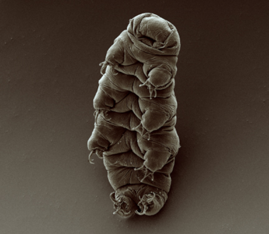
Scanning electron micrograph of an adult tardigrade. Source: Wikimedia Commons
University of Wyoming researchers have gained further insight into how tardigrades survive extreme conditions and shown that proteins from the microscopic creatures expressed in human cells can slow down molecular processes.
This makes the tardigrade proteins potential candidates in technologies centred on slowing the aging process and in long-term storage of human cells.
The new study, published in the journal Protein Science, examines the mechanisms used by tardigrades to enter and exit from suspended animation when faced by environmental stress.
Led by Senior Research Scientist Silvia Sanchez-Martinez in the lab of UW Department of Molecular Biology Assistant Professor Thomas Boothby, the research provides additional evidence that tardigrade proteins eventually could be used to make life-saving treatments available to people where refrigeration is not possible — and enhance storage of cell-based therapies, such as stem cells.
Measuring less than half a millimetre long, tardigrades can survive being completely dried out; being frozen to just above absolute zero; heated to more than 150°C; survive radiation of several thousand times a human’s lethal dose; and even survive the vacuum of outer space.
They survive by entering a state of suspended animation called biostasis, using proteins that form gels inside of cells and slow down life processes, according to the new UW-led research.
Co-authors of the study are from institutions including the University of Bristol in the United Kingdom, Washington University in St. Louis, the University of California-Merced, the University of Bologna in Italy and the University of Amsterdam in the Netherlands.
Sanchez-Martinez, who came from the Howard Hughes Medical Institute to join Boothby’s UW lab, was the lead author of the paper.
“Amazingly, when we introduce these proteins into human cells, they gel and slow down metabolism, just like in tardigrades,” Sanchez-Martinez says.
“Furthermore, just like tardigrades, when you put human cells that have these proteins into biostasis, they become more resistant to stresses, conferring some of the tardigrades’ abilities to the human cells.”
Importantly, the research shows that the whole process is reversible: “When the stress is relieved, the tardigrade gels dissolve, and the human cells return to their normal metabolism,” Boothby says.
“Our findings provide an avenue for pursuing technologies centred on the induction of biostasis in cells and even whole organisms to slow aging and enhance storage and stability,” the researchers concluded.
Previous research by Boothby’s team showed that natural and engineered versions of tardigrade proteins can be used to stabilize an important pharmaceutical used to treat people with hemophilia and other conditions without the need for refrigeration.
Tardigrades’ ability to survive being dried out has puzzled scientists, as the creatures do so in a manner that appears to differ from a number of other organisms with the ability to enter suspended animation.
Researchers from the Nuffield Department of Clinical Neurosciences at the University of Oxford have used data from UK Biobank participants to reveal that diabetes, traffic-related air pollution and alcohol intake are the most harmful out of 15 modifiable risk factors for dementia.
The researchers had previously identified a ‘weak spot’ in the brain, which is a specific network of higher-order regions that not only develop later during adolescence, but also show earlier degeneration in old age.
They showed that this brain network is also particularly vulnerable to schizophrenia and Alzheimer’s disease.
In this new study, published in Nature Communications, they investigated the genetic and modifiable influences on these fragile brain regions by looking at the brain scans of 40 000 UK Biobank participants aged over 45.
The researchers examined 161 risk factors for dementia, and ranked their impact on this vulnerable brain network, over and above the natural effects of age.
They classified these modifiable risk factors into 15 broad categories: blood pressure, cholesterol, diabetes, weight, alcohol consumption, smoking, depressive mood, inflammation, pollution, hearing, sleep, socialisation, diet, physical activity, and education.
Prof Gwenaëlle Douaud, who led this study, said: “We know that a constellation of brain regions degenerates earlier in aging, and in this new study we have shown that these specific parts of the brain are most vulnerable to diabetes, traffic-related air pollution – increasingly a major player in dementia – and alcohol, of all the common risk factors for dementia.”
“We have found that several variations in the genome influence this brain network, and they are implicated in cardiovascular deaths, schizophrenia, Alzheimer’s and Parkinson’s diseases, as well as with the two antigens of a little-known blood group, the elusive XG antigen system, which was an entirely new and unexpected finding.”
Prof Lloyd Elliott, a co-author from Simon Fraser University in Canada, concurs: ‘In fact, two of our seven genetic findings are located in this particular region containing the genes of the XG blood group, and that region is highly atypical because it is shared by both X and Y sex chromosomes.
This is really quite intriguing as we do not know much about these parts of the genome; our work shows there is benefit in exploring further this genetic terra incognita.’
Importantly, as Prof Anderson Winkler, a co-author from the National Institutes of Health and The University of Texas Rio Grande Valley in the US, points out: “What makes this study special is that we examined the unique contribution of each modifiable risk factor by looking at all of them together to assess the resulting degeneration of this particular brain ‘weak spot’. It is with this kind of comprehensive, holistic approach – and once we had taken into account the effects of age and sex – that three emerged as the most harmful: diabetes, air pollution, and alcohol.”
This research sheds light on some of the most critical risk factors for dementia, and provides novel information that can contribute to prevention and future strategies for targeted intervention.