Patients treated with medication for benign prostatic hyperplasia at the time of a severe acute heart attack experienced fewer serious complications from the attack, according to a study from the University of Gothenburg.
Men likely suffer from more extensive heart attacks than women. Studies have shown that male sex hormones, such as testosterone, intensify inflammation during an acute heart attack and lead to a larger area of damage.
Inflammation is a key factor in the extent of heart damage when an acute heart attack is treated with balloon angioplasty, one of the most common methods for rapidly opening blocked vessels that supply blood to the heart muscle.
Protected after heart attack
The registry study, published in JAMA Network Open, shows that Swedish patients treated with finasteride for benign prostatic hyperplasia faced a lower risk of serious complications following an acute heart attack compared to patients matched to be as similar as possible in all respects other than the use of this specific drug.
Finasteride belongs to the class of drugs known as 5-alpha-reductase inhibitors, which reduce the size of an enlarged prostate. The drug blocks an enzyme that converts testosterone into dihydrotestosterone, a more potent and biologically active form of testosterone.
Among those treated with finasteride, the risk of serious complication was 20.8 percent, compared to 24.3 percent among the matched controls. Serious complications included, for example, cardiac arrest, severe signaling disturbances affecting heart rhythm and pumping ability, severely impaired left ventricular function, or death within 30 days.
Greater insight into the impact
The link between finasteride treatment and less severe complications following a heart attack aligned with the researchers’ hypothesis. However, no significant difference was observed for patients treated with hormone medication for prostate cancer.
Hannah Colldén, a pharmacist and researcher at the University of Gothenburg, comments:
“This result contributes to the exciting field of research into how sex hormones can affect the heart, for instance during a heart attack, but it has no direct impact on patient treatment at present.”
The study is based on the SWEDEHEART quality registry and national registers, including all men who suffered a severe acute heart attack, specifically an ST-elevation myocardial infarction (STEMI), and underwent balloon angioplasty. Those treated with drugs affecting male sex hormones (androgen-modulating agents) were matched with similar individuals who had also suffered a heart attack but did not receive that treatment.
Analysis of more than 14 000 patient-drug pairs found biosimilar use was associated with lower costs and declining use of original branded biologic drugs
Biologic drugs play an important role in treating cancers such as breast cancer and lymphoma, but their high costs can add to the financial burden of cancer care. Now, lower-cost versions known as biosimilars are gaining ground, and a new study led by researchers at the UCLA Health Jonsson Comprehensive Cancer Center suggests their growing use is associated with lower costs for both insurers and patients.
The findings, published in JAMA Oncology, show that patients with cancer who exclusively used biosimilars had average monthly costs that were $3820 (R61 502) lower for insurers and $39.50 (R636) lower out of pocket compared with patients who exclusively used the original branded biologic drugs. The results provide evidence that market competition enabled by entry of biosimilars may help reduce the cost of cancer biologic drugs.
Why it matters
Cancer treatment can place a significant financial burden on patients, with financial hardship associated with medication nonadherence and poorer health outcomes. The high and rising cost of anticancer medications, particularly biologic drugs, which are complex medicines made using living cells, is one contributor to this burden. Previous research has estimated that financial toxicity may affect as many as half of patients with cancer in the United States.
Biosimilars were introduced as one potential way to address the high cost of biologic medicines. Unlike generic versions of conventional drugs, biosimilars are highly similar to, but not identical to, their originator products and have no clinically meaningful differences from them. A federal policy enacted in 2009 was designed to establish an abbreviated regulatory pathway for biosimilars in the US market to increase competition, lower prices and improve access to biologic treatments. By the end of 2024, 13 biosimilars were available for three major cancer biologics: bevacizumab, rituximab and trastuzumab. These drugs are used to treat cancers including breast cancer, lymphoma, colorectal cancer, lung cancer and ovarian cancer.
Previous studies have found that biosimilar competition in other therapeutic areas can lower prices and increase use of these lower-cost alternatives. However, less was known about how biosimilar entry across these three major cancer drugs affected the prices and market share of the original branded biologics and, importantly, whether those changes translated into lower costs for insurers and patients. The new study sought to address this gap by examining the economic impact of biosimilar use in real-world cancer care.
What the study did
Researchers conducted a retrospective cohort study using health insurance claims data to examine the use and costs of three cancer biologics after biosimilars became available for each. They analysed data from 14,655 patient-drug pairs involving people with cancer who initiated bevacizumab, rituximab or trastuzumab between 2020 and 2023. The study included patients with commercial insurance and Medicare-related coverage and tracked their treatment and costs for 12 months after they began treatment with a biologic.
The researchers grouped patients based on whether they exclusively used a biosimilar, exclusively used the originator, switched from the originator to a biosimilar, or switched from a biosimilar to the originator. They compared monthly costs paid by insurers and patients’ out-of-pocket payment, calculated as the sum of deductibles, copayments and coinsurance. They also examined changes in the average sales price and market share of the originators and their biosimilars from before biosimilar entry through 2024 to assess how the introduction of biosimilars affected the market dynamics.
What they found
Among the patient-drug pairs, 59.4% of patients exclusively used a biosimilar during the first 12 months of treatment, while 32.5% exclusively used the originator. About 6.9% of patients switched from the originator to a biosimilar, and 1.2% switched from a biosimilar to the originator. This suggests that biosimilars were adopted primarily by patients starting treatment rather than through switching patients who were already receiving the originator.
Patients who exclusively used biosimilars were associated with substantially lower monthly costs for insurers. After adjusting for differences among patients and treatment patterns, average monthly payer costs were $8,959 (R144 230) for patients who exclusively used biosimilars, compared with $12 779 (R205 751) for those who exclusively used the originator, a difference of $3820 (R61 502) per month. Patients who exclusively used biosimilars also had lower average monthly out-of-pocket costs, $118.90 (R1950) compared with $158.40 (R2550) for those who exclusively used the originator, a savings of $39.50 (R636) per month.
The researchers also found that biosimilar entry was associated with lower prices and declining market share for the originators. The average sales price of the three originators declined by 3.8% per year after biosimilars entered the market, while their market share decreased by about 30% annually among patients with commercial insurance and 31.5% in Medicare Part B. The average sales price of the 12 biosimilars included in the study declined by 12.4% per year.
What this means for patients
The findings suggest that biosimilars could help reduce some of the financial burden associated with cancer treatment for patients while creating savings for the healthcare system.
“Although the savings were substantially greater for insurers than for patients, even modest reductions in out-of-pocket costs may be meaningful for people facing the financial challenges of cancer care – savings that can be used toward other treatment-related expenses or cover living expenses,” said Tina Shih, PhD, director of the Cancer Health Economics Research Program at the UCLA Health Jonsson Comprehensive Cancer Center, professor of Health Economics in the Department of Radiation Oncology, and senior author of the study.
The study also points to treatment initiation as an important opportunity to expand biosimilar use. Because most patients in the study who received biosimilars started treatment with them, rather than switching from an original biologic, decisions about which drug to use when treatment begins may play an important role in increasing access to lower-cost and equally efficacious alternatives.
“The FDA has continued to take regulatory actions aimed at further unlocking biosimilar competition recently,” said Xiaoyu Liu, PhD, first author of this study. Our study findings offered further evidence to support patients, providers and insurers in their consideration of biosimilars.”
A tool developed using data on more than 3.5 million adults in Sweden can identify individuals at high risk of hip fracture using registry data, with no in-person assessment
A machine-learning tool built from Swedish national health registry data can predict hip fracture risk with high accuracy and no in-person assessment, and identifies far more at-risk individuals than current clinical screening practices, according to a study published August 27thin the open access journal PLOS Medicine by Kristian Axelsson and Mattias Lorentzon of the University of Gothenburg, Sweden, and colleagues.
Hip fractures are associated with substantial disability, illness, and death in older adults, but existing risk prediction tools typically require in-person patient assessment, including measurements like body mass index and lifestyle information, making large-scale screening difficult.
Researchers analysed nationwide registry data from 3 542 647 individuals aged 50 and older in Sweden, following them for up to ten years. During the study period, 142 327 of the participants sustained a hip fracture. Using more than 100 000 variables drawn from diagnoses, medications, procedures, and demographic and socioeconomic data, the research team developed and tested a deep-learning approach called FRACTURE-ML.
When tested on data from a separate group of people, not included in the original model development, FRACTURE-ML showed good discrimination of people who went on to fracture their hip from those who didn’t with an area under the curve (AUC) of 0.89 one year ahead, and only slightly worse with AUC 0.85 when predicting five years ahead. A simplified version using just 35 variables performed nearly as well. Compared with the current screening methods used in Swedish clinical practice, FRACTURE-ML identified nearly seven times more people at risk of hip fracture within two years (sensitivity 0.84 versus 0.12), with only a modest reduction in specificity (0.79 versus 0.98).
Because the model relies solely on registry data, it lacks information on lifestyle factors such as smoking and alcohol use, which may also affect fracture risk. The authors note that validation in other countries and studies testing real-world implementation are still needed.
“The findings show that it is possible to predict hip fracture risk at the population level without direct patient interaction,” lead author Kristian Axelsson says. “This approach could help target preventive measures more efficiently and potentially reduce the number of hip fractures.”
Mattias Lorentzon adds, “FRACTURE-ML accurately identified people at high risk of hip fracture using routinely collected healthcare and population data, without requiring an in-person clinical assessment. This could make large-scale screening more efficient and help preventive care reach people before a hip fracture occurs.”
“Hip fractures have serious consequences for independence, health and survival. A tool that can identify high-risk individuals directly from existing data could support earlier intervention and potentially reduce the burden of hip fractures across the population,” the authors say.
“One important finding was that a reduced model using only 35 predictors performed nearly as well as the much larger machine-learning model. This suggests that strong predictive performance may be achievable with a comparatively practical and interpretable tool.”
“By using information already available in national registers, FRACTURE-ML could help shift hip-fracture care from reacting after an injury to preventing the injury in the first place.”
“Machine learning performed very well, but carefully developed traditional statistical models achieved similar accuracy. The key advance may therefore be less about a particular algorithm and more about making better use of comprehensive, routinely collected data.”
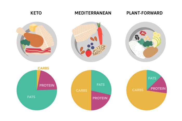
WashU Medicine researchers led a clinical trial testing three diets with different proportions of carbohydrates, fats and proteins and found all of them improved metabolic health, but a very low-carb ketogenic diet had additional benefits for liver health and blood sugar control. Credit: Katie Gertler/WashU Medicine
There is no shortage of popular diets to try, and they can generally produce weight loss if followed to the letter. But are all diet plans created equal in terms of reducing the cardiometabolic risks that come with obesity, such as Type 2 diabetes and liver disease?
A randomised clinical trial from Washington University School of Medicine in St. Louis suggests they aren’t, even when they lead to identical amounts of weight loss. Comparing three commonly recommended diet plans, the researchers found that losing weight on any of them improved overall metabolic health in adults with obesity who also had elevated blood sugar and excess fat in their liver – which are important risk factors for developing diabetes. But limiting carbohydrates through a ketogenic diet offered additional benefits for liver health and blood sugar control.
“For patients with obesity, prediabetes and fatty liver disease, weight loss induced by a very low-carbohydrate diet provides additional therapeutic effects on glucose and lipid metabolism that should further help prevent the progression to more severe metabolic diseases than weight loss alone,” said Samuel Klein, MD, the Danforth Professor of Medicine and Nutritional Science at WashU Medicine and the study’s senior author. “But all three diets – despite vastly different macronutrient makeups, from very low carbohydrates to very high carbohydrates – successfully improved metabolic health through weight loss alone.”
Which diet best improves metabolic and liver health?
Obesity affects roughly four in 10 Americans, most of whom also face metabolic health risks, including insulin resistance, which leads to prediabetes, and fat buildup in the liver. If left untreated, these problems can progress to Type 2 diabetes, chronic liver disease and cardiovascular events, among other irreversible conditions. While weight loss is the gold standard for reducing obesity-related health risks, it hasn’t been clear which type of diet, in terms of its protein, fat and carb content, works best to improve metabolic health.
To explore that question, the researchers, including first author Max C. Petersen, MD, PhD, an assistant professor of medicine in the John T. Milliken Department of Medicine at WashU Medicine, and Gordon I. Smith, PhD, an associate professor of medicine in the department, randomly assigned 55 adults with metabolically unhealthy obesity – meaning obesity with prediabetes and fatty liver – to follow one of three diets for around five months: a low-carbohydrate, high-fat ketogenic diet; a high-carbohydrate, low-fat, plant-forward diet; or a Mediterranean diet balanced between the two. Participants received 100% of their food throughout the study and attended weekly meetings with a study dietitian to support adherence to the assigned diet.
Across all three groups, participants lost an equal amount of weight, shedding about 10% of their total starting weight, and boosted insulin sensitivity in muscle cells by roughly 50% from baseline. The comparable restoration of insulin sensitivity across diet groups suggests that the weight loss itself was the important factor in combatting muscle insulin resistance – not the combination of fat and carbohydrates used to get there.
“Many metabolically unhealthy patients also are candidates for GLP-1 medicines, which have been very useful tools for helping people lose weight. But our results show that choice of diet remains important because it has an impact on specific health outcomes that go beyond weight loss alone.”
Max C. Petersen, MD, PhD, WashU Medicine
But this wasn’t the case for liver health. The researchers found that sensitivity to insulin in liver cells – which regulates how well the liver suppresses glucose production – improved two to three times more on the ketogenic diet compared with the other diets, though all three groups saw improvement. They also found that the ketogenic diet reduced fat inside the liver by 67% compared to 45% for the other two diets after five months.
“Fatty liver disease affects about 75% of adults with obesity worldwide and has become the fastest-growing cause of chronic liver disease and liver cirrhosis,” said Petersen. “Our study shows that for people with obesity and fatty liver disease, a low-carbohydrate ketogenic diet could help reduce that statistic.”
The ketogenic diet also provided greater improvements in blood sugar control than the other plans did, lowering 24-hour blood glucose measurements by 20% from baseline compared to 8% on the other diets. Insulin levels in the blood throughout the day also decreased by 74% on the low-carbohydrate diet compared to 44% and 27% on the Mediterranean and high-carbohydrate diets, respectively. This sharp decline in insulin reflects the decreased need for insulin to regulate blood glucose when consuming a very-low carbohydrate diet, so the pancreas doesn’t need to overproduce insulin to get a response.
Half of the participants on the low-carbohydrate diet reversed their prediabetes, compared to 29% of participants on the Mediterranean diet and 7% on the high-carbohydrate diet.
“Weight loss – even just a moderate amount – is universally beneficial in people who are metabolically unhealthy,” said Petersen. “Many metabolically unhealthy patients also are candidates for GLP-1 medicines, which have been very useful tools for helping people lose weight. But our results show that choice of diet remains important because it has an impact on specific health outcomes that go beyond weight loss alone.”
In future studies, the researchers are interested in understanding the fundamental mechanisms responsible for the metabolic benefits of weight loss, including the effects of GLP-1 medicines.
When Raghav Sehgal arrived at Yale as a graduate student several years ago, he wanted to explore the use of powerful computer science tools in cancer research. With an engineering background and a computational biology startup already under his belt, he was well prepared.
But as Sehgal began his Ph.D. studies, his thesis adviser at the Yale School of Medicine posed a larger question: If you want to solve cancer or any age-related disease, such as cardiovascular disease, why not solve aging itself?
“That put me on a different path,” said Sehgal, an associate research scientist in psychiatry with specialties in computational biology and bioinformatics. “As researchers, we have a long way to go in understanding the aging process and whether the steps we take to manage it really work.
What works and what doesn’t
Improved blood-based DNA tests indicate that these interventions seem to be the most effective at slowing the biological aging process:
Prescribed medications for metabolic control and weight management, including metformin and semaglutide.
Healthy diet and exercise combined. Researchers looked at popular diets including, the Mediterranean, low-fat, and low-carb plans.
Additional research is needed to further validate the findings.
Today, Raghav’s research focuses on the biological systems that drive human aging and how to best measure aging, as well as why aging gives rise to diseases like cancer, diabetes, and dementia. And while his research might not necessarily solve aging, he does explore whether the aging process can be reversed through interventions.
Sehgal is the first author of a new paper from the lab of Albert Higgins-Chen, assistant professor of psychiatry in the Yale School of Medicine. In the paper, Higgins-Chen’s research team took a significant step toward determining which existing anti-aging measures have quantifiable impacts.
Using a new class of DNA-based blood tests, they found that lifestyle interventions and certain drug treatments indeed appear to slow the aging process, while over-the-counter supplements don’t have much effect. All members of the Higgins-Chen lab collaborated in the work, including researchers Daniel Borrus, Jenel F. Armstrong, John Gonzalez, Jessica Kasamoto, and Yaroslav Markov. The paper appears in the journal Nature Medicine.
“For the first time we’ve shown that certain therapies have measurable impacts,” said Sehgal. “This wasn’t previously possible, because we didn’t have enough data to say these biomarkers are consistently responsive to these interventions.” Biomarkers are measurable indicators that reflect a person’s functional or biological age rather than just the number of years lived.
For their analysis, the researchers primarily used a type of blood-based biomarker known as epigenetic clocks, which estimate epigenetic or biological age by measuring the pattern of methyl groups (basic chemical structures that act like tags) attached to a person’s DNA. As people age, these chemical tags change, previous research has shown. Successful anti-aging interventions can reverse the tags. Computer algorithms are then able to use the changes observed in these tags to estimate a person’s epigenetic “age.”
In their work, the researchers combined data from 51 anti-aging intervention studies – which examined a range of strategies, from supplements to medical procedures – and measured more than 110 DNA methyl biomarkers (including 16 major epigenetic clocks). They then compared participants’ biological ages before and after various interventions.
“What we did was pretty unique,” Sehgal said. “We already know that certain things might prolong health span and lifespan. There is data from retrospective analysis as well as from animal models. We took all that knowledge along with the real-world clinical studies to identify which interventions in humans were slowing down aging across the board in these known biomarkers.”
The researchers tested four categories of intervention: pharmacological drugs, lifestyle-based changes, over-the-counter supplements, and medical procedures. They found that lifestyle interventions, such as a combination of a healthy diet (whether it’s Mediterranean, low-carb, or low-fat) and exercise, consistently decreased epigenetic age.
Pharmacologic interventions – including metformin and semaglutide, prescription medications used for metabolic control and weight management, and anti-TNF therapies, which use biologic medications to target an immune system protein that triggers harmful inflammation –decreased epigenetic age the most. (TNF, or tumour necrosis factor, is an immune-system protein that helps fight inflammation-related injury.)
On the other hand, over-the-counter supplements and certain medical procedures didn’t have an effect in decreasing epigenetic age, their analysis showed. Other key findings revealed that the newer biological age clocks outperformed older models, and that biomarkers changed more in people with diseases than in healthy volunteers.
Going forward, the next step would be expanded testing measuring interventions and impact in a widespread, diverse group of volunteers, Sehgal said.
“If these new biomarkers are eventually validated to predict long-term health, scientists will be able to evaluate anti-aging therapies much faster,” he added. “Instead of waiting decades for evidence from clinical trials, we’ll be able to see which interventions are effective and in which people in a few years or even months.”
Senior author Higgins-Chen warned that researchers need to be cautious in interpreting intervention studies.
“We have developed a rigorous, systematic approach to evaluating and comparing how biomarkers change across a wide variety of interventions, and that is a critical step in the right direction,” he said. “However, the true meaning of that change must be fully understood.”
Which biomarker changes are due to the intervention, and which result from hidden confounders? What is the time course of the change during an intervention – and after the intervention ends? What are the biological mechanisms underlying the change?
“And most importantly – what does the change mean for our long-term risk of age-related disease and functional decline?” Higgins-Chen said. The lab is continuing to investigate these questions in additional studies led by researchers Borrus, Armstrong and Grace Zou. Chen’s lab is also affiliated with the Department of Pathology, the Computational Biology and Bioinformatics Program, and the Y-Age Yale Center for Research on Aging.
A one-year follow-up analysis of a landmark clinical trial suggests that patients with severe strokes experienced better long-term outcomes after undergoing endovascular thrombectomy compared with medical management alone, according to findings published in JAMA.
The study evaluated ischemic stroke patients presenting with large-core infarcts, meaning a significant amount of brain tissue had suffered damage. These patients are historically considered less likely to benefit from mechanical clot removal because of the amount of brain damage that has already occurred by the time treatment begins, said Sameer A. Ansari, MD, PhD, professor and chief of Interventional Neuroradiology in the Department of Radiology, who was a co-author of the study.
While the original trial did not show meaningful benefit at 90 days, the newly reported one-year data show functional gains among patients who received thrombectomy. The results of the current study suggest longer-term follow-up was needed to fully understand the benefits of the procedure, Ansari said.
“At one year, it’s quite evident based on the statistical analysis that there’s significant benefit with thrombectomy,” said Ansari, also a professor in the Ken and Ruth Davee Department of Neurology and of Neurological Surgery.
In the study, investigators found that 23.6 percent of patients treated with endovascular thrombectomy achieved functional independence one year after their stroke, compared with 6.8 percent of patients who received medical management. Patients in the thrombectomy group were also significantly more likely to regain the ability to walk independently, with 35.4 percent achieving independent walking versus 18 percent in the medical-management group.
The multicentre trial enrolled patients at 47 US stroke centres who presented within 24 hours of symptom onset and had large strokes identified using noncontrast computed tomography (CT) imaging.
Patients who underwent thrombectomy also reported better quality of life one year after stroke, while one-year mortality rates were similar between the treatment groups.
The findings add to growing evidence from other large core interventional stroke trials that patients with extensive brain injury can still benefit from rapid thrombectomy to restore blood flow.
One possible explanation for the delayed benefit, Ansari said, is that recovery from severe stroke continues long after the standard 90-day assessment period used in most clinical trials.
“We know that people continue to improve after a stroke for the next one to two years,” Ansari said. “Three months has been an arbitrary standard used in prior stroke trials to assess benefit and functional outcomes after targeted interventions.”
Restoring blood flow may also support long-term neuroplasticity, the brain’s ability to reorganise and form new neural connections after injury, Ansari said. The study reported that patients who received thrombectomy continued to improve between 90 days and one year, while outcomes remained static or declined in the medical-management group.
While more research is needed to fully understand which large core stroke patients benefit most from thrombectomy, the results of the study may provide additional confidence that aggressive intervention can yield meaningful recovery for patients, even when improvement is not immediately apparent.
“What we’re seeing across multiple randomised controlled trials, endovascular thrombectomy remains a powerful intervention across a spectrum of stroke presentations,” Ansari said. “It gives clinicians more confidence to treat patients even with severe brain injury and to counsel their loved ones that the outcome benefit may require time to heal, hard work with physical therapy/rehabilitation, and family support.”
The study was supported by unrestricted grants from Medtronic, Cerenovus, Penumbra, Stryker and Genentech.
The North-West University (NWU) has reached a decisive milestone in establishing the Desmond Tutu School of Medicine following the accreditation of its Bachelor of Medicine and Bachelor of Surgery (MBChB) programme by the Higher Education Quality Committee (HEQC) of the Council on Higher Education (CHE).
The CHE confirmed the accreditation in correspondence dated 21 August 2026. The six-year, 940-credit programme will be offered in contact mode at the Potchefstroom Campus.
For Prof. Binu Luke, inaugural chief director of the Desmond Tutu School of Medicine, the decision represents both an institutional achievement and the culmination of an immense collective effort.
“The accreditation of our MBChB programme by the Council on Higher Education is a major milestone in the development of the programme,” he says.
“More than excitement, there is a sense of relief because accreditation by the CHE is a crucial step in our progress towards establishing the school.”
The achievement follows an extensive process of planning, coordination and documentation involving teams across the university. According to Prof. Luke, the application required detailed evidence of how the school and its medical programme would function, covering everything from its curriculum and governance to its clinical training arrangements, staffing, finances and infrastructure.
“There is certainly excitement because this is the result of extensive hours of planning, organisation and implementation across our five workstreams,” he says.
“These workstreams cover defining the ‘North-West doctor’, preparing the clinical training platform, human resources and financial matters, infrastructure, governance, and curriculum development and mapping.”
Translating years of preparation into a comprehensive accreditation application was itself a demanding undertaking.
“Documenting all these activities, completing the relevant forms and providing the required portfolio of evidence was no mean feat,” says Prof. Luke.
“Credit must go to the core team, as well as to all the relevant entities within the university that supported this project.”
The CHE’s correspondence confirms the programme’s full accreditation and that all stipulated conditions have been met. The process of registering the qualification with the South African Qualifications Authority (SAQA) on the National Qualifications Framework (NQF) is now under way.
The accreditation process included the submission of detailed information about clinical rotation modules, assessment practices, exit-level outcomes and opportunities for articulation within and across the university.
Prof. Luke compares the establishment of the medical school to the journey from conception to birth, with accreditation representing the point at which the vision has become viable and the university must carry it through to completion.
“If we compare this process to pregnancy and delivery, from conception to a full-term birth at approximately 40 weeks, this milestone represents the point of viability, which we would regard as about 28 weeks,” he says.
“From now on, there is no turning back. We must continue and deliver the fully developed school in January 2028.”
The Desmond Tutu School of Medicine is set to become South Africa’s 11th medical school and plans to welcome its first cohort of students in 2028. Its development forms part of the NWU’s commitment to addressing the country’s healthcare workforce needs and strengthening healthcare delivery, particularly in underserved communities.
Several important steps must still be completed before the first students begin their studies. Among these is professional accreditation by the Health Professions Council of South Africa (HPCSA).
“There are still several activities that we need to complete. The next major milestone is accreditation by the HPCSA, which is essential for our graduates ultimately to enter professional practice,” says Prof. Luke.
“We have planned the HPCSA accreditation visit for February 2027, and we hope to receive its approval so that we can begin enrolling students in the middle of 2027, with the first cohort commencing its studies in 2028.”
The university will also finalise agreements with the University of the Witwatersrand and the National Health Laboratory Service. These partnerships will support the academic, clinical and laboratory components required to train future doctors.
The school’s curriculum and clinical training model are being designed to produce doctors who are clinically competent, ethically grounded and responsive to the realities of South Africa’s healthcare system. Its approach will be guided by the values associated with its namesake, Archbishop Desmond Tutu, including compassion, justice, integrity and service.
For Prof. Luke, the CHE’s decision is not the end of the journey, but confirmation that a vision years in the making has entered a new and irreversible phase.
“This is a crucial step forward,” he says. “There is no going back. We must now continue the work and deliver the school in January 2028.”
According to Prof. Awie Kotzé, executive dean of the Faculty of Health Sciences, the milestone ushers in a new era for both the NWU and the faculty.
His work towards establishing the medical school began in 2017. It will become the sixth school within the faculty and enable the NWU to offer a comprehensive range of clinically oriented qualifications, with several additional programmes expected to be introduced in the near future.
They are high in sugar, low in nutrients, and too thin, finds analysis of UK products. Current labelling as complementary food source needs regulatory review, say researchers.
Image made with AI
Commercial baby foods in spouted pouches – intended to boost nutritional intake from the age of 6 months onwards – are often more like sugary drinks and smoothies than nutritious food, finds an analysis of UK products, published online in Archives of Disease in Childhood.
They are often higher in sugar and lower in nutrient density than breast milk or formula, as well as frequently lacking the texture needed to help babies learn to eat solid foods, the analysis shows.
These pouches are currently labelled as a complementary food to be used alongside breast milk or formula, but the findings prompt the researchers to question whether this labelling is appropriate and to call for a regulatory review of these products.
Breast milk alone supplies all the key nutrients needed up to the age of around 6 months. After that, complementary foods are essential for establishing healthy food preferences, optimal growth, and long term health, as well as for helping babies learn how to eat solid foods, explain the researchers.
Complementary foods should have an increasingly solid texture and be more energy and nutrient dense than breast milk, they add. But while spouted pouches have become the dominant packaging for commercial baby foods, it’s not clear if their content fulfils these criteria, particularly as the contents need to be thin enough to pass through the spout.
To find out, the researchers reviewed the commercial baby foods available in 6 large hypermarkets, serving families in Glasgow, Scotland in October and November 2024.
In all, they found 298 products from 16 manufacturers, of which 178 (60%) were spouted pouches.
All these pouches were labelled as suitable for children under 12 months; two-thirds were aimed at babies aged 6 months or younger—counter to official recommendations. Of the other products, 46 (38%) were aimed at infants aged 12 months or more.
Foods were categorised relative to breast milk for energy, protein, and sugar content, and defined as suitable for complementary feeding if they were: a purée or solid food which would enable weaning; more energy and protein dense than breast milk; had a sugar content no higher than breast milk.
Food texture was rated by two assessors for 284 (95%) of all the products. Half of the spouted pouches (49%) were liquids (score 1–3, where 0 is thin, and 4 is very thick) compared with 7% of the other products.
Only 10% of the spouted pouches were soft foods, compared with more than three-quarters (77%) of the other products.
“The texture and manner of complementary feeding is important,” emphasise the researchers. “While suckling is an innate characteristic, the oromotor skills required for the manipulation of solid food in the mouth must be learnt experientially.
“For that reason, recommendations suggest that first foods offered should progress from purées to lumpy, mashed, and finger foods. For pouches specifically, not only do they deliver a thin food that can be swallowed with little oromotor manipulation, but they are also often consumed by sucking from the spout, rather than from a spoon,” they add.
Overall, the available baby foods had an average energy content of 66.5 kcal/100 g, with only 35% above that for breast milk (65–70 kcal/100 g).
Only half (52%) of spouted pouches had a protein content higher than the range for breast milk..
In common with most of the other baby foods, only around a third of the spouted pouches had more energy per weight than breast milk, even though 61% had higher sugar content than breast milk. This compares with only 8% of the other baby foods.
“High sugar intakes in early childhood have long-term health implications, both in terms of dental health and overweight,” point out the researchers.
In all, only 14 (8.5%) of the spouted pouches were defined as being completely suitable for a complementary food in that they met all the desired criteria; 48 (29%) met none. By contrast, 37 (32%) of the other wet baby foods met all the desired criteria; only 2 (2%) met none.
The researchers acknowledge various limitations to their findings. For example, they had no data on actual consumption, and the assessment of nutrient content was limited to the information provided by the manufacturers, with iron content, which is a key nutrient, rarely stated.
But they nevertheless conclude: “Products delivered in [spouted] pouches generally do not meet the desired characteristics of complementary foods. Many are more similar to sweet fruit smoothies, which are not recommended at all for babies or preschool children.
“Our findings suggest that regulators should consider whether a change in product categorisation and labelling is needed to make clear which products are foods and which are drinks, to discourage the use of [these pouches] and to enable parents to make informed choices in line with public health recommendations for feeding infants and young children.”
Specially trained pharmacists will be allowed to dispense HIV treatment and prevention medicines without a doctor’s script. (Photo: Unsplash)
By Catherine Tomlinson for Spotlight
Ten months after an enabling court judgment, some pharmacists in South Africa can finally start applying for permits that allows them to dispense HIV prevention pills without a script from a doctor.
Specially trained pharmacists can now begin applying for permits to dispense antiretroviral medicines for HIV prevention without a doctor’s prescription. The long-awaited permitting process was announced to pharmacists in a South African Pharmacy Council (SAPC) e-Note on 13 August.
PIMART is a form of task-shifting that allows pharmacists to provide some limited HIV services that are currently only provided by doctors and nurses.
It is intended that, under the PIMART programme, pharmacists that have completed a dedicated training programme and have received a special permit from the Director-General of Health will be authorised to provide first-line antiretroviral treatment to people with uncomplicated HIV without a doctor’s script.
They will also be allowed to dispense HIV prevention medicines without a doctor’s script – this includes both pre-exposure prophylaxis (PrEP) and post-exposure prophylaxis (PEP). PrEP is taken prior to sex to prevent potential infection while PEP is taken shortly after a possible HIV exposure to prevent infection.
PIMART’s long road to implementation
The SAPC first proposed PIMART to the health department in 2018. The proposal was in response to a request from the department for pharmacists to take on a larger role in the country’s HIV response.
In 2021, Spotlight reported the imminent launch of PIMART services in South Africa’s pharmacies, but then a court challenge against PIMART by a group of general practitioners blocked the programme’s implementation.
Four years later, in October 2025, a ruling by the Supreme Court of Appeal finally cleared the way for SAPC to launch PIMART. Following the court ruling, Vincent Tlala, CEO and Registrar of the SAPC, told media that the SAPC would issue a memo in November 2025 inviting pharmacists to apply for PIMART permits. But this would in fact only materialise in August 2026.
The SAPC’s 13 August memo to pharmacists, said that certain pharmacists can now begin to apply for temporary PIMART permits to provide PEP, PrEP, and HIV testing services – but not yet HIV treatment services. The temporary permits will be valid for 24 months.
Also, only pharmacists that completed the South African HIV Clinicians Society’s (SAHCS) previous PIMART training course can apply for the limited-scope, temporary PIMART permits.
Pharmacists that have not completed this course will have to wait for a new training course to be accredited and launched before they can apply for permits to provide PIMART services.
How long will it take to get a permit?
Tlala told Spotlight that the granting of the limited-scope, temporary PIMART permits by the Director-General and their recording by the SAPC may take a few months.
“The Department of Health processes applications for permits within 90 days (maximum),” said Tlala, adding that “[t]he PIMART permit recording process with the South African Pharmacy Council then takes between 72 working hours and 14 days, depending on the completeness of the application.”
The permits will only be granted to pharmacists who have already completed the Southern African HIV Clinicians Society’s (SAHCS) continuous professional development (CPD) PIMART course. Pharmacists that did not complete this course will need to wait for an updated PIMART course to become available before they can undertake the required training to be permitted.
Fiona Story, CEO of SAHCS, told Spotlight that nearly 800 pharmacists have completed the course.
Taken together, all of this suggests that toward the end of the year, a few hundred pharmacists in South Africa might be dispensing HIV prevention medicines without doctors’ scripts.
When will the updated training course be available?
Currently, there is no PIMART training course available for pharmacists, as the previous PIMART training course provided by SAHCS is on pause, while the updated course awaits accreditation. Based on dates provided by SAHCS, the updated course might only be available early next year.
However, once the updated course is available, it is anticipated that pharmacists that complete it will be able to apply for full-scope PIMART permits that allow them to dispense first-line antiretrovirals to people with uncomplicated HIV without a doctor’s script, in addition to PEP and PrEP.
“The CPD-accredited PIMART course is no longer available as it was reviewed and updated in line with the requirements for accreditation as a supplementary training course,” Story told Spotlight.
She said that the SAHCS is seeking accreditation of its updated PIMART training course from the SAPC. “SAHCS anticipates the PIMART supplementary training course accreditation process will conclude before the end of 2026,” said Story. She added: “SAHCS has prepared to launch the PIMART supplementary training course as soon as accreditation has been received.”
When will pharmacists be able to dispense HIV treatment without a doctor’s script?
The timeline for when pharmacists will be able to provide the full scope of services intended under PIMART, including HIV treatment for uncomplicated cases, remains unclear. Before this can happen, the SAPC must accredit the SAHCS’ updated supplementary training course and the SAHCS must launch this course.
After this, the permits granted to pharmacists by the Director-General will need to be expanded to include the provision of first-line antiretroviral therapy for treatment of uncomplicated HIV.
In the meantime, the around 800 pharmacists who completed the SAHCS’s previous PIMART training course can now start applying for permits to provide PrEP, PEP, and HIV testing services.
Hepatitis B remains one of the world’s most underdiagnosed infections.[1]
Untreated hepatitis B can remain silent for years while increasing the risk of severe liver disease and preventable infant infection.[1,2]
Integrated point-of-care testing during antenatal care visits presents an important opportunity to identify undiagnosed infections and support measures aimed at reducing transmission to newborns.[2,3]
A pregnant woman attending her first antenatal care (ANC) appointment will often be screened for conditions that could affect her health and the health of her unborn child, including HIV and syphilis, but not always for hepatitis B virus (HBV) infection. This is not because hepatitis B is rare; in fact, quite the opposite. Millions of people worldwide are living with chronic hepatitis B infection, yet many remain unaware of their infection because the disease can progress silently for years without symptoms.[1] As a result, infection is often diagnosed only after serious liver complications develop.[1]
Late diagnosis increases healthcare costs, may limit treatment options and contributes to preventable illness and death.[1] For pregnant women, delayed diagnosis creates an additional risk of vertically transmitting the virus to their babies during childbirth.[2] That missed opportunity is why hepatitis B needs to be seen not only as a liver disease, but as a maternal and child health priority. For healthcare companies such as Abbott, there is an opportunity to help health systems make every ANC touchpoint work harder by bringing reliable, rapid and integrated testing closer to the point of care.
The central question is no longer whether hepatitis B can be detected, prevented or managed. It can. The question is whether health systems are using routine moments of care, especially pregnancy, to find the infection early enough to enable interventions that can improve outcomes for mothers, infants and families.
Hepatitis B is often silent, but its consequences are not.[1]
ANC offers a critical window to prevent mother-to-child transmission through timely immunoprophylaxis.[2,3]
Integrated testing can help expand access while reducing practical barriers for patients and frontline healthcare workers.[3]
Africa has the tools to change the trajectory; now systems must scale them.[1,3]
The cost of not knowing
Hepatitis B remains one of the world’s most significant infectious diseases despite being preventable and treatable. According to the World Health Organization’s (WHO) Global Hepatitis Report 2026, an estimated 240 million people were living with chronic hepatitis B in 2024, and the WHO African Region accounted for 68% of new hepatitis B virus infections globally.[1]
Behind these numbers are people who may be asymptomatic for years while the virus quietly damages the liver. When hepatitis B is not detected, the first sign of illness may be cirrhosis, liver cancer or liver failure.[1] For families, delayed diagnosis may contribute to repeated clinic visits, rising treatment costs, lost income and the emotional burden of managing a condition that could have been identified much earlier.
“Hepatitis B challenges health systems because it is often invisible until the consequences are severe,” says Dr Evans Mathebula, Senior Medical Affairs Manager at Abbott’s rapid diagnostics business in Africa. “If we wait for symptoms, we have waited too long. The priority should be to make hepatitis B screening routine, accessible and connected to care, particularly at moments when people are already engaging with the health system.”
This is especially important in maternal care. Pregnancy represents one of the most important opportunities to identify previously undiagnosed hepatitis B infection. For many women, ANC is among the few periods of regular engagement with the healthcare system.[2] This visit already serves as an important screening and health education opportunity and can facilitate preventive interventions. Many African countries have made important progress in preventing mother-to-child transmission of HIV. That progress shows what is possible when testing, treatment, policy and community trust are aligned. Hepatitis B now needs the same systems discipline, because the antenatal care clinic may be one of the few reliable points of contact between a pregnant woman and the health system.
This approach aligns with the WHO’s triple elimination initiative, which encourages countries to eliminate mother-to-child transmission of HIV, syphilis and hepatitis B together, rather than through fragmented disease-specific programmes.[3] It recognises a simple reality: healthcare systems across Africa continue to face constraints, workforce pressures and laboratory challenges. In this context, adding new screening priorities must be practical and sustainable. Additionally, pregnant women should not have to navigate three separate pathways when one integrated screening moment can support identification of multiple risks and enable timely care during a single encounter.[3]
An integrated antenatal panel that screens for HIV, syphilis and hepatitis B from a single finger-prick is one example of how health systems can simplify access while maintaining a comprehensive standard of care.[3] For mothers, it can help reduce the risk of missed results and clarify next steps. For healthcare workers, it can support faster decision-making at the point of care when linked to appropriate follow-up pathways. For newborns, timely identification of maternal hepatitis B can support measures that reduce the risk of exposure and enable timely preventive action.
“The most powerful public health interventions are those that fit naturally into the realities of existing care,” adds Mathebula. “The easier it is for healthcare workers to identify hepatitis B during a single antenatal visit, through the use of an integrated panel, the sooner patients can be considered for appropriate follow-up care. We are not only improving diagnosis; we are giving clinicians the information they need to support mothers, help reduce the risk of infant infection and strengthen confidence in the health system.”
A hopeful path to elimination
For too long, hepatitis B has remained overshadowed by other infectious diseases despite its scale and impact. The tools needed to prevent transmission, diagnose infection and support long-term management already exist.[1] What remains is the challenge of finding the millions of people who are living with this virus without knowing it, starting with those accessing maternal and primary healthcare.
To move from awareness to action, policymakers and healthcare leaders should prioritise routine hepatitis B screening in antenatal care, support point-of-care testing in decentralised settings, strengthen linkage to treatment and ensure newborns receive timely protection.[1,3] These are practical steps that can turn a silent infection into a manageable health issue and help prevent transmission to the next generation.
Eliminating hepatitis B as a public health threat will require more than technology alone. It will require political will, domestic investment, continued education to increase awareness, trusted community engagement and health systems designed around people’s real lives. But the path is clear: if one antenatal care visit can identify risk, enable timely care and support measures that help reduce a child’s risk of infection, then it is one of the most powerful opportunities public health has to change two lives at once.
References:
[1] World Health Organization. (2026). Global hepatitis report 2026. Geneva: World Health Organization.
[2] Afolabi, I. B., Phillips, J. C., El-Chaar, D., Bainbridge, S. A., & Phillips, K. P. (2026). Preventing vertical transmission of hepatitis B in sub-Saharan Africa: protocol for a systematic review and meta-analysis on vaccination uptake and determinants among pregnant women. BMJ Open, 16(1), e113229. https://doi.org/10.1136/bmjopen-2025-113229
[3] World Health Organization & United Nations Children’s Fund. (2025). Country guidance for planning triple elimination of mother-to-child transmission of HIV, syphilis and hepatitis B virus programmes. Geneva: World Health Organization.