Socioeconomic status had greater effects on child health than parental smoking or alcohol consumption
Photography by Drew Hays on Unsplash
Family socioeconomic position may be a more important determiner of children’s health than parental behaviours such as smoking, drinking or caffeine consumption, according to a study published July 23rd in the open access journal PLOS Medicine by Gemma Sharp from the University of Exeter, UK, and colleagues.
According to the Developmental Origins of Health and Disease (DOHaD) hypothesis, prenatal and early childhood environmental exposures can affect a child’s health long-term. Most research, however, has focused on maternal behaviours and has not included fathers, the environment in which the parents live, and other factors. To better understand how environmental influences might impact child health, the authors of this study analysed data from four large studies of parents and children in the United Kingdom and Norway, including more than 230 000 participants, running several models to produce high confidence in the results. They looked at parental health behaviours including smoking, drinking, and caffeine consumption, as well as their socioeconomic position. They associated these variables with 72 different child health outcomes measured at six different time points, including size at delivery, body mass, hyperactivity, social communication, aggression, depressive symptoms, and more.
The authors found that maternal behaviours did not have larger effects than those of their partners. While 6% of the results found links between smoking and child health, 3% showed links for alcohol and 0.4% for caffeine, 15% of the child health effects were associated with the socioeconomic position of the child’s family. While the results are associations and based on observational data of families in the United Kingdom and Norway, the authors note that efforts to improve child health might be more effective if they are aimed at socioeconomic inequalities, rather than individual parent behaviour.
Gemma Sharp adds, “Our study suggests that the social and economic circumstances children grow up in may have a greater influence on their health than specific parental behaviours during pregnancy. By analysing data from more than 230 000 participants across four long-term studies, we found that socioeconomic disadvantage was more consistently linked to poorer child health outcomes than smoking, alcohol, or caffeine use by either parent.”
“One of the most interesting findings was that we didn’t see consistently stronger effects for mothers than for fathers. We often assume that a mother’s behaviours during pregnancy will have a larger impact on child health because of direct effects on the developing baby. However, we found that mothers’ and fathers’ smoking, alcohol, and caffeine use showed remarkably similar patterns of association with child health outcomes. This also points towards the importance of the wider family environment and social circumstances, rather than pregnancy-related behaviours alone, in shaping children’s health.”
Globally, more people are using GLP-1 and other weight-loss medicines, and some are experiencing severe hair loss while taking them. But just because it is happening during treatment does not necessarily mean the treatment caused it. According to Dr Kashmal Kalan, Medical Director at Alvi Armani, “Hair shedding during treatment isn’t always caused by the medicine itself. In many cases, it may be the body’s response to losing weight too quickly. Your body reacts as though food is scarce and thinks: ‘Hair isn’t essential. Let’s save energy’.”
Rapid weight loss can increase the risk of temporary hair shedding, whether it follows GLP-1 treatment, bariatric surgery, a very low-calorie diet, or illness. The trigger is often the speed and extent of the weight loss rather than the treatment itself. Eating much less can also leave patients short of nutrients that hair needs. “Think of hair like a houseplant. If you don’t water it enough with the right nutrients, it grows poorly. It doesn’t necessarily die permanently but simply pauses its growth.”
The concern is becoming more relevant as weight-loss medicine use grows in South Africa. Discovery Bank and Visa’s latest SpendTrend report found that 14% of surveyed higher-income South Africans use prescribed weight-management medication. Clinical research has also found that GLP-1 medicines reduced energy intake on a controlled test day by nearly a quarter compared with placebo, which helps explain why nutritional adequacy may become more important when appetite drops.
Dr Kalan says Alvi Armani is seeing more patients who report sudden hair shedding weeks or months after starting weight loss treatment and assume the medicine is directly responsible. The consultation must then establish when the shedding began, how quickly the patient lost weight, how their eating patterns changed, and whether another medical cause needs to be investigated.
The potential causes behind rapid hair loss
A review of nearly half a million adults on GLP-1 treatment found vitamin D deficiency in 7.5% of patients at six months, climbing to 13.6% by twelve months. That’s why bloodwork and not a shopping list of supplements is the first step in any hair loss consultation at Alvi Armani, whether GLP-1-related or not. Standard testing includes vitamin D, B12, ferritin, and thyroid function as standard.
“Ferritin, the protein that stores iron, is one marker I monitor closely. A level below 30 ng/mL, generally considered indicative of low iron stores in adults, is often enough to cause shedding on its own, even though most labs still call that number normal. Most of these patients feel completely fine elsewhere, so there’s no reason for a routine GP visit to pick it up. By the time the hair’s already falling out, we go back and look for what that visit potentially missed.”
Some patients may also reach for gut-health supplements because they assume hair loss comes from issues in the gut. However, Dr Kalan notes, probiotics will only address the problem if a digestive condition is actually contributing to it. Research remains limited, with the largest randomised trial to date finding reduced shedding among the probiotic group, but no meaningful improvement in hair density or thickness.
“If someone has a diagnosed digestive condition, that’s worth treating, and probiotics may have a place. Outside of that, I’d rather see patients pursue tests that can identify what’s missing than a supplement with no clear indication, strain, or dose.”
Dr Kalan encourages anyone on a GLP-1 medication to take shedding seriously if it continues past three months, worsens noticeably, or comes with fatigue or other symptoms that don’t add up. “These medications genuinely change lives for the right patient. If your registered health professional recommends continuation, stay on it. Just make sure your body is still getting what it needs.”
Research suggests that more than half of children with autism spectrum disorder, a neurodevelopmental condition, experience motor impairments. An analysis in Developmental Medicine & Child Neurology indicates that sports-based interventions may improve motor skills in children with autism spectrum disorder, with martial arts and aquatic training demonstrating the most consistent benefits.
The analysis was based on data from 11 clinical trials of various sports. Martial arts demonstrated consistently large improvements in balance, total motor skills, and object control skills, with especially strong effects in Tai Chi Chuan and kata programs. Aquatic training demonstrated large improvements across balance, locomotor skills, and object control skills. Gymnastics and trampoline interventions demonstrated large improvements in balance and bilateral coordination, with additional effects on total motor scores in trampoline studies. Table tennis produced broad gross motor improvements, and Australian football had a large effect on object control skills and had a medium effect on balance and total motor skills. Across all sports categories, balance was the most consistently improved motor domain.
The authors noted that many of the studies had significant limitations, however, and higher-quality studies are needed.
“Organized sports-based interventions may offer benefits beyond recreation for children with autism by supporting motor skill development,” said corresponding author Sonia Khurana, PT, PhD, of Old Dominion University. “While our review identified promising effects, particularly for martial arts and aquatic training, larger and more rigorous studies are needed to confirm these benefits and guide evidence-based recommendations.”
Study indicates the need for a revised definition of functional high-risk multiple myeloma
Depiction of multiple myeloma. Credit: Scientific Animations
Researchers have found that with new treatments for multiple myeloma, a serious type of blood cancer, clinicians should use different criteria for identifying patients with poor odds of survival. The study is published by Wiley online in CANCER, a peer-reviewed journal of the American Cancer Society.
Functional high-risk (FHR) multiple myeloma, which affects a subset of patients with blood cancer, is commonly defined as a multiple myeloma that progresses within 18 months of starting treatment, and it comes with a survival prognosis of less than 2 years after progression. Recently, however, the use of a 4-part treatment regimen – including anti-CD38 antibodies, proteasome inhibitors, immunomodulatory agents, and dexamethasone – followed by autologous stem cell transplantation, has helped to slow the cancer’s progression.
To update the definition of FHR multiple myeloma in the current era of combination therapy, investigators from the University of Alabama at Birmingham and the CoMMiT consortium analysed information on 310 patients with newly diagnosed multiple myeloma who received this therapy and were followed for a median of 3.5 years.
Survival analyses indicated that cancer progression within 36 months of the onset of combination therapy identified patients whose survival was likely under 2 years from the time of progression. Adding another treatment called T-cell redirecting therapy helped slow progression. This “FHR36” patient population, corresponding to 16.4% of all treated patients, should be prioritised as candidates for early use of T-cell redirecting therapy and for clinical trials of medications with novel mechanisms of action.
“The findings will help physicians choose therapies for this important minority of patients who have disease progression in the first 3 years of diagnosis and identify an important population in greater need for treatment innovations to be addressed in the next generation of clinical trials,” said senior author Luciano J. Costa, MD, PhD, of the University of Alabama at Birmingham.
Cambridge researchers have shown how commonly-used sweeteners slow the growth of certain gut bacteria. Isosteviol – a compound derived from stevia (a common sweetener) – when combined with the anti-depressant duloxetine significantly impaired two important gut bacteria linked to regulating blood sugar and gut health and may affect the body’s immune responses.
Sweeteners are often marketed as metabolically neutral, but our study challenges this idea
Sonja Blasche
The scientists say more research is needed to understand the real-world health impacts of this laboratory study, one of the first to assess the direct impact of sweeteners on gut bacteria, particularly when they are combined with other substances.
Sweeteners are widely used in a range of food and drinks, including soft drinks, sweets, desserts, snacks and cereals. While marketed as healthier alternatives to sugar, there is increasing evidence of links to diseases such as type 2 diabetes, obesity and cancer.
Despite their pervasive use, there have been very few studies that look at the direct interactions between sweeteners and gut bacteria – the vast community of microorganisms that live in the digestive tract and play a crucial role in keeping our bodies healthy.
Professor Kiran Patil from the Medical Research Council (MRC) Toxicology Unit at the University of Cambridge said: “Most of what we know about the potential impact of sweeteners on our health comes from animal research or from population studies. While these studies have indicated involvement of the microbiome in mediating the effect of sweeteners, it’s difficult to know how sweeteners act in the body – is it through direct interactions with our gut bacteria?”
“Answering this is further complicated by the fact that we rarely ever take sweeteners by themselves – we take them with drinks, in snacks, or even in medication to mask bitterness,” added Dr Sonja Blasche, a lead author of the study, also the MRC Toxicology Unit.
In research published in Molecular Systems Biology, Dr Blasche and colleagues looked at how artificial and low‑calorie sweeteners affect the bacteria living in our gut, and how these effects change when sweeteners are consumed together with other common substances such as caffeine, flavourings or medicines.
The researchers grew each of 25 gut bacterial species – including beneficial, neutral, and potentially harmful bacteria – in the lab. They then exposed each culture individually to 39 common, commercially-used sweeteners, some of which are artificial, others natural, and measured how well the bacteria multiplied.
Around three‑quarters of the sweeteners changed how at least one bacterial species grew. Some sweeteners slowed down or stopped the growth of certain bacteria linked to a healthy gut.
The researchers then tested each sweetener in combination with common compounds such as caffeine, vanillin (vanilla extract), advantame (an artificial sweetener) and eight commonly-used drugs to assess whether this had any impact on the gut bacteria. They found over 100 interactions where sweeteners acted differently when combined with other substances. In 34 cases, combinations made the effects stronger, while in 68 cases the effects were weaker.
Most striking was the combination of isosteviol (a compound derived from stevia, a sweetener widely used in the food and beverage industry) and the antidepressant duloxetine. This combination strongly suppressed Roseburia intestinalis and Parabacteroides merdae, two gut bacteria that play important roles in maintaining a healthy digestive system. In the US in 2023, over 4.2 million patients were prescribed duloxetine.
As no gut bacterium exists alone, but rather as part of a ‘community’ within the gut, the researchers created a synthetic community containing all 25 bacteria. After allowing it to grow over time, they tested the community against a variety of sweetener and drug combinations, looking at which species increased or decreased and whether the overall diversity changed.
By mimicking in this simplified way what might happen in the human gut, they showed that the combination of isosteviol and duloxetine reduced microbial diversity. A diverse microbiome is considered important for good gut health. The sweetener-drug combination also altered which bacterial species thrived or declined.
Further analysis showed that the effect of the isosteviol-duloxetine combination on the community increased toxicity towards certain host cells and interfered with other cells that play a role in the body’s inflammation and immune responses.
Dr Blasche said: “Sweeteners are often marketed as metabolically neutral, but our study challenges this idea. We found that they can directly affect gut bacteria, particularly when mixed with other compounds such as medication and food additives. These common combinations could have unintended effects on our gut microbiome.”
The researchers stress that, as their experiments were carried out in the lab, not tested in humans, more research needs to be done before it is possible to conclude that there will be direct health effects in people.
Professor Patil, the study’s senior author, added: “Our study suggests that artificial sweeteners don’t just pass through the body passively — they can interact with gut microbes, and these effects can be amplified or altered by other substances like medications. These findings can help guide new studies towards understanding how sweeteners might influence health in unexpected ways.”
The research was funded by the European Union’s Horizon 2020 programme and the UK Medical Research Council.
The discovery may explain side effects from taking statins, opening the door to future therapies that could make statins easier to tolerate.
Photo by Towfiqu Barbhuiya on Unsplash
Millions of people rely on statins to lower cholesterol and reduce the risk of heart attack and stroke. But for some, the drugs come with an unwelcome trade-off: muscle pain, weakness and exercise intolerance that can make it difficult to continue treatment.
Now, researchers at McMaster University have uncovered a biological pathway that may explain why those side-effects occur, opening the door to future therapies that could make statins easier to tolerate while maintaining their life-saving cardiovascular benefits.
Published in Science Advances, the study identifies an immune and metabolic mechanism that drives statin-induced muscle damage, challenging longstanding assumptions about how these side-effects develop.
“Statins are among the most effective medications we have for reducing cardiovascular disease risk and preventing early death,” said senior author Jonathan Schertzer, a professor in McMaster’s Department of Biochemistry and Biomedical Sciences.
“Unfortunately, muscle side-effects lead some people to reduce their dose or stop taking the medication altogether. We wanted to understand why this happens and whether it might be possible to separate the side effects from the benefits.”
Statin-associated muscle symptoms affect an estimated seven to 29 per cent of people who take the drugs. While researchers have long known that statins can sometimes cause muscle problems, the biological mechanisms behind those effects have remained unclear.
Led by first authors Nazli Robin and Nicole Barra of the Schertzer Lab at McMaster, the team found statins can disrupt how muscle cells produce energy, triggering an immune response that damages muscle tissue. In experiments using muscle cells and mouse models, researchers were able to prevent much of that damage by blocking the immune response.
“One of the most exciting findings of the research is that the mechanism causing muscle side-effects appears to be separate from the mechanism that lowers cholesterol,” said Schertzer. “That suggests it may one day be possible to target the side-effects without interfering with the cardiovascular benefits that make statins so valuable.”
The study also revealed an unexpected link between metabolism and immunity. Researchers found that changes in muscle-cell metabolism triggered an immune response within the cells themselves, providing new insight into how inflammation can contribute to drug side-effects.
Although additional research will be needed before the findings can be translated into therapies for patients, the discovery identifies several potential targets for future drug development aimed at preventing statin intolerance.
“These findings give us a clearer understanding of why some patients experience muscle symptoms and provide promising directions for making these important medications safer and more effective in the future,” added Schertzer.
Stimulating a nerve in the ear may alleviate lung inflammation in mice
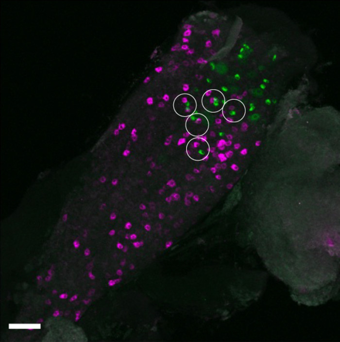
In the mouse vagal ganglion, sensory neurons labeled from the auricular skin (green) and the lung (magenta) are located in close proximity (white circles). These findings suggest a possible anatomical basis for how sensation from the auricular skin may influence airway immune responses via nerves that directly supply the lung. (Rintaro Shibuya, Kim Lab, Icahn School of Medicine at Mount Sinai.)
Neuroimmunology, the study of interactions between the nervous and immune systems, is a rapidly growing field enabling new approaches for monitoring and treating inflammatory diseases. In a recent study, scientists showed that in mice, stimulating a nerve in the external ear may help to ease inflammation in the lungs. Based on these findings, which are published in Immunity, the researchers are designing a clinical trial to test a novel device for treating asthma.
“We are always looking for new therapeutics and devices that can kickstart the body and get it back to doing what it needs to do,” says senior and corresponding author Brian S. Kim of the Icahn School of Medicine at Mount Sinai in New York. “This research suggests a new way to target the body’s inflammatory pathways.”
Neuroimmunology took off after research revealed how the vagus nerve – the ‘information superhighway’ that connects the brain to major organs and controls their functions – helps to regulate the immune system.
“The vagus nerve is hardwired to be a homeostatic organ,” says Kim. “You can think of it like a rheostat that integrates everything and keeps it in check.”
For this study, the authors leveraged the unique characteristics of the auricular vagus nerve, the only branch of the vagus nerve that reaches the surface of the skin. This nerve is found in the cymba conchae, the small, bowl-shaped depression in the upper part of the outer ear.
“Given the strong connection between the lungs and the vagus nerve, we sought to use the lungs as a test case to study these mechanisms and see whether manipulating the auricular branch could modulate inflammation,” says Kim.
The team studied these connections in mouse models using several approaches, including chemogenetics and optogenetics, to see what happened when the auricular vagus nerve was stimulated in the presence of an allergen. Their work showed that stimulation of the nerve increased levels of a neurotransmitter protein called CGRPβ in the airway. This, in turn, reduced inflammation in the lung. When the nerve fibres were instead inhibited, airway disease was exacerbated.
“Our findings reveal a previously unrecognised neuroimmune reflex linking the skin and the lung,” says first author Rintaro Shibuya of Kyoto University. “I hope this work inspires new ways of thinking about vagus nerve biology and future bioelectronic and neuroimmune therapies for inflammatory diseases.”
Although the research is in early stages, the team says this approach has many potential applications for treating diseases characterized by inflammation, including pulmonary fibrosis, inflammatory bowel disease, and rheumatoid arthritis.
“We still don’t know the extent to which the effects of stimulating the auricular vagus nerve go beyond the airway, but it’s something we will continue to study in the lab,” says Kim.
Increased brain ageing was associated with dementia, addiction and psychiatric disorders like schizophrenia
Generated with Gencraft AI
People with dementia, mild cognitive impairment, alcohol addiction, or psychiatric disorders such as schizophrenia show increased brain ageing, each in specific patterns within the brain, according to a study published July 21st in the open access journal PLOS Medicine by Shile Qi from the Nanjing University of Aeronautics and Astronautics, China, and colleagues.
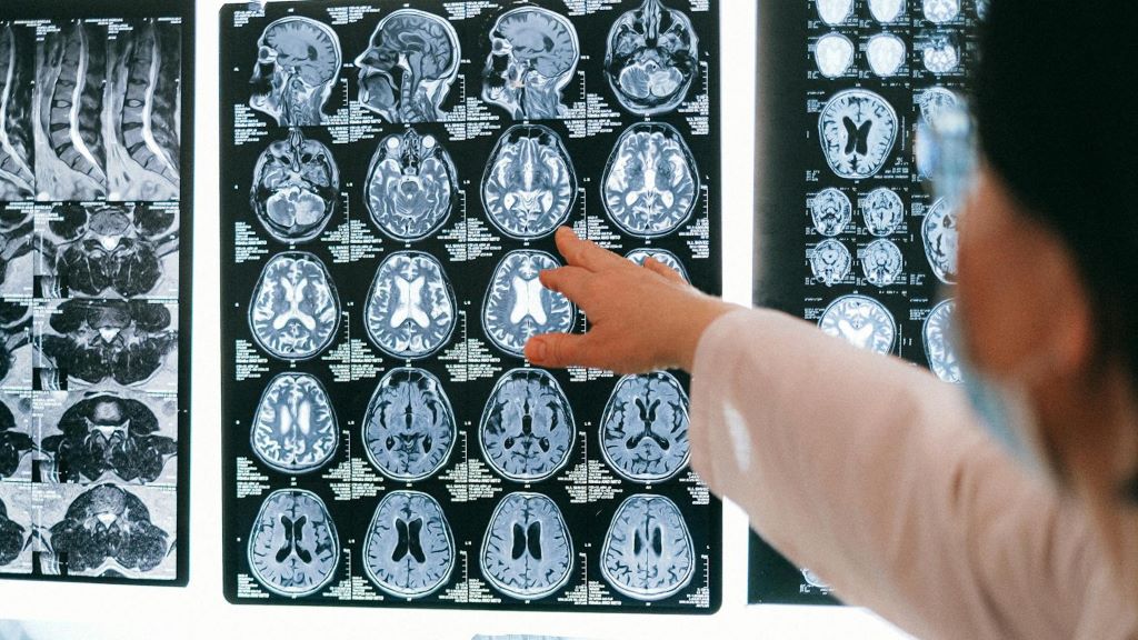
Some conditions can make the brain age faster. Scientists calculate how old the brain is relative to the body using the predictive age difference (PAD), the difference between chronological age and the age predicted by brain imaging, where a positive PAD indicates that ageing is accentuated or increased. To better understand how brain disorders and divergences might affect brain ageing, the authors of this study collected structure magnetic resonance imaging (MRI) data from 45 900 controls across several brain imaging banks, and compared them with those of 2698 patients with different brain conditions and differences, including attention-deficit/hyperactivity disorder (ADHD), autism spectrum disorder (ASD), alcohol or tobacco addiction, Alzheimer’s disease (AD), mild cognitive impairment (MCI), schizophrenia, bipolar disorder or major depressive disorder.
The authors found that neurodegenerative disorders of AD and MCI had the largest association with a high PAD. Addiction and psychiatric disorders were also associated with increased PAD. In contrast, there were no differences in PAD between people with ADHD or ASD and controls.
The researchers also looked at PAD values in specific areas of the brain, and examined which genes showed increased expression in people with different brain conditions. The prefrontal cortex showed higher PAD across brain disorders. Higher PAD in the frontal and temporal lobes was associated with psychiatric disorders, while high PAD in the frontal and occipital cortex was associated with dementia. Addiction was connected with high PAD in the default mode network, and in the salience network and the putamen and thalamus. There were also differences in gene transcription that associated with specific conditions and divergences. While the results are correlational, and not causal, and while some conditions such as psychiatric disorders and addiction have high co-occurrence, the author suggest that understanding more about PAD could help provide biomarkers for commonly occurring brain disorders.
The authors add, “Different neurological disorders appear to leave different signatures on the brain ageing clock, which may help researchers better understand the neural and biological pathways involved in these conditions.”
Vaccine approach yields high numbers of HIV-neutralising antibodies in non-human primates
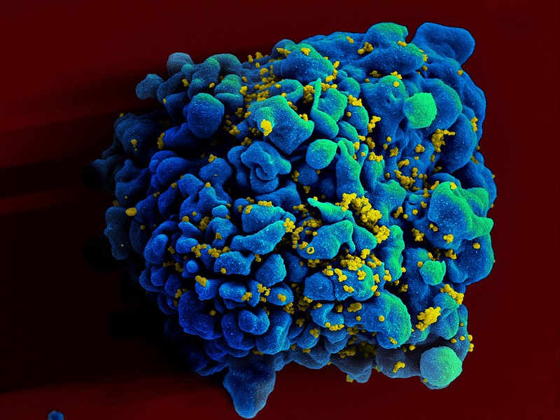
Colourised scanning electron micrograph of HIV (yellow) infecting a human T9 cell (blue). Credit: NIH
A new HIV vaccine developed by La Jolla Institute for Immunology (LJI), Scripps Research scientists, and IAVI has the potential to protect humans from developing HIV infection and AIDS. This HIV vaccine is the first to generate a high number of ‘broadly neutralising’, virus-fighting antibodies in primates.
“This feels like a huge success,” says LJI Professor and Chief Scientific Officer Shane Crotty, PhD, who co-led the research with Scripps Research Professor William Schief, PhD. “We constructed a successful vaccine from the ground up, which required a deep understanding of the immune system.”
This groundbreaking research, published inNature, is the result of 14 years of collaboration between La Jolla Institute for Immunology and Scripps Research, as part of the Scripps Consortium for HIV/AIDS Vaccine Development (CHAVD). “This has been one of those Apollo moon mission-type projects, where there is an exceptional goal and the team has to accomplish a myriad of discoveries and inventions along the way,” says Crotty.
Outsmarting HIV
The new vaccine works by intervening in a process called B cell maturation. B cells make antibodies. Like many immune cells, B cells have an early ‘naïve’ stage before they are ready to make antibodies. B cells start to mature once they get the signal that a pathogen, such as a virus, is trying to attack. B cells see pieces of that pathogen’s molecular structure and start producing antibodies that can bind to that structure and halt infection.
It can take a little while for B cells to find the right “bullseye” on a pathogen. But B cells keep trying. As they mature, B cells tweak their antibody production, refining antibody structures to bind to a pathogen in just the right, vulnerable spots.
Scientists describe B cell development as a training process or bootcamp. In most cases, the body is left with a well-honed B cell army.
HIV is hard to beat because it doesn’t give B cells a chance to develop effective antibodies. The first problem is that HIV disguises itself from the immune system. The virus is wrapped in an ever-shifting cloak of sugar molecules, called glycans. This lets HIV sneak undetected past human cells, which are also covered in glycans.
The second big problem is that HIV mutates very quickly. “The worldwide diversity of HIV mutations is extraordinary. Even the diversity within one individual person living with HIV is dramatic,” says LJI Instructor Patrick Madden, Ph.D., who served as study co-first author with Jon Steichen, Ph.D., an institute investigator at Scripps Research.
The third problem is that HIV changes its shape when it infects human cells. Even if B cells get a glimpse of its viral structure – snap! – the structure changes.
Taken together, these problems rarely give B cells a chance to hone their antibody responses against HIV. Even if a B cell manages to make neutralising antibodies, the virus can mutate or change its shape, rendering those antibodies useless.
The LJI and Scripps Research teams spent years hunting for ‘broadly neutralising’ antibodies that can actually bind to HIV and recognise key viral structures, even if the rest of the virus mutates. These antibodies are very, very rare, but they can be found in blood samples from a small number of people living with HIV.
An effective HIV vaccine would need to prompt the immune system to make these same broadly neutralising antibodies. “How could we flip the whole immune response on its head so the rare responses become the common responses? That was a critical challenge we faced,” says Crotty.
Testing the new vaccine
It was time to go back to B cell bootcamp. The scientists studied what made the HIV-fighting B cells special. Then they reversed the process to see exactly how those B cells matured. By looking back at the maturation process, the researchers could track how the B cells changed when they saw specific pieces of the HIV structure.
The team discovered that B cells matured to make broadly neutralising antibodies after they got an early look at parts of HIV’s outer “envelope” protein. Because these viral sites sparked an immune response, scientists would call them “antigens.”
An effective HIV vaccine would likely need to include models of these antigens. The antigens would work like mugshots of America’s most wanted. If B cells saw those antigens early and often, they would get really good at recognising and even neutralising HIV. “We were trying to mimic the progression of those neutralising antibodies,” says Madden.
In a feat of molecular engineering, the Schief Lab developed vaccine molecules that resembled the real HIV antigens. The scientists then worked with Emory National Primate Research Center, to test this potential HIV vaccine in a non-human primate species called rhesus macaques.
The researchers first administered a “priming” vaccine meant to activate each animal’s naïve B cells. The animals then received a series of “shepherding” booster shots to help their B cells develop along the right path.
“This series of vaccinations will guide, or ‘walk’, a B cell from its naïve state to its broadly neutralising state,” says Madden.
This new type of vaccine approach is called “germline targeting” because it targets naïve B cells in their ‘germline’ or naïve form, before they begin their training process.
The scientists found that around 44 percent of the animals went on to produce broadly neutralising antibodies against HIV in their blood. These antibodies were impressively abundant.
“We succeeded in taking ultra-rare antibody responses and turning them into common responses by the end of the vaccination process,” adds Crotty. In other research recently published, they reported a new strategy to accelerate related vaccine antibody responses [See Nature Immunology paper].
The team didn’t test whether these antibodies could prevent infection, but it’s significant that these antibodies could be found in the blood, where they could encounter and potentially block HIV.
Bringing the HIV vaccine to humans
The Crotty Lab plans to investigate how they might change the booster shot regimen to make the HIV vaccine even more effective. “It was incredible to get those results, but of course we’d like to see a response in 100 percent of the animals,” says Madden.
Importantly, the antibodies found in the animal subjects resembled the exact kinds of broadly neutralising antibodies seen in those rare humans who made their own neutralising antibodies. It’s clear that our immune systems can make these powerful antibodies, given the right training.
“We believe this vaccine approach is even more likely to succeed in humans, because of the immunogenetics,” Crotty says.
The priming immunogen used in this study was evaluated in humans in the HVTN 144 trial and is currently being tested in the Phase 1 trial IAVI G004. IAVI, Scripps Research, the HIV Vaccine Trials Network, and partners are now advancing plans to further evaluate the full immunisation regimen in a future human clinical study.
Sometimes, doctors cannot find the cause of intracranial hypertension, a syndrome characterised by excess pressure inside the skull. Patients’ symptoms are effectively relieved by a procedure that dilates a vein in the brain… yet no one truly understands why. At the Paris Brain Institute, Stéphanie Lenck, Jean-Léon Thomas, and their colleagues have identified the mechanism behind this phenomenon. Their work, published in the journal Nature Neuroscience, sheds new light on the links between venous flow and the drainage of brain fluids, opening new therapeutic avenues.
Idiopathic intracranial hypertension (IIH) is a rare and poorly understood neurological disorder that affects 1 to 20 people per 100 000 – most often women of childbearing age. It is characterised by increased pressure inside the skull, in the absence of a tumour or any other identifiable cause. Patients typically present with a narrowing of the venous sinuses – large veins located in the dura mater, the fibrous membrane that envelops the brain and spinal cord – as well as poor circulation of brain fluids.
Patients live with chronic headaches, often accompanied by visual disturbances, tinnitus, dizziness, and severely disabling cognitive difficulties.
“When intracranial pressure is so high that it damages the optic nerve, patients receive emergency treatment to preserve their sight. However, other symptoms—sleep disorders, attention difficulties, and chronic fatigue—are often overlooked, even though they profoundly affect quality of life, sometimes for years. We still do not know why these symptoms vary so much from one patient to another and do not always match abnormalities seen on MRI,” explains Stéphanie Lenck (AP-HP), an interventional neuroradiologist at the Pitié-Salpêtrière Hospital and a researcher at the Paris Brain Institute.
Understanding the mechanisms of the disease is therefore essential for refining diagnosis and improving patient care in the short, medium, and long term.
A Closer Look at Meningeal Lymphatic Vessels
One effective treatment for IIH involves dilating narrowed venous sinuses using a stent, a small mesh tube that restores blood flow and venous pressure in the brain. In most cases, after a brief adjustment period, intracranial pressure drops and symptoms subside. Yet the mechanism behind this improvement has remained unclear: what is the relationship between veins and intracranial hypertension? Why is this intervention so effective?
To find out more, Stéphanie Lenck, Jean-Léon Thomas, and their colleagues followed 16 female patients with IIH and 20 healthy female volunteers, all of whom underwent thorough MRI examinations. In parallel, the researchers developed and studied a mouse model with altered cerebral venous flow.
Their findings are unequivocal: the circulation of brain fluids, including cerebrospinal fluid, is closely dependent on blood flow in the venous sinuses.
The brain’s drainage and immune surveillance rely on a network of specialized vessels: the meningeal lymphatics. They line the perisinus, an anatomical space between the outer layer of the dura mater, which is attached to the skull, and the inner layer, which runs alongside the venous sinuses. These delicate, highly environment-dependent vessels only function when blood flow in the large dural veins is normal.
Jean-Léon ThomasYale University, Inserm
When the venous sinuses are narrowed, venous pressure rises upstream, and fluid circulation in the perisinus is impaired. Brain fluids accumulate, causing the brain and skull of patients to swell under pressure. In mice, the lymphatic vessels can regress until they disappear entirely.
Because meningeal lymphatic vessels also regulate brain inflammation and immune surveillance, this regression is likely to have multiple consequences for the brain. These findings, therefore, open new avenues of research.
“Our findings could shed light on the mechanisms of sleep, a period during which the glymphatic system is activated. This waste-clearance system of the central nervous system is closely connected to the meningeal lymphatic vessels,” the researcher adds.
Draining the Brain: A Well-Oiled Machine
For the past decade, we have known that brain drainage and detoxification are partly driven by cerebrospinal fluid, which circulates through two complementary transport routes: the glymphatic system and the meningeal lymphatic vessels. This brain-cleansing system is activated during sleep, promoting a daily detoxification of the tissues.
Essential for immune surveillance and the removal of waste, including toxic proteins, these two pathways could influence the course of several neurological conditions, such as neurodegenerative diseases, multiple sclerosis, and glioblastomas. A better understanding of how cerebral clearance works could therefore lead to new therapeutic approaches.
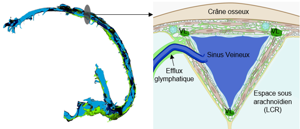
Dural venous sinuses. Left: general side view. Right: frontal section at the level of the superior sagittal sinus. The blood leaving the brain flows through the venous sinuses (in blue). These sinuses are surrounded by the perisinus, a thin region of the dura mater where small lymphatic vessels (LV) run. In this region, glymphatic fluid – responsible for clearing waste from the brain – drains before reaching the cervical lymph nodes via the lymphatic vessels.
Science, Health, and Prejudice
Idiopathic intracranial hypertension occurs mainly in obese or overweight women, following rapid weight gain. It is still unknown why. This uncertainty contributes to the stigmatization of patients, who are sometimes unfairly blamed for being responsible for their own illness. Such prejudice can lead to inadequate care, an underestimation of the severity of symptoms and a downplaying of patients’ complaints, who may then withdraw and develop depressive symptoms.
The disease remains largely underdiagnosed, probably because it is poorly understood. Too often, only the objective symptoms are considered, such as a cerebrospinal fluid leak or papilledema—that is, a swelling of the optic nerve. Yet IIH should be regarded as a neurological condition in its own right, one that has a major impact on brain function… and not as a mere collection of isolated symptoms. Patients describe their illness very well. Let us listen to them! They are highly committed and have contributed enormously to our team’s advances, notably through the AFHTIC patient association.
Stéphanie LenckInterventional neuroradiologist at the Pitié-Salpêtrière Hospital and a researcher at the Paris Brain Institute
There is probably a genetic predisposition to the disease, to which hormonal factors contribute. Moreover, in women, the meningeal lymphatic network is less developed than in men, making them more vulnerable to damage to these vessels. This may explain their over-representation in IIH – but also in other diseases, such as meningiomas.
But what link could there be between weight gain and the onset of intracranial hypertension?
“Lymphatic vessels play a key role in the transport of fats. In cases of excess weight, their function may be disrupted. There is a lead worth exploring here,” the researcher concludes.