Tag: antibodies

Unprecedented Success for HIV Vaccine in Preclinical Study

Vaccine approach yields high numbers of HIV-neutralising antibodies in non-human primates
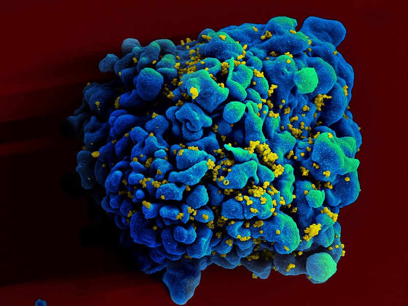
Colourised scanning electron micrograph of HIV (yellow) infecting a human T9 cell (blue). Credit: NIH

A new HIV vaccine developed by La Jolla Institute for Immunology (LJI), Scripps Research scientists, and IAVI has the potential to protect humans from developing HIV infection and AIDS. This HIV vaccine is the first to generate a high number of ‘broadly neutralising’, virus-fighting antibodies in primates.

“This feels like a huge success,” says LJI Professor and Chief Scientific Officer Shane Crotty, PhD, who co-led the research with Scripps Research Professor William Schief, PhD. “We constructed a successful vaccine from the ground up, which required a deep understanding of the immune system.”

This groundbreaking research, published in Nature, is the result of 14 years of collaboration between La Jolla Institute for Immunology and Scripps Research, as part of the Scripps Consortium for HIV/AIDS Vaccine Development (CHAVD). “This has been one of those Apollo moon mission-type projects, where there is an exceptional goal and the team has to accomplish a myriad of discoveries and inventions along the way,” says Crotty.

Outsmarting HIV

The new vaccine works by intervening in a process called B cell maturation. B cells make antibodies. Like many immune cells, B cells have an early ‘naïve’ stage before they are ready to make antibodies. B cells start to mature once they get the signal that a pathogen, such as a virus, is trying to attack. B cells see pieces of that pathogen’s molecular structure and start producing antibodies that can bind to that structure and halt infection.

It can take a little while for B cells to find the right “bullseye” on a pathogen. But B cells keep trying. As they mature, B cells tweak their antibody production, refining antibody structures to bind to a pathogen in just the right, vulnerable spots.

Scientists describe B cell development as a training process or bootcamp. In most cases, the body is left with a well-honed B cell army.

HIV is hard to beat because it doesn’t give B cells a chance to develop effective antibodies. The first problem is that HIV disguises itself from the immune system. The virus is wrapped in an ever-shifting cloak of sugar molecules, called glycans. This lets HIV sneak undetected past human cells, which are also covered in glycans.

The second big problem is that HIV mutates very quickly. “The worldwide diversity of HIV mutations is extraordinary. Even the diversity within one individual person living with HIV is dramatic,” says LJI Instructor Patrick Madden, Ph.D., who served as study co-first author with Jon Steichen, Ph.D., an institute investigator at Scripps Research.

The third problem is that HIV changes its shape when it infects human cells. Even if B cells get a glimpse of its viral structure – snap! – the structure changes.

Taken together, these problems rarely give B cells a chance to hone their antibody responses against HIV. Even if a B cell manages to make neutralising antibodies, the virus can mutate or change its shape, rendering those antibodies useless.

The LJI and Scripps Research teams spent years hunting for ‘broadly neutralising’ antibodies that can actually bind to HIV and recognise key viral structures, even if the rest of the virus mutates. These antibodies are very, very rare, but they can be found in blood samples from a small number of people living with HIV.

An effective HIV vaccine would need to prompt the immune system to make these same broadly neutralising antibodies. “How could we flip the whole immune response on its head so the rare responses become the common responses? That was a critical challenge we faced,” says Crotty.

Testing the new vaccine

It was time to go back to B cell bootcamp. The scientists studied what made the HIV-fighting B cells special. Then they reversed the process to see exactly how those B cells matured. By looking back at the maturation process, the researchers could track how the B cells changed when they saw specific pieces of the HIV structure.

The team discovered that B cells matured to make broadly neutralising antibodies after they got an early look at parts of HIV’s outer “envelope” protein. Because these viral sites sparked an immune response, scientists would call them “antigens.” 

An effective HIV vaccine would likely need to include models of these antigens. The antigens would work like mugshots of America’s most wanted. If B cells saw those antigens early and often, they would get really good at recognising and even neutralising HIV. “We were trying to mimic the progression of those neutralising antibodies,” says Madden.

In a feat of molecular engineering, the Schief Lab developed vaccine molecules that resembled the real HIV antigens. The scientists then worked with Emory National Primate Research Center, to test this potential HIV vaccine in a non-human primate species called rhesus macaques.

The researchers first administered a “priming” vaccine meant to activate each animal’s naïve B cells. The animals then received a series of “shepherding” booster shots to help their B cells develop along the right path. 

“This series of vaccinations will guide, or ‘walk’, a B cell from its naïve state to its broadly neutralising state,” says Madden.

This new type of vaccine approach is called “germline targeting” because it targets naïve B cells in their ‘germline’ or naïve form, before they begin their training process.

The scientists found that around 44 percent of the animals went on to produce broadly neutralising antibodies against HIV in their blood. These antibodies were impressively abundant. 

“We succeeded in taking ultra-rare antibody responses and turning them into common responses by the end of the vaccination process,” adds Crotty. In other research recently published, they reported a new strategy to accelerate related vaccine antibody responses [See Nature Immunology paper].

The team didn’t test whether these antibodies could prevent infection, but it’s significant that these antibodies could be found in the blood, where they could encounter and potentially block HIV.

Bringing the HIV vaccine to humans

The Crotty Lab plans to investigate how they might change the booster shot regimen to make the HIV vaccine even more effective. “It was incredible to get those results, but of course we’d like to see a response in 100 percent of the animals,” says Madden. 

Importantly, the antibodies found in the animal subjects resembled the exact kinds of broadly neutralising antibodies seen in those rare humans who made their own neutralising antibodies. It’s clear that our immune systems can make these powerful antibodies, given the right training.

“We believe this vaccine approach is even more likely to succeed in humans, because of the immunogenetics,” Crotty says.

The priming immunogen used in this study was evaluated in humans in the HVTN 144 trial and is currently being tested in the Phase 1 trial IAVI G004. IAVI, Scripps Research, the HIV Vaccine Trials Network, and partners are now advancing plans to further evaluate the full immunisation regimen in a future human clinical study.

Source: La Jolla Institute

Two New Antibodies to Treat Inflammatory Autoimmune Diseases

Antibodies by Pikisuperstar on Freepix

An international research group directed by UMC Utrecht have developed and characterised two first-in-class antibodies that specifically block the high-affinity IgG receptor FcγRI. Their findings open new perspectives for therapeutic modulation of FcγRI-driven inflammation in autoimmune diseases such as rheumatoid arthritis (RA), systemic lupus erythematosus (SLE) and immune thrombocytopenia (ITP).

FcγRI, also known as CD64, is a high-affinity receptor on myeloid cells that binds to the Fc region of immunoglobulin G (IgG) antibodies. It plays a key role in immune defence by triggering cellular functions such as phagocytosis and cytokine production. In a normal immune response, FcγRI is activated by immune complexes – clusters of antibodies bound to pathogens, which mark them for clearance. In autoimmune diseases, however, the immune system mistakenly targets the body’s own tissues (such as joint proteins in RA, nuclear antigens in SLE, or platelets in ITP), which results in the production of autoantibodies that form immune complexes. These misdirected complexes activate FcγRI unnecessarily, driving chronic inflammation and subsequent tissue damage.

The study, led by Prof Jeanette Leusen, PhD from the Antibody Therapy research group at the Center for Translational Immunology (UMC Utrecht) and carried out by PhD candidate Tosca Holtrop MSc, was a true team effort, in collaboration with experts from Kiel University (Germany), Leiden University Medical Center, Utrecht University, and Friedrich-Alexander University Erlangen-Nürnberg (Germany).

Discovery of first-in-class antibodies

For over 30 years, scientists have tried to generate antibodies against the IgG-binding domain of CD64, but the receptor’s extremely high affinity for IgG made this impossible with earlier technologies. Combining the UMAB unique immunisation method with novel phage display antibody libraries, the team bypassed this challenge by excluding the Fc region of the antibodies. This allowed them to discover two unique Fc-silent antibodies, C01 and C04, that bind purely via their Fab domains to FcγRI. Crystal structural analysis confirmed that C01 binds precisely within the IgG-binding site on EC2, making binding mutually exclusive.

High affinity for FcγRI

Quantitative binding studies revealed that both antibodies have higher affinity for FcγRI than human IgG, allowing them to efficiently displace IgG or pathogenic immune complexes up to 60 percent and block binding up to 90 percent. Importantly, neither antibody triggered FcγRI activation, a critical distinction from earlier anti-FcγRI antibodies, which could inadvertently trigger receptor clustering and cytokine release.

Subsequently. an in vitro model for immune thrombocytopenia was set-up, where C01 and C04 effectively inhibited opsonized platelets from binding to immune cells from ITP patients. In a preclinical in vivo ITP model, the antibodies significantly reduced IgG-dependent platelet depletion. The antibodies were also tested in an in vitro rheumatoid arthritis models, where they effectively inhibited patient-derived autoantibody–immune complex binding to monocytes, macrophages, and neutrophils from healthy donors.

Promising therapeutic candidate drugs

The study demonstrates that direct Fab-mediated inhibition of FcγRI is feasible and effective, opening a new avenue for treating autoimmune diseases characterised by IgG-autoantibody complexes. By preventing immune complex–driven activation without triggering the receptor, C01 and C04 represent a promising next step toward targeted, inflammation-sparing immunotherapy. “I think we found the needle in the haystack, after searching over a decade and thanks to a true team effort,” says Jeanette Leusen, principal investigator of the study. “Each research partner contributed a critical piece, from antibody discovery and structure determination to patient sample testing and preclinical models. Only together could we bring this to fruition. These antibodies not only provide a unique tool for studying FcγRI biology, but also hold promise as therapeutic candidates in autoimmune and infectious diseases.”

Source: University Medical Centre Utrecht

Solving Africa’s Hidden Snakebite Problem with a New Universal Antivenom

Photo by Nivedh P on Unsplash

In Sub-Saharan Africa, more than 300 000 people are bitten by venomous snakes annually, 3000 of whom die – but with the underreporting that goes hand in hand with the lack of healthcare infrastructure, the real number could be as much as five times higher. Many more face amputations. Even if patients manage to make it to a clinic or hospital in time, there is no guarantee that there will be any anti-venom available to treat them. As a South African case study shows, just having antivenom in the right place is a problem even in Western Cape’s relatively well-developed healthcare system, with antivenom’s three-year shelf life and cold chain failures posing a major problem for rural healthcare centres.

But now, scientists have developed a new kind of antivenom that is effective for 17 different snake species, including mambas, cobras and a rinkhals. The study, published in Nature, makes use of a nanobody-based cocktail that targets common mechanisms across venoms – and which is also more effective at preventing the tissue damage that leads to amputations.

A huge obstacle to creating broad-spectrum antivenoms is the enormous diversity of venomous snakes and the complexity of their venoms – a single species’ venom may contain 100 toxins from multiple different protein families. Listen to our podcast to hear a deep dive into Africa’s snakebite burden and how the international team of researchers accomplished their feat:

New Antibodies Potentially Effective Against All SARS-CoV-2 Variants

Image by Fusion Medical on Unsplash

SARS-COV-2 has been very good at mutating to keep infecting people – so good that most antibody treatments developed during the pandemic are no longer effective. Now a team led by Stanford University researchers may have found a way to pin down the constantly evolving virus and develop longer-lasting treatments.

The researchers discovered a method to use two antibodies, one to serve as a type of anchor by attaching to an area of the virus that does not change very much and another to inhibit the virus’s ability to infect cells. This pairing of antibodies was shown to be effective against the initial SARS-CoV-2 virus that caused the pandemic and all its variants through omicron in laboratory testing. The findings are detailed in the journal Science Translational Medicine.

“In the face of an ever-changing virus, we engineered a new generation of therapeutics that have the ability to be resistant to viral evolution, which could be useful many years down the road for the treatment of people infected with SARS-CoV-2,” said Christopher O. Barnes, the study’s senior author, an assistant professor of biology.

An overlooked option

The team led by Barnes and first author Adonis Rubio, a doctoral candidate in the Stanford School of Medicine, conducted this investigation using donated antibodies from patients who had recovered from COVID-19. Analysing how these antibodies interacted with the virus, they found one that attaches to a region of the virus that does not mutate often.

This area, within the Spike N-terminal domain, or NTD, had been overlooked because it was not directly useful for treatment. However, when a specific antibody attaches to this area, it remains stuck to the virus. This is useful when designing new therapies that enable another type of antibody to get a foothold and attach to the receptor-binding domain, or RBD, of the virus, essentially blocking the virus from binding to receptors in human cells.

An illustration of the bispecific antibodies the Stanford-led research team developed to neutralise the virus that causes COVID-19. Named “CoV2-biRN,” these two antibodies work together by attaching to different areas of the virus.
The bispecific antibodies target two areas of the virus: One attaches to the “NTD,” or Spike N-terminal domain, an area on the virus that does not change very much. This allows the second antibody to attach to the “RBD,” or receptor-binding domain, essentially preventing the virus from infecting human cells. | Christopher O. Barnes and Adonis Rubio using Biorender stock images

The researchers designed a series of these dual or “bispecific” antibodies, called CoV2-biRN, and in laboratory tests they showed high neutralisation of all the variants of SARS-CoV-2 known to cause illness in humans. The antibodies also significantly reduced the viral load in the lungs of mice exposed to one version of the omicron variant.

More research, including clinical trials, would have to be done before this discovery could be used as a treatment in human patients, but the approach is promising – and not just for the virus that causes COVID-19.

Next, the researchers will work to design bispecific antibodies that would be effective against all coronaviruses, the virus family including the ones that cause the common cold, MERS, and COVID-19. This approach could potentially also be effective against influenza and HIV, the authors said.

“Viruses constantly evolve to maintain the ability to infect the population,” Barnes said. “To counter this, the antibodies we develop must continuously evolve as well to remain effective.”

Source: Stanford University

Antibodies in Breastmilk Protect Infants Against Rotavirus

Photo by Wendy Wei: https://www.pexels.com/photo/mother-breastfeeding-her-child-3074935/

A study led by researchers at the found that breast milk provides protection against rotavirus, a common gastrointestinal disease in infants. Babies whose mothers had high levels of specific antibodies in their breast milk were able to fend off the infection for a longer period than infants whose mothers had lower levels. The researchers also uncovered an unexpected relationship between BMI and antibody levels.

Published in the Journal of Clinical Investigation, the University of Rochester Medical Center-led study also found significant differences in antibody profiles in breast milk between mothers in high-income countries (HICs) and low- and middle-income countries (LMICs). Researchers analysed human milk samples from 695 women in Finland, the US, Pakistan, Peru, and Bangladesh, and measured specific IgA and IgG antibodies, which are common antibodies produced in breast milk, against 1607 proteins from 30 pathogens.

The research, led by Dr Kirsi Jarvinen-Seppo, MD, PhD, professor at UR Medicine Golisano Children’s Hospital (GCH), tracked antibody levels and kinetics over time to analyse antibody responses to a wide range of respiratory, diarrhoeal and sepsis pathogens in human milk. The primary aim of the study, funded by the Bill and Melinda Gates Foundation, was to understand the protective properties of these antibodies and how they vary across different geographic and economic regions.

“We would expect to find differences in antibody levels in different countries, due to different diseases circulating among areas of the world, but this is one of the first times that there’s been a head-to-head comparison for dozens of pathogens across several continents,” said Jarvinen-Seppo. “It was encouraging to see such a clear link between higher antibody levels and a delay to rotavirus infection, and this was consistently observed among an independent validation cohort.”  

Other notable findings from the study:

  • Milk from women in LMICs had higher levels of IgA and IgG antibodies against various intestinal and respiratory pathogens compared to milk from HICs. This difference was particularly notable for pathogens such as Shigella and pneumococcus, which are major contributors to morbidity and mortality in young children.
  • Higher body mass index (BMI) was associated with lower antibody levels, which went against expectations.

“The variation in antibody profiles between regions highlights the impact of economic and environmental factors on maternal immunity,” said Jarvinen-Seppo.

In addition to Rotavirus findings, the discovery that a higher BMI was associated with lower antibody counts in breast milk was also unexpected.

“We had anticipated that underweight mothers might have lower antibody levels due to poorer nutritional status,” said Jarvinen-Seppo. “Due to rising obesity rates worldwide, this could be a significant finding, but this is preliminary and additional research is needed since this is the first time this has been measured.”

“While the data on rotavirus protection is compelling, the geographical and BMI-related variations highlight areas where further research is essential. The study sets the stage for additional investigations that could lead to better understanding and interventions for improving infant health globally,” said Jarvinen-Seppo.

Source: University of Rochester Medical Center

COVID Vaccination and Antibody Responses Found to be Long-lasting

Image by Fusion Medical on Unsplash

A long-term analysis conducted by leading microbiologists at the Icahn School of Medicine at Mount Sinai reveals that antibody responses induced by COVID vaccines are long-lasting. The study results, published online in the journal Immunity, challenge the idea that mRNA-based vaccine immunity wanes quickly.

The emergence of SARS-CoV-2 in late 2019 sparked the global pandemic that is now in its fifth year. Vaccines that were developed at record speed have saved millions of lives. However, the emergence of SARS-CoV-2 variants and waning immunity have decreased the effectiveness of the vaccines against symptomatic disease. The common perception now is that mRNA-based vaccine-induced immunity wanes quickly. However, this assumption is largely based on data from short-term studies that include a very limited number of data points following peak responses.

The Mount Sinai research team’s analysis of more than 8000 samples collected over a three-year period in New York City examined how antibody responses to the virus’s spike protein changed after infections, during the primary immunisation series, during monovalent and bivalent booster vaccination, and during breakthrough infections.

They found that upon primary immunisation, participants with pre-existing immunity (those who had previously been infected with the virus) mounted higher antibody responses faster and achieved higher steady-state antibody titres than individuals who had not been previously infected. The waning of antibody response was characterised by two phases: an initial rapid decay from the strong peak after vaccination, followed by a stabilisation phase with very slow decay, suggesting that antibody levels were very long-lasting. Booster vaccination equalised the differences in antibody concentration between participants with and without pre-existing immunity. Breakthrough infections increased antibodies to similar levels as an additional vaccine dose in individuals who had not previously been infected.

This investigation represents one of the most extensive and in-depth assessments of the longevity of SARS-CoV-2 immune responses to date. Its major conclusion is that changes in the virus that allow it to evade immunity, rather than waning immunity, are the major reason for breakthrough infections.

“Ours is one of the longest-running COVID-19 studies out there,” said Viviana Simon, MD, PhD, Professor of Microbiology, Medicine and Pathology, Molecular and Cell-Based Medicine, at Icahn Mount Sinai and lead author of the paper. “Following the same group of people monthly over time is rare and powerful because you can compare immune responses on an individual level. SARS-CoV-2 continues to evolve, so this research is important to provide an understanding about the impact of new variants and new vaccine doses on a healthy immune system, and to guide all of us to make the best choices to maintain protection against the virus that continues to circulate in our communities.”

This in-depth analysis was made possible through the Protection Associated with Rapid Immunity to SARS-CoV-2 (PARIS) study, an observational, longitudinal cohort of health care workers of the Mount Sinai Health System that was initiated in April 2020. At that time, the densely populated New York metropolitan area was hit with an exponential increase in severe SARS-CoV-2 infections, and essential workers in the health care system were at high risk for infection. In response to the crisis, a team of leading virologists, physician-scientists, and pathologists at Mount Sinai established a specific and sensitive SARS-CoV-2 binding enzyme-linked immunosorbent assay to accurately measure the SARS-CoV-2 antibody titres. This test was used to measure immune responses in the PARIS cohort in order to determine how quickly the antibody defences were mounted and much these changed over the months and years of follow up.

In addition to showing the impact on a person’s individual antibody response to vaccines based on the type of vaccine received and whether or not they were infected before receiving the first dose, the PARIS study made possible the development of a mathematical model that can be used to predict and characterize antibody responses of both individual people and populations.

“People have pandemic fatigue and vaccine uptake has slowed, especially after the vaccines started to be charged to insurance,” said Komal Srivastava, MS, Director of Strategy and Operation of the Mount Sinai Center for Vaccine Research and Pandemic Preparedness and co-first author of the paper. “We were pleasantly surprised to see that the booster doses promoted a large antibody response regardless of a person’s personal infection history, so we are hopeful that our study findings will encourage people to get their vaccine boosters when eligible and to stay engaged in research. Our work also showcases the impact of viral evolution over time and why it’s critical to keep studies like this going, despite the pandemic fatigue.”

According to the research team, the PARIS model has broad applications for studying the kinetics of antibodies produced to different COVID vaccines in diverse populations. They stress much more work remains to analyse side effects, applications of the antibody model and continued research about new vaccines and viral variants.

“This study adds an essential piece of data to understand the intricate immune response elicited by SARS-CoV-2 infection and COVID-19 vaccination,” says Juan Manuel Carreno Quiroz, PhD, Assistant Professor in the Department of Microbiology and co-first author of the paper. “In light of the emerging viral variants, which predominantly induce a cross-reactive antibody response against the spike protein, it will be exciting to characterise in depth the role of these antibodies – in particular the non-neutralising ones – in protection against the most recent circulating viral variants. Likewise, monitoring the induction of variant-specific antibodies after multiple exposures by breakthrough infections and by administration of updated COVID-19 vaccines, such as the XBB.1.5 monovalent booster, will be key to understand the evolution of the antibody response over time.”

Source: The Mount Sinai Hospital / Mount Sinai School of Medicine

Epstein–Barr Virus Antibodies may Trigger Multiple Sclerosis

Source: CC0

Researchers at Karolinska Institutet have found further links between Epstein–Barr virus and multiple sclerosis. A study published in Science Advances shows that some individuals have antibodies against the virus that mistakenly attack a protein in the brain and spinal cord.

Many years ago, the Epstein–Barr virus (EBV), which infects most people early in life and then usually lies dormant was linked to multiple sclerosis (MS) but the reason remained a mystery. Increasing evidence, including two papers published in Science and Nature last year, suggests that EBV infection precedes MS and that antibodies against the virus may be involved. However, the molecular mechanisms seem to vary between patients and remain largely unknown.

“MS is an incredibly complex disease, but our study provides an important piece in the puzzle and could explain why some people develop the disease,” says Olivia Thomas, postdoctoral researcher at the Department of Clinical Neuroscience, Karolinska Institutet and shared first author of the paper. “We have discovered that certain antibodies against the Epstein-Barr virus, which would normally fight the infection, can mistakenly target the brain and spinal cord and cause damage.”

The researchers analysed blood samples from more than 700 patients with MS and 700 healthy controls. They found that antibodies that bind to a certain protein in the Epstein-Barr virus, EBNA1, can also bind to a similar protein in the brain and spinal cord called CRYAB, whose role is to prevent protein aggregation during conditions of cellular stress such as inflammation. These misdirected, cross-reactive antibodies may damage the nervous system and cause severe symptoms in MS patients, including problems with balance, mobility and fatigue. The antibodies were present in about 23 percent of MS patients and 7% of control individuals.

“This shows that, whilst these antibody responses are not required for disease development, they may be involved in disease in up to a quarter of MS patients,” says Olivia Thomas. “This also demonstrates the high variation between patients, highlighting the need for personalised therapies. Current therapies are effective at reducing relapses in MS but unfortunately, none can prevent disease progression.”

“We are now expanding our research to investigate how T cells fight EBV infection and how these immune cells may damage the nervous system in multiple sclerosis and contribute to disease progression,” says joint first author of the paper Mattias Bronge, affiliated researcher at the Department of Clinical Neuroscience, Karolinska Institutet.

Source: Karolinska Institutet

COVID Vaccination Protection Wanes Faster in People with Obesity

Antibodies by Pikisuperstar on Freepix

According to new research from the Universities of Cambridge and Edinburgh, COVID vaccination protection in people with severe obesity wanes faster than in people of normal weight. The study suggests that people with obesity are likely to need more frequent booster doses to maintain their immunity.

Previous studies on COVID vaccines have suggested that antibody levels may be lower in vaccinated people who have obesity and that they may remain at higher risk of severe disease than vaccinated people with normal weight. The reasons for this have, however, remained unclear.

This study, published in the journal Nature Medicine, shows that the ability of antibodies to neutralise the virus (their ‘neutralisation capacity’) declines faster in vaccinated people who have obesity. The findings have important implications for vaccine prioritisation policies around the world.

During the pandemic, people with obesity were more likely to be hospitalised, require ventilators and to die from COVID. In this study, supported by the NIHR Bioresource and funded by UKRI, the researchers set out to investigate how far two of the most extensively used vaccines protect people with obesity compared to those with a normal weight, over time.

A team from the University of Edinburgh looked at real-time data tracking the health of 3.5 million people in the Scottish population as part of the EAVE II study. They looked at hospitalisation and mortality from COVID in adults who received two doses of COVID vaccine (either Pfizer-BioNTech or AstraZeneca).

They found that people with severe obesity (a BMI > 40kg/m2) had a 76% higher risk of severe COVID outcomes, compared to those with a normal BMI. A modest increase in risk was also seen in people with obesity (30-39.9kg/m2), which affects a quarter of the UK population, and those who were underweight. ‘Break-through infections’ after the second vaccine dose also led to hospitalisation and death sooner (from 10 weeks) among people with severe obesity, and among people with obesity (after 15 weeks), than among individuals with normal weight (after 20 weeks).

University of Edinburg leader Prof Sir Aziz Sheikh said: “Our findings demonstrate that protection gained through COVID vaccination drops off faster for people with severe obesity than those with a normal body mass index. Using large-scale data assets such as the EAVE II Platform in Scotland have enabled us to generate important and timely insights that enable improvements to the delivery of COVID vaccine schedules in a post-pandemic UK.”

The University of Cambridge team studied people with severe obesity attending the Obesity clinic at Addenbrooke’s Hospital in Cambridge, and compared the number and function of immune cells in their blood to those of people of normal weight.

They studied people six months after their second vaccine dose and then looked at the response to a third ‘booster’ vaccine dose over time. The Cambridge researchers found that six months after a second vaccine dose, people with severe obesity had similar levels of antibodies to the COVID virus as those with a normal weight – but those antibodies were less effecctive.

The antibodies’ neutralisation capacity was reduced in 55% of individuals with severe obesity were found to have unquantifiable or undetectable ‘neutralising capacity’ compared to 12% of people with normal BMI.

“This study further emphasises that obesity alters the vaccine response and also impacts on the risk of infection,” said first author Dr Agatha van der Klaauw. “We urgently need to understand how to restore immune function and minimise these health risks.”

The researchers found that antibodies produced by people with severe obesity were less effective at neutralising the SARS-CoV-2 virus, potentially because the antibodies were not able to bind to the virus with the same strength.

When given a third (booster) dose of a COVID vaccine, neutralisation capacity was restored in both the normal weight and severely obese groups. But the researchers found that immunity again declined more rapidly in people with severe obesity, putting them at greater risk of infection with time.

Antibodies can Prevent Bacteria from Infiltrating Cells

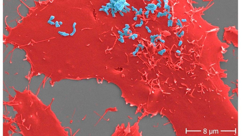
Adhesion of Bartonella henselae to human cells. B. henselae  (strain Marseille) bacteria (light blue) in an early stage infection process (30 min) to human HeLa-229 cells (red). Adhesion to host cells is mediated by specific interactions between B. henselae  surface proteins and components of the host extracellular matrix including molecules such as fibronectin or collagen. Scale bar: 8 μm.

Using Bartonella henselae bacteria, the cause of cat scratch disease, researchers have demonstrated for the first time that antibodies can prevent certain surface proteins of bacterial pathogens from entering host cells. The findings are important for the development of new drugs against highly resistant infectious agents.

Infections pose a significant threat to human health, especially when pathogens manage to colonise the organism and subsequently cause severe infections. The first step in such an infection always consists of the pathogens attaching themselves to the host cells’ surface. From here, the infections spread, resulting, for example, in infections of deeper tissue layers and organs.

A group of scientists surrounding Prof. Volkhard Kempf from Frankfurt University Hospital’s Institute of Microbiology and Hospital Hygiene has now succeeded in blocking this adhesion mechanism in a bacterium, thereby preventing the infection of host cells. For this purpose, the researchers examined the pathogen Bartonella henselae, usually causing cat scratch disease. Transmitted by cats, the disease mainly affects young children, whose symptoms include swollen and hardened lymph nodes around the site of infection, usually after a scratch or bite injury caused by infected cats.

Bartonella bacteria infect so-called endothelial cells, which line the blood vessels. Via their surface protein Bartonella adhesin A (BadA), they attach themselves to a protein (fibronectin) of the so-called “extracellular matrix,” a network of protein fibers that lie on top of the endothelial cells.

Breaking BadA

To determine which parts of the BadA protein are important in the bacterial adhesion process, the researchers equipped Bartonella bacteria with various genetically modified BadA variants, among others, and then analysed the extent to which these variants were still able to bind fibronectin. Once it was clear which BadA segments were responsible for the binding, the team produced antibodies against them, demonstrating for the first time that such antibodies can prevent infection by such bacteria.

Prof. Volkhard Kempf explains: “Bartonella henselae is not a very dangerous pathogen, and in most cases, cat scratch disease does not require any specific medical treatment. However, for us Bartonella henselae is a very important model organism for far more dangerous pathogens such as Acinetobacter baumannii, a serious pathogen that usually causes wound infection or pneumonia and often shows resistance to several last-choice antibiotics. The BadA protein of Bartonella henselae belongs to the so-called ‘trimeric autotransporter adhesins’, which are also responsible for adhesion to human cells in Acinetobacter and a number of other pathogens. A drug-induced blocking of these adhesins is therefore a promising novel and future approach to combat dangerous bacterial infections.”

The researchers published their findings in Diagnostics.

Source: Goethe University Frankfurt

New Omicron Variant Escapes Antibodies and Monoclonal Therapy

Source: Fusion Medical Animation on Unsplash

The emerging Omicron variant BA.2.75.2 largely evades neutralising antibodies in the blood and resists several monoclonal antibody antiviral treatments, according to a study published in the journal The Lancet Infectious Diseases. The findings suggest a risk of increased COVID infections in the northern hemisphere’s winter, unless the new updated bivalent vaccines help to boost immunity in the population.

“While antibody immunity is not completely gone, BA.2.75.2 exhibited far more dramatic resistance than variants we’ve previously studied, largely driven by two mutations in the receptor binding domain of the spike protein,” said the study’s corresponding author Ben Murrell, assistant professor at the Department of Microbiology, Tumor and Cell Biology, Karolinska Institutet.

The study shows that antibodies in random serum samples from 75 blood donors in Stockholm were approximately only one-sixth as effective at neutralising BA.2.75.2 compared with the now-dominant variant BA.5. The serum samples were collected at three time points: in November 2021 before the emergence of Omicron, in April 2022 after a large wave of infections in the country, and at the end of August to early September after the BA.5 variant became dominant.

The researchers found that only one of the clinically available monoclonal antibody treatments that were tested, bebtelovimab, managed to effectively neutralise the new variant.

BA.2.75.2 is a mutated version of another Omicron variant, BA.2.75. Since it was first discovered earlier this fall, it has spread to several countries but so far represents only a minority of registered cases.

A possible increase in infections

“We now know that this is just one of a constellation of emerging variants with similar mutations that will likely come to dominate in the near future,” Ben Murrell says, adding “we should expect infections to increase this winter.”

Some questions remain. It is unclear whether these new variants will drive an increase in hospitalisation rates. Also, while current vaccines have, in general, had a protective effect against severe disease for Omicron infections, there is not yet data showing the degree to which the updated COVID vaccines provide protection from these new variants. “We expect them to be beneficial, but we don’t yet know by how much,” Ben Murrell says.

Source: Karolinska Institutet