Researchers at The Ottawa Hospital and the University of Ottawa were able to reverse certain behavioural symptoms in a mouse model of autism by fixing a problem in the brain blood vessels. Their findings, published in Neuron, present a promising new treatment target for these symptoms.
Autism is a neurodevelopmental condition with widely varying characteristics. Some of the behavioural symptoms can make life more challenging. While many people with autism have found ways to manage these symptoms, no drug treatment exists.
“The road from discovery to clinical trials is long, but we’re excited by the possibility that our findings might one day improve the daily lives of people with autism,” said senior author Dr Baptiste Lacoste, senior scientist at The Ottawa Hospital and professor at the University of Ottawa.
Dr Lacoste’s team previously discovered that blood vessels in the brain don’t work properly in mouse models with a 16p11.2 deletion, one of the most common genetic mutations seen in autism. They were the first to look at what was happening in the blood vessels of the brain.
The researchers found the problem began in cells lining the blood vessels, called the endothelial cells. These cells make sure blood quickly gets to the parts of the brain that are active. This responsive blood supply is needed for proper brain function.
However, the endothelial cells in these mice don’t respond quickly enough. This happens early in brain development and causes behavioral symptoms later in life, including hyperactivity, repetitive movements, and motor learning impairment.
In this new study, driven by former PhD student Dr Julie Ouellette, the research team looked at what was wrong with these endothelial cells and whether it could be fixed.
The team led by Dr Lacoste found that the endothelial cells had half the normal level of a molecule called ATP. ATP is usually considered an energy molecule, but in this case the cell was missing its target, called a P2Y2 receptor, on its surface.
By activating this P2Y2 receptor, the researchers could restore the cell’s function, increase blood flow in the brain and reverse the behavioral symptoms in adult mice. This was achieved using a drug known to activate P2Y2 and currently approved for humans in Japan and South Korea to treat dry eye syndrome.
“It’s as if these cells are asleep, and now we can wake them up,” says Dr Lacoste. “And we may only need to treat them once to wake them up permanently. We will test that further, but it’s an encouraging feature for a future treatment.”
This study only looked at adult mice. This means targeting P2Y2 could reverse behavioural symptoms that were already well established. Next, the team plans to treat mice earlier in life to see if early treatment has additional benefits.
The team has also filed a patent application for using P2Y2 activation in the blood vessels to treat autism symptoms. They are interested in exploring drug development with the aim of eventual clinical trials.
Sometimes, doctors cannot find the cause of intracranial hypertension, a syndrome characterised by excess pressure inside the skull. Patients’ symptoms are effectively relieved by a procedure that dilates a vein in the brain… yet no one truly understands why. At the Paris Brain Institute, Stéphanie Lenck, Jean-Léon Thomas, and their colleagues have identified the mechanism behind this phenomenon. Their work, published in the journal Nature Neuroscience, sheds new light on the links between venous flow and the drainage of brain fluids, opening new therapeutic avenues.
Idiopathic intracranial hypertension (IIH) is a rare and poorly understood neurological disorder that affects 1 to 20 people per 100 000 – most often women of childbearing age. It is characterised by increased pressure inside the skull, in the absence of a tumour or any other identifiable cause. Patients typically present with a narrowing of the venous sinuses – large veins located in the dura mater, the fibrous membrane that envelops the brain and spinal cord – as well as poor circulation of brain fluids.
Patients live with chronic headaches, often accompanied by visual disturbances, tinnitus, dizziness, and severely disabling cognitive difficulties.
“When intracranial pressure is so high that it damages the optic nerve, patients receive emergency treatment to preserve their sight. However, other symptoms—sleep disorders, attention difficulties, and chronic fatigue—are often overlooked, even though they profoundly affect quality of life, sometimes for years. We still do not know why these symptoms vary so much from one patient to another and do not always match abnormalities seen on MRI,” explains Stéphanie Lenck (AP-HP), an interventional neuroradiologist at the Pitié-Salpêtrière Hospital and a researcher at the Paris Brain Institute.
Understanding the mechanisms of the disease is therefore essential for refining diagnosis and improving patient care in the short, medium, and long term.
A Closer Look at Meningeal Lymphatic Vessels
One effective treatment for IIH involves dilating narrowed venous sinuses using a stent, a small mesh tube that restores blood flow and venous pressure in the brain. In most cases, after a brief adjustment period, intracranial pressure drops and symptoms subside. Yet the mechanism behind this improvement has remained unclear: what is the relationship between veins and intracranial hypertension? Why is this intervention so effective?
To find out more, Stéphanie Lenck, Jean-Léon Thomas, and their colleagues followed 16 female patients with IIH and 20 healthy female volunteers, all of whom underwent thorough MRI examinations. In parallel, the researchers developed and studied a mouse model with altered cerebral venous flow.
Their findings are unequivocal: the circulation of brain fluids, including cerebrospinal fluid, is closely dependent on blood flow in the venous sinuses.
The brain’s drainage and immune surveillance rely on a network of specialized vessels: the meningeal lymphatics. They line the perisinus, an anatomical space between the outer layer of the dura mater, which is attached to the skull, and the inner layer, which runs alongside the venous sinuses. These delicate, highly environment-dependent vessels only function when blood flow in the large dural veins is normal.
Jean-Léon ThomasYale University, Inserm
When the venous sinuses are narrowed, venous pressure rises upstream, and fluid circulation in the perisinus is impaired. Brain fluids accumulate, causing the brain and skull of patients to swell under pressure. In mice, the lymphatic vessels can regress until they disappear entirely.
Because meningeal lymphatic vessels also regulate brain inflammation and immune surveillance, this regression is likely to have multiple consequences for the brain. These findings, therefore, open new avenues of research.
“Our findings could shed light on the mechanisms of sleep, a period during which the glymphatic system is activated. This waste-clearance system of the central nervous system is closely connected to the meningeal lymphatic vessels,” the researcher adds.
Draining the Brain: A Well-Oiled Machine
For the past decade, we have known that brain drainage and detoxification are partly driven by cerebrospinal fluid, which circulates through two complementary transport routes: the glymphatic system and the meningeal lymphatic vessels. This brain-cleansing system is activated during sleep, promoting a daily detoxification of the tissues.
Essential for immune surveillance and the removal of waste, including toxic proteins, these two pathways could influence the course of several neurological conditions, such as neurodegenerative diseases, multiple sclerosis, and glioblastomas. A better understanding of how cerebral clearance works could therefore lead to new therapeutic approaches.
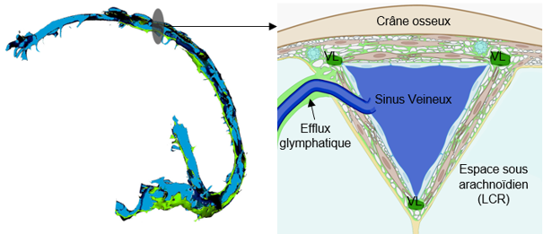
Dural venous sinuses. Left: general side view. Right: frontal section at the level of the superior sagittal sinus. The blood leaving the brain flows through the venous sinuses (in blue). These sinuses are surrounded by the perisinus, a thin region of the dura mater where small lymphatic vessels (LV) run. In this region, glymphatic fluid – responsible for clearing waste from the brain – drains before reaching the cervical lymph nodes via the lymphatic vessels.
Science, Health, and Prejudice
Idiopathic intracranial hypertension occurs mainly in obese or overweight women, following rapid weight gain. It is still unknown why. This uncertainty contributes to the stigmatization of patients, who are sometimes unfairly blamed for being responsible for their own illness. Such prejudice can lead to inadequate care, an underestimation of the severity of symptoms and a downplaying of patients’ complaints, who may then withdraw and develop depressive symptoms.
The disease remains largely underdiagnosed, probably because it is poorly understood. Too often, only the objective symptoms are considered, such as a cerebrospinal fluid leak or papilledema—that is, a swelling of the optic nerve. Yet IIH should be regarded as a neurological condition in its own right, one that has a major impact on brain function… and not as a mere collection of isolated symptoms. Patients describe their illness very well. Let us listen to them! They are highly committed and have contributed enormously to our team’s advances, notably through the AFHTIC patient association.
Stéphanie LenckInterventional neuroradiologist at the Pitié-Salpêtrière Hospital and a researcher at the Paris Brain Institute
There is probably a genetic predisposition to the disease, to which hormonal factors contribute. Moreover, in women, the meningeal lymphatic network is less developed than in men, making them more vulnerable to damage to these vessels. This may explain their over-representation in IIH – but also in other diseases, such as meningiomas.
But what link could there be between weight gain and the onset of intracranial hypertension?
“Lymphatic vessels play a key role in the transport of fats. In cases of excess weight, their function may be disrupted. There is a lead worth exploring here,” the researcher concludes.