Even when controlling for age and family background, COVID’s impact was evident
Photo by Kelly Sikkema on Unsplash
The COVID pandemic disrupted children’s ability to self-regulate, according to research from three UK universities just published in the journal Child Development.
The study by Lancaster University, East Anglia and Durham reveals that the pandemic hampered children’s ability to regulate their behaviour, stay focused and adapt to new situations – skills known collectively as executive functions.
The greatest impact was seen among pupils who were in reception when the first lockdowns began – a crucial stage at four or five when youngsters normally learn to socialise, follow routines and navigate the busy world of the classroom. Primary school in the UK then begins at Grade 1, starting at age five or six.
These children showed less growth in their self-regulatory and cognitive flexibility scores over time compared to a second group of children who were in preschool when the pandemic started.
The research team say these children may still be feeling the effects years later.
How the research happened
Scientists were already running a long-term study tracking youngsters from toddlerhood to early school years when the COVID pandemic hit.
They followed 139 children aged between two-and-a-half and six-and-a-half years old over several years, including 94 families who joined the study before Covid struck.
This meant that they had a rare baseline of children’s abilities before the pandemic began, which allowed them to track exactly how development changed during and after the lockdowns.
Using a standardised assessment called the Minnesota Executive Function Scale, they were able to measure the same cognitive skills at regular intervals.
Dr Eleanor Johns from Lancaster University’s Department of Psychology said: “We began this study to understand how children’s executive function develops across early childhood, and we saw clear, steady growth between 2.5 and 6.5 years of age. However, because our longitudinal study spanned the COVID-19 pandemic, we also had a unique opportunity to examine how this unprecedented disruption affected the children we were already following.
“We found that children who had just started school when the first lockdown began showed a slower rate of growth in executive function compared to those who were preschool age. Starting school is a major developmental transition, as children learn new routines, adapt to classroom rules, and develop self-regulation alongside their peers. When schools closed almost overnight, those opportunities were suddenly removed.”
The research revealed that:
Individual differences in executive function abilities were remarkably stable. Children who had stronger skills at two-and-a-half years old tended to remain ahead at six-and-a-half years.
Children from lower socio-economic households consistently scored lower, echoing long-standing research on the impact of maternal education and home environment.
Even when controlling for age and family background, COVID’s impact was evident. Children who were in reception at the start of the pandemic made more modest improvements in executive function compared to those still in preschool.
Dr Johns said: “Our findings suggest that the structured school environment and regular interaction with peers play a crucial role in supporting the development of executive function. When those experiences were disrupted, children’s executive function developed more slowly than that of younger children who were still in preschool.”
The researchers say their work highlights a generation of children who may need more support from teachers, schools and health services in coming years.
Before antibiotics and antiseptics, healers across ancient Egypt, Greece, and China reached for honey to treat wounds. Archaeological evidence shows humans have been harvesting and collecting honey for thousands of years – and for much of that time, we understood it to be more than just food.
Today, honey sits in most kitchen cupboards as a perfectly ordinary pantry staple. But honey has never entirely shed its medicinal reputation. And modern research shows us why: it possesses genuine antimicrobial properties, capable of killing or inhibiting a wide range of bacteria, including drug-resistant strains.
This matters now more than ever. Antimicrobial resistance – where bacteria evolve to survive drugs designed to kill them – is one of the defining public health crises of our time. Infections caused by these resistant microbes are becoming harder and more expensive to treat, creating an urgent need for alternative therapies.
Our new study, published in the journal MicrobiologyOpen, shows honeys from Australia’s native flora might be a big part of the solution.
What did we do?
We analysed 56 honey samples collected from more than 35 apiaries across New South Wales. Many samples came from landscapes recovering from the 2019–2020 bushfires. Most were derived from native Australian plants such as eucalyptus, leptospermum and melaleuca.
We tested the honeys against two common bacterial pathogens: Staphylococcus aureus (golden staph) and E. coli – both among the six leading causes of deaths associated with antibiotic resistance. For each sample we measured the minimum concentration needed to stop bacterial growth. The lower the concentration, the more potent the honey.
We also carried out comprehensive chemical profiling, measuring sugars, organic acids, amino acids, enzymes and a wide range of plant-derived compounds. Statistical and machine-learning analyses helped us identify which chemical features best explained antibacterial strength.
What did we find?
More than three-quarters of the honey samples stopped bacterial growth even when the honeys were diluted to 10% or less. This places Australian native flora honeys alongside some of the world’s most potent varieties.
The most striking factor was floral diversity.
Honeys from mixed floral sources – where bees foraged across multiple native plant species rather than a single species – were consistently the most antimicrobial.
This potency wasn’t due to any single compound but to a chemically rich combination.
Multiple bioactive factors – substances that have a measurable effect on living cells or tissues – worked together to inhibit bacteria. These included naturally produced hydrogen peroxide, plant-derived phenolic compounds (naturally occurring chemicals that plants produce as part of their own defence systems), and antioxidants.
When bacteria encounter honey, this combination acts on several fronts at once. The low moisture content draws water out of bacterial cells, while the acidity disrupts their metabolism. Hydrogen peroxide damages their cellular structures, and phenolic and antioxidant compounds interfere with their ability to function and reproduce.
The strength of mixed floral honeys may also reflect the health of the bees themselves.
Access to diverse forage keeps colonies well nourished. And healthier bees produce more biologically active honey as their enzymes help integrate and activate the plant compounds into a complex antimicrobial mixture.
What does this mean for antimicrobial resistance?
Honey won’t replace antibiotics for serious or systemic infections.
But for topical applications – chronic wounds, burns, or surgical site infections – it is a genuinely promising option. Because honey attacks bacteria through multiple simultaneous mechanisms, resistance is far less likely to emerge than with single-target drugs. Our team is now exploring these applications in more detail.
Australia is particularly well-placed to lead in bioactive honey production. Around 70% of Australian honey comes from native plants. These plants are found not only in forests but also across farmland, regional landscapes, and urban green spaces.
Our findings show that prioritising floral diversity over monoculture isn’t just good for ecosystems – it produces more potent honey. With the beekeeping industry under serious pressure from bushfires, floods, and now the varroa mite, protecting and restoring florally-rich landscapes is critical: for bee health, for industry resilience, and for expanding our natural antimicrobial toolkit.
In the meantime, the next jar of Australian honey you buy may just be doing more good than you realise.
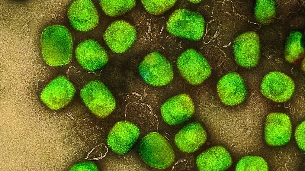
Researchers have discovered new diagnostic and prognostic markers for multiple sclerosis.
This is a pseudo-colored image of high-resolution gradient-echo MRI scan of a fixed cerebral hemisphere from a person with multiple sclerosis.
Credit: Govind Bhagavatheeshwaran, Daniel Reich, National Institute of Neurological Disorders and Stroke, National Institutes of Health
Researchers from the Max Planck Institute of Biochemistry and the Technical University of Munich (TUM) have discovered new diagnostic markers for multiple sclerosis (MS), a disease which affects 3 million people worldwide.
Using mass spectrometry, about 1500 proteins were analysed simultaneously per sample in the cerebrospinal fluid (CSF) of 5000 patients. The study uncovered a set of marker proteins that improve differentiation of MS from other inflammatory brain diseases where classical MS markers are negative. Additionally, the study identified changes in the CSF proteome that may potentially predict disease progression. This approach could also open up new avenues for the diagnosis of other diseases. The study was published in Cell.
Unspecific neurological symptoms can lead to lengthy or even inaccurate diagnoses of diseases, which is why improved protein markers are needed for swift and clear diagnosis
Using a new mass spectrometry method, approximately 1500 proteins were analyzed per cerebrospinal fluid sample across 5000 patients, and up to 2000 proteins in a further improved method
A new set of disease markers enable improved differentiation of multiple sclerosis (MS) from other inflammatory brain diseases, in particular for patients lacking the classical markers
The proteome of the cerebrospinal fluid (CSF) at diagnosis is informative for various aspects of disease evolution in a patient, such as long-term disability, risk of conversion from relapsing to progressive disease course and time to conversion
The method also has the potential to discover other proteins that could be used as markers for the diagnosis of other neurological diseases
Biomarker needs for multiple sclerosis
Imagine living with unexplained neurological symptoms: numbness, visual disturbances, fatigue, but not receiving a clear diagnosis for months or years. Non-specific neurological symptoms can make diagnosis difficult because, despite modern imaging techniques, there are no reliable molecular biomarkers for many neurological diseases.
Professor Bernhard Hemmer, head of the Department of Neurology at TUM University Hospital, explains: “The diagnosis of neurological diseases such as multiple sclerosis is based on a combination of imaging techniques using magnetic resonance imaging (MRI) and cerebrospinal fluid (CSF) analysis. While MRI reveals inflammatory changes in the brain and spinal cord, CSF shows chronic immune activity in the nervous system. In most cases, this combination enables a reliable diagnosis. In individual cases, however, differentiation can be challenging. This can lead to lengthy and less reliable diagnoses and is associated with uncertain and delayed treatment decisions. For this reason, we need new biomarkers to better diagnose the various diseases. In addition to diagnostic challenges, predicting disease progression, particularly disability accumulation, to guide optimal treatment, remains a major unmet need in MS”.
Proteomic study of cerebrospinal fluid across neurological diseases
In order to find new biomarkers, neurologists Bernhard Hemmer and Christiane Gasperi, both experts in MS research at TUM, have joined forces with Professor Matthias Mann, a world-leading expert in proteomics research. Matthias Mann, director at the MPI of Biochemistry, explains: “We have been developing the technology for measuring proteins using mass spectrometry in our laboratory together with colleagues for decades. Now we can reliably and accurately measure proteins in body fluids. However, for a long time researchers could only measure tens to hundreds of samples and only those proteins with the highest concentration in a body fluid. These proteins often turned out not to be the best markers for diseases. To go one step further, we combined the latest advances in mass spectrometry hardware, software, and sample preparation and adapted the workflow to cerebrospinal fluid.”
In this study, CSF samples from more than 5000 people with a wide range of neurological diseases were analysed. Jakob Bader, first author of the study and postdoctoral researcher in the field of proteomics research, explains: “Proteomics is a scientific discipline that aims to characterise a biological system by measuring all proteins, or at least as many as possible. For our study, it is essential to cover as many proteins as possible in order to increase the likelihood of measuring and later finding real disease markers in our analyses. The great advantage of this proteomic approach is that the identity of the markers does not have to be known beforehand. This saves years of research work in which individual candidates are examined one after the other.”
To avoid misinterpreting random differences between people as disease markers, it is essential to have a sufficient number of patients. Similarly, it is only possible to determine whether a marker is specific to a particular disease by considering the many other relevant diseases in parallel. “The breakthrough was achieving both objectives simultaneously: Analysing thousands of proteins while studying thousands of patients across many neurological diseases.” Jakob Bader adds.
A systematic analysis of disease effects and possible confounders
The 5,000 CSF samples came from a wide range of neurological disorders, including stroke, brain cancer, infections, autoimmune diseases such as MS, and others. Additionally, patient samples were analysed from individuals who had provided CSF samples for the diagnosis of severe headache disorders but in whom no neurological disease was found. This allowed the researchers to use these samples as controls. Systematic comparison of these disorders revealed shared and specific protein deviations from the controls.
For diagnostic use, an elevated protein concentration rarely points unambiguously to a single disorder. The study further revealed that disease-unspecific other effects like a person’s age, sex, and in particular degradation of the barriers insulating the brain from the CSF have a very large impact on the composition of this fluid, which complicates the quest for disease markers.
Biomarkers for a hard-to-identify form of multiple sclerosis
To showcase the potential of proteomic analysis for biomarker discovery, the researchers focused on the search for diagnostic markers for MS, a challenging task but with a direct medical need. Physician Christiane Gasperi says: “In approximately 10% of MS patients, diagnosis of the disease is particularly difficult because they lack the typical MS marker of so-called oligoclonal bands of antibodies that are specific to the CSF and not found in the blood. This complicates and potentially delays the diagnosis.”
She continues: “However, for our patients, a quick and clear diagnosis of the disease is of enormous importance. While current therapies cannot cure MS, they can slow its progression and reduce the long-term disability. That makes it crucial to start treatment early. At the same time, these therapies can have significant side effects, so treatment decisions require a high level of diagnostic certainty. When this confidence is not reached yet, therapy is often delayed. Thus, MS patients really benefit from an early intervention that depends on a clear and early diagnosis.”
To find better markers, the researchers applied an enhanced version of the proteomic method to measure about 2000 proteins in samples of MS and other inflammatory diseases of the CNS, which can mimic MS, and thus pose the greatest diagnostic challenges. This let them identify a set of 22 proteins that distinguishes MS from these inflammatory diseases with better accuracy than other parameters in the CSF that are currently measured in clinical practice. Christiane Gasperi comments: “It is particularly encouraging that we have found a combination of marker proteins that help in the diagnosis of this particularly difficult-to-identify form of MS.”
Predicting disease progression at diagnosis
Beyond improving diagnosis, the study also addressed a second major challenge: Some patients remain relatively stable for many years, while others accumulate disability more rapidly or transition from the relapsing disease course that is typical early on to a progressive course where disability accumulates persistently. At the time of diagnosis, it is very difficult to predict which trajectory a patient will follow. This uncertainty complicates treatment decisions and can be deeply unsettling for those newly diagnosed.
By analyzing hundreds of MS patient samples, the researchers showed that the CSF proteome at the time of diagnosis was associated with the level of disability years later. In addition, these patterns reflected a higher risk for patients to convert from the relapsing to the progressive disease course, as well as shorter times until such conversion occurred. Bernhard Hemmer explains: “Our findings suggest that important aspects of future disability and disease course are reflected in the proteome from the very beginning. This demonstrates that the biological information required for a prognostic test is already present at diagnosis.”
He summaries the study: “For diagnosis, we were able to define and validate a focused protein panel that improves differentiation in difficult cases. Additionally, we found that the overall protein pattern in the CSF at the time of diagnosis is linked to how the disease develops years later. Together, these findings bring us closer to more precise diagnosis and a more individualized treatment strategy from the very beginning.
An avenue for efficient biomarker discovery in neurology
Matthias Mann sees broader potential: “Proteins control almost all biological processes in the body and have long been the most important group of diagnostic markers. Nevertheless, we are probably only at the beginning here. With the methodology established here, we can now analyse the proteome in the CSF of many patients with an unprecedented number of proteins. This technological progress changes how we should search for biomarkers. Comprehensive proteome analysis of large patient collectives promise to be the most efficient path to new and better biomarkers. Beyond MS, this approach opens up prospects for many other diseases of the central nervous system – from Alzheimer’s and Parkinson’s to brain tumours and other neurological disorders.”
Research in Aging Cell indicates that blood levels of particular small non-coding RNAs, which regulate gene expression, may influence how long a person lives.
Investigators evaluated 828 small non-coding RNAs in blood samples from 1,271 community-dwelling older adults 71 years of age and older who were participating in an ongoing study. They then used machine learning to develop a model that could predict survival at 2, 5, and 10 years based on baseline small non-coding RNAs, age, and clinical variables (demographics, lifestyle, mood, physical function, standard clinical laboratory tests, lipid and metabolite levels, and medical conditions).
The test worked especially well for predicting survival over the next 2 years. “One surprising finding involved a group of small non-coding RNA molecules called piRNAs”, said co–corresponding author Virginia Byers Kraus, MD, PhD, of the Duke Molecular Physiology Institute. Scientists have long known that piRNAs help protect DNA in reproductive cells, but their role in the rest of the body is still a mystery. In this study, nine piRNAs, all reduced in longer-lived individuals, were identified as potential therapeutic targets to prolong longevity.
“These results suggest that simple blood tests measuring piRNAs might one day help doctors better understand health and aging – and possibly even guide new treatments to help people live longer, healthier lives,” said Dr Byers Kraus.
The study reveals that the combination of antibiotics reduces mortality in patients with high-risk Staphylococcus aureus bacteriaemia by half, if applied in a personalised way
A study led by researchers from the Infectious Diseases Service of the Bellvitge University Hospital (HUB), the Bellvitge Biomedical Research Institute (IDIBELL) and the University of Barcelona (UB) shows for the first time that the combination of antibiotics can significantly improve the prognosis of patients with high-risk Staphylococcus aureus bacteriaemia, if it is applied selectively and in a personalised way.
The article has been published in the prestigious scientific journal The Lancet Regional Health – Europe and is the result of the collaboration of a dozen Spanish hospitals.
A reanalysis that redefines the strategy against bacteriaemia
Bacteraemia for S. aureus is a frequent and serious infection, with mortality reaching 30%. Its management requires prolonged intravenous antibiotherapy, the removal of possible infected devices and a thorough evaluation to rule out complications such as endocarditis or metastatic focus. In recent years, several clinical trials evaluating the combination of antibiotics had failed to demonstrate clear benefits in the global set of patients, a fact that the authors of the present study attributed to the lack of stratification according to risk.
For this reason, the study has carried out a reanalysis of the individualised data of two previously performed randomised clinical trials, differentiating patients according to their risk profile. This stratification was done through the FEN-AUREUS classification – a recently developed clinical tool that allows estimating the risk of mortality with information available during the first 24 hours of evolution – and the complication criteria of the Infectious Diseases Society of America (IDSA).
A personalised medicine according to individual risk
The re-evaluated trials include, on the one hand, a study with 155 patients from 18 Spanish hospitals that compared the use of daptomycin with the combination of daptomycin and phosphomycin; and, on the other, a study with 215 patients from 19 hospitals that compared cloxacillin in monotherapy with the combination of cloxacillin and phosphomycin. In both cases, the initial conclusions had shown no significant benefits of combined therapy in the overall set of patients, beyond a reduction in the duration of bacteraemia in patients treated with phosphomycin.
After the new risk group analysis, the work shows that low-risk and uncomplicated patients do not obtain significant benefits from combined therapy. On the other hand, high-risk patients showed remarkable therapeutic success at eight weeks (69.2% vs. 25.8%) and lower mortality at 60 days (23.1% vs. 45.2%).
According to first author Dr Francesc Escrihuela-Vidal, “the integration of risk and complications criteria can help identify patients who can really benefit from combined therapy.”.
In the same vein, co-author Dr Jordi Carratalà points out that “the results represent a real paradigm shift in the approach to this infection: we move from a uniform strategy for all patients to a precision medicine based on individual risk”. In addition, it highlights that this approach allows to avoid unnecessary intensive treatments in low-risk patients and will contribute to improving the design of future clinical trials.
Researchers have found that sucrose can relieve newborn babies’ pain during common hospital procedures
Photo by Christian Bowen on Unsplash
A new Cochrane review has found that sucrose can help with pain relief in newborn babies during common hospital procedures, such as venepuncture. This involves drawing blood with a needle, typically for testing.
Newborns, especially preterm infants in neonatal intensive care units (NICUs), undergo numerous painful procedures. Because of their immature pain regulation, they can experience these procedures intensely. Preventing and treating procedural pain in hospitalized newborns is important, as repeated untreated pain has been associated with poorer physical growth and potential effects on brain development.
Accessible, low-cost solutions such as sucrose – a sweet sugar solution placed in a baby’s mouth shortly before needle procedures – have been used for decades. However, evidence specific to some procedures, such as venepuncture, has been limited.
Despite sucrose being recommended in multiple guidelines for procedural pain relief in infants, its use in clinical settings remains inconsistent.
Low-cost, safe intervention
The new review examined 29 clinical trials involving more than 2700 preterm and full-term babies undergoing venepuncture in hospital. It found that sucrose probably reduces pain during and immediately after the needle procedure when compared to no treatment, water or standard care. The findings also suggest that sucrose works especially well when combined with non-nutritive sucking, such as a pacifier or dummy.
“Newborn babies undergo frequent needle procedures in hospital without any pain relief or comforting measures, even though older children and adults rarely have these procedures done without pain care.
The evidence shows that a small amount of sucrose given just before the procedure is a simple, fast and effective way to reduce that pain. Our review helps clinicians use this evidence more confidently and consistently in practice.”
—Mariana Bueno, University of Toronto
None of the studies included in the review reported immediate side effects from sucrose when used in the small amounts required for pain relief. However, the studies focused on short-term effects, and more research is needed to understand any potential long-term effects of repeated use in babies who spend extended time in neonatal care.
“Parents may be surprised to learn that something as simple as a few drops of sugar solution can make a real difference to their baby’s comfort during blood tests.
This is a low-cost, safe intervention that works within minutes, and it can be especially helpful when other comforting methods like skin-to-skin contact or breastfeeding aren’t possible.”
—Ligyana Candido, University of Ottawa
Treated like other medications
Although sucrose is already widely used in neonatal units, the researchers found considerable variation in how it is given, including differences in dose and timing.
Bueno added:
“What stood out to me when doing this review was the wide variation in how sucrose was given to newborns.”
The authors suggest the findings can help inform clearer clinical protocols and more consistent practice.
They also highlight that sucrose should be used purposefully for painful procedures and documented appropriately, rather than being given routinely to settle a crying baby.
“To ensure safety and clinical consistency, sucrose must be administered under formal medication protocols that define specific timing and dosage for painful procedures.”
— Jiale Hu, Virginia Commonwealth University
The review authors say future research should focus on comparing effective comfort measures such as skin-to-skin contact, breastfeeding and sucrose with each other, rather than continuing to compare them to no treatment, and on understanding any potential long-term effects of repeated use in babies who spend extended time in neonatal care.
An international, randomised, double‑blind, placebo‑controlled phase 3 study, the largest of its kind for mpox, found that tecovirimat did not improve clinical outcomes for adults with clade II mpox compared with placebo, while demonstrating a similar safety profile. Results of the STOMP/A5418 trial, published in the New England Journal of Medicine underscore both the urgent need for alternative therapeutics and the critical importance of randomised trials during public health emergencies.
This Phase 3 study randomised 412 participants (344 with laboratory‑confirmed mpox), to receive either tecovirimat or a matching placebo for 14 days. Randomisation was stratified by early versus later symptom onset and by the presence of severe pain. Participants had active skin or mucosal lesions and self‑reported daily symptoms, pain scores, and lesion status through Day 29, with confirmatory clinical assessments at scheduled visits. Biospecimens, including lesion swabs, oral and rectal swabs, and blood samples, were collected at multiple time points to assess viral DNA clearance. The primary endpoint was time to clinical resolution of all lesions, and key secondary endpoints included pain reduction, complete lesion healing, and virologic response.
Conducted across seven countries at 49 sites, the phase 3 study showed that tecovirimat did not shorten the time to lesion resolution, reduce pain, or speed viral clearance compared with placebo. These results align with interim findings released in December 2024, which led the trial’s independent Data and Safety Monitoring Board to halt further enrolment due to statistical futility.
“In the midst of a global public health emergency, the ACTG team rapidly conducted this randomized controlled trial to deliver a clear answer for patients and clinicians,” said William A. Fischer II, MD, associate professor of pulmonary and critical care medicine at the UNC School of Medicine, and director of emerging pathogens research at the UNC Institute for Global Health and Infectious Diseases. “These findings advance our understanding of mpox and help the field refocus efforts on identifying safe, effective and accessible treatment strategies, particularly for people at highest risk of severe disease.”
Although the trial did not demonstrate efficacy, tecovirimat demonstrated a favourable safety profile, with no major safety concerns identified – an important confirmation as thousands of patients worldwide have already received the drug under expanded access protocols.
“The STOMP trial provides essential evidence at a critical time and demonstrates why randomized controlled trials are an indispensable part of outbreak response,” said Joe Eron, MD, chief of infectious diseases and chair of the ACTG network. “But now we must keep going to find safe and effective treatment for people as this virus continues to circulate globally.”
The study’s conclusions are expected to influence clinical practice and public health guidance worldwide. With mpox still causing outbreaks in multiple regions, researchers emphasise that developing and evaluating new antiviral candidates remains a top priority.
If someone living with HIV is not on antiretroviral therapy, the virus can cause inflammation in, among other places, the brain. Photo by Anna Shvets
By Biénne Huisman
Antiretroviral therapy has shifted HIV from a fatal to a chronic condition. But neuropsychiatrists say it is imperative for people living with the virus to start treatment immediately as the “duration of untreated exposure” may cause irreversible brain damage and impact long-term cognitive health.
It has been recognised for decades that cognitive impairment is a potential complication of HIV infection. Questions over how likely and how serious this potential complication is have become more urgent over time as the population of people living with HIV ages – ageing after all also increases the risk of cognitive decline.
There were around 1.75 million people over the age of 50 living with HIV in South Africa in 2024, according to Thembisa, the leading mathematical model of HIV in the country. This is just over 20% of the estimated eight million HIV positive people in the country. A study published in the Lancet medical journal also has the number at around 20% in sub-Saharan Africa.
This is a delicate field of enquiry as researchers walk a tightrope to avoid “the burden of double stigma”, while conceptualising the necessary tools to best diagnose brain problems and suitable interventions.
Within as little as two weeks
At Groote Schuur Hospital’s Neuroscience Institute, Professor John Joska, director of the University of Cape Town’s (UCT’s) HIV Mental Health Research Unit, explains that HIV can enter the brain within as little as two weeks after the initial infection – primarily through infected white blood cells, such as lymphocytes. If a person is not on antiretroviral therapy, the virus can cause inflammation in the brain and possibly also tissue damage.
“The brain is a protected compartment,” says Joska. “A theory as to how the virus, which is a protein particle, gets into the brain is through infected lymphocytes. This doesn’t directly infect nerve cells, what we call neurons. It infects other supporting tissues and cells in the brain, causing an inflammation which damages typically the white matter of the brain. Over time, that inflammation can cause loss of neurons, but indirectly.”
While antiretroviral therapy is crucial for clearing and suppressing HIV in all body compartments, including in the brain, he says that it does not reverse damage that occurred before the treatment was started.
“Today, people with HIV are living near normal lifespans,” he says. “The question is, will the fact that they’ve had HIV, with some duration of untreated exposure and potential loss of brain tissue, cause them to be at higher risk than the average person for developing dementias of old age – which really are mainly Alzheimer’s disease or vascular dementia.” It is these longer-term effects that are the main concern when it comes to the impact of HIV on the brain.
Part of the problem is that South Africa not only has an ageing population of people living with HIV, but many of these people would only have started treatment quite long after they contracted the virus. One key reason for this is the South African government’s reluctance to make antiretroviral treatment available in the early 2000s. It has been estimated that those delays resulted in over 300 000 avoidable deaths – they may also be contributing to brain health issues now and in the future.
From efavirenz to dolutegravir
Apart from HIV itself, some of the medicines used to treat the infection have also had an impact on the brain.
In 2019, the standard HIV treatment in South Africa changed from a three-drug combination containing an antiretroviral drug called efavirenz, to a combination containing the drug dolutegravir. This shift had mental health benefits, as evidenced in research lead by Joska’s fellow UCT Neuro-HIV researcher, Associate Professor Sam Nightingale.
Joska says: “The study looked at the period from 2017 to 2020 and the switch from efavirenz to dolutegravir based treatment. It was well known that efavirenz caused, certainly for the first two months, a bunch of psychotropic or psychological issues like nightmares or anxiety, even psychosis for some people. But our findings showed people who switched to dolutegravir actually do very well. They look more like people without HIV after eight months. So dolutegravir has been a huge advantage, not only because it’s robust, but because it’s neuro-protective.”
New models for HIV and cognitive impairment
A shift is underway in how experts are thinking about cognitive impairment in people with HIV. Some neuropsychiatrists, including Joska, are recommending a shift away from the 2007 HIV-Associated Neurocognitive Disorders model, arguing that its cognitive test scores do not adequately account for variables such as education and socioeconomic background, and that it can overdiagnose impairment. The argument is set out in an article, lead-authored by Nightingale, that was published in the journal Nature Reviews Neurology in 2023.
The authors argue that a label of cognitive impairment might cause a “double burden of stigma” for people living with HIV – affecting self-esteem, inciting fear and prompting further discrimination against persons already subject to stigma as it stands. To illustrate the point, they point out how, up until recently, people with HIV in the United Kingdom could not become airline pilots due to concerns over cognitive impairment. However, following a campaign by a pilot living with HIV, the United Kingdom’s Civil Aviation Authority removed the ban in 2022.
Nightingale and his colleagues argue that traditional test scores be used in conjunction with real-life symptoms and medical evidence of brain problems. It introduces the conceptual model of HIV-Associated Brain Injury, which refers specifically to damage caused by the virus. This distinguishes it from other causes of cognitive impairment such as depression, substance abuse, diabetes and cardiovascular disease. As Spotlight previously reported, HIV is also associated with an increased risk of depression, though this is at least partially driven by social factors.
Lower cognitive function associated with late diagnosis
At the 2026 Conference on Retroviruses and Opportunistic Infections hosted in Denver in the United States in late February, these issues were tabled at a discussion titled “When I’m 64: Neurodegeneration, Epigenetic Aging, and Cognition in Older People With HIV.”
Professor John Joska is the director of the University of Cape Town’s HIV Mental Health Research Unit. (Photo: Biénne Huisman/Spotlight)
In his presentation, Professor Alan Winston of Imperial College London, also a member of the International HIV-Cognition Working Group, and a frequent co-author alongside Joska and Nightingale, relayed existing research findings that on average, people living with HIV have lower cognitive function – including memory, attention span and executive function like planning – compared to people who don’t have HIV of the same age. He said that this manifests as an increased risk of lower grade early dementia.
Like Joska, Winston stressed that the most deteriorated cognitive function in people living with HIV is associated with untreated HIV and late HIV diagnosis. He reiterated that starting HIV treatment soon after diagnosis is protective, and that viral suppression is associated with better cognition. In groups of patients with HIV well controlled on dolutegravir-based HIV treatment, cognition appears similar to HIV negative groups, he said.
HIV clinicians need to pay better attention to the brain
In an impassioned presentation, Dr Shibani Mukerji, Associate Professor of Neurology at Harvard Medical School, argued that protecting the brain is an overlooked frontier in effective HIV treatment, and that clinicians need to pay more attention to it.
“By the time patients and clinicians notice cognitive decline – generally and in HIV – the damage to the brain is done and lives are affected negatively. People don’t raise cognitive concerns early enough due to stigma, fear, [and] lack of recognition of the issues. It is seen as ‘just getting old’,” she said.
Mukerji emphasised the need to prioritise brain health. “HIV doctors and treatment programmes are focused, almost exclusively, on viral load as the marker of successful treatment. They may be thinking laterally and consider TB and other infections, maybe cardiovascular disease – but they are definitely not paying enough attention to brain health. HIV doctors aren’t aware enough of brain health issues in people living with HIV, and even when they are, they often don’t feel comfortable diagnosing or managing it, so it is under recognised and under diagnosed.”
The perception that there is no way to manage or treat cognitive decline –generally and in people living with HIV – is wrong, she said, adding that optimising physical, mental and social health is critical for brain health.
“Almost half of dementia risk [in people in general] is linked to preventable causes,” she told conference delegates, along with a slide listing preventable causes including loss of hearing, social isolation, cardiovascular disease and depression.
She explained: “If someone has cognitive decline and for example you improve their hearing – if they have hearing issues – and you work on their social isolation, and treat their vascular disease, and treat their depression, you can see a marked improvement in their cognition.”
Ending her presentation with a twist of humour, Mukerji’s last slide referred to the session’s title, a reference to the Beatles song on aging “When I am 64”. She printed the song’s lyrics: “When I get older, losing my hair, many years from now…”, closing her talk by saying: “It’s okay to stand up and sing, in fact your doctor might prescribe it.”
Cyclone Gezani caused extensive damage across the region, displacing thousands and severely affecting homes, public infrastructure, and healthcare facilities
Following the devastating cyclone that struck Madagascar’s east coast, Mercy Ships (www.MercyShips.org) has joined with national disaster response efforts in Toamasina (Tamatave) through the provision of essential relief supplies, in coordination with the government’s disaster management authorities.
Cyclone Gezani caused extensive damage across the region, displacing thousands and severely affecting homes, public infrastructure, and healthcare facilities.“In moments like these, partnerships and solidarity matter most,” said Nicholas Ahadjie, Country Director of Mercy Ships in Madagascar. “We are committed to supporting the national response and ensuring that assistance reaches communities where the needs are greatest.”
As part of its own immediate response, Mercy Ships has delivered 537 bags of rice, 1000 roofing sheets, and 1000 ready-to-eat meals. These supplies arrived in Toamasina and were officially handed over to the government’s Designated Disaster Response Coordination Body for distribution to affected communities.
The roofing materials will enable families, schools, and community health facilities to begin urgent repairs. The rice will be distributed to households impacted by the storm that still have functional cooking facilities, while ready-to-eat meals will provide immediate support to individuals and displaced families.Although the Mercy Ships hospital vessel Africa Mercy®, is currently undergoing scheduled maintenance in South Africa, preparations are underway for her return to Madagascar. Sometime this May, she is expected to resume surgical services and medical training programs in collaboration with the Ministry of Health.
“Our presence in Madagascar is on-going,” added Nicholas Ahadjie. “While the ship is in maintenance, our engagement with partners continues. We stand with the Malagasy people today and remain dedicated to strengthening healthcare capacity for the future.
”For several years, Mercy Ships has partnered with Madagascar to provide free specialised surgeries, professional medical training, and infrastructure support. The recent disaster will not stop the organisation’s ongoing support for the Malagasy people as it continues to help reinforce their national health systems.Distributed by APO Group on behalf of Mercy Ships.
ABOUT MERCY SHIPS:
Mercy Ships operates hospital ships that deliver free surgeries and other healthcare services to those with little access to safe medical care. An international faith-based organisation, Mercy Ships has focused entirely on partnering with African nations for the past three decades. Working with in-country partners, Mercy Ships also provides training to local healthcare professionals and supports the construction of in-country medical infrastructure to leave a lasting impact. Each year, more than 2500 volunteer professionals from over 60 countries serve on board the world’s two largest non-governmental hospital ships, the Africa Mercy® and the Global Mercy™. Professionals such as surgeons, dentists, nurses, health trainers, cooks, and engineers dedicate their time and skills to accelerate access to safe surgical and anaesthetic care. Mercy Ships was founded in 1978 and has offices in 16 countries as well as an Africa Service Center in Dakar, Senegal. For more information, visit www.MercyShips.org and follow @MercyShips on social media.
Researchers at WashU Medicine have identified a potent pathway that begins in the brain and leads to loss of all body fat without reducing food intake. The study is reported in Nature Metabolism.
The team – led by senior author Erica L. Scheller, DDS, PhD, an associate professor in the Division of Bone and Mineral Diseases in the Department of Medicine; Xiao Zhang, PhD, a former graduate student in Scheller’s lab who is now a postdoctoral fellow at the University of Pennsylvania School of Medicine; and Sree Panicker, a graduate student in Scheller’s lab – was inspired by a unique population of fat cells located deep within the skeleton.
“About 70% of our bone marrow is filled with fat that doesn’t respond to diet or exercise,” said senior author Scheller. “We wanted to figure out why.”
The team found that these special cells, called constitutive bone marrow adipocytes, expressed high levels of proteins that inhibit fat breakdown. This causes resistance to fat loss in day-to-day settings. “We call these cells stable adipocytes,” said Zhang, the study’s first author. In mice, sustained injection of leptin, a hormone, into the brain was able to unlock the stable adipocytes by putting the body into a state of low glucose and insulin. This reduced the inhibitors of fat breakdown, causing complete loss of body fat within days, even though the mice were still eating normally.
This pathway is so powerful that the scientists caution against using it in humans until it is better understood. Stable adipocytes occur in places like the bone marrow, in the hands and feet, and around important glands. In severe wasting disorders, loss of fat within these cells is associated with bone fractures and reduced quality of life. Scheller’s team hopes to prevent this loss and preserve health in patients with severe wasting disorders by defining the mechanisms of stable fat loss. Conversely, methods to activate fat loss from stubborn adipocytes may support future treatments for obesity. This work was funded by the National Institutes of Health (NIH).