Month: January 2026

Scientists Find Hidden Diversity Among T. Gondii

UC Riverside study reshapes understanding of toxoplasmosis and identifies new paths for treatment

Toxoplasma gondii. Source: Wikimedia CC0

A University of California, Riverside team of scientists has found that Toxoplasma gondii, a common parasite affecting up to one-third of the global population, is far more complex than previously believed. The findings, published in Nature Communications, offer new insight into how T. gondii causes disease and why it has been so difficult to treat.

Humans commonly contract toxoplasmosis by eating undercooked meat or through exposure to contaminated soil or cat faeces. The parasite is best known for its ability to hide in the body by forming tiny cysts in the brain and muscles. 

Most people who are infected never notice any symptoms. However, the parasite remains in the body for life as cysts, which can contain hundreds of parasites. The parasites they lodge can become active again later, however, especially in people with weakened immune systems, leading sometimes to serious problems affecting the brain or eyes. Most people who are infected never notice any symptoms. Infection during pregnancy can cause serious complications for developing babies with limited immune systems. 

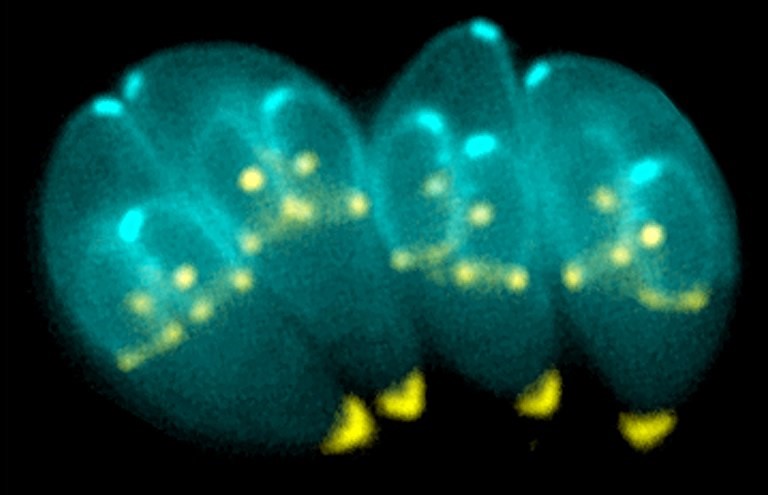
Until now, scientists believed that the cysts contained a single, uniform type of parasite lying dormant until it reactivated. But using advanced single-cell analysis techniques, the UC Riverside team discovered that each cyst contains multiple distinct subtypes of parasites, each with different biological roles.

“We found the cyst is not just a quiet hiding place – it’s an active hub with different parasite types geared toward survival, spread, or reactivation,” said Emma Wilson, a professor of biomedical sciences in the UCR School of Medicine who led the study. 

Wilson explained that cysts form slowly under immune pressure and are encased in a protective wall, housing hundreds of slow-replicating parasites called bradyzoites. Although microscopic, cysts are relatively large for intracellular pathogens, reaching up to 80 microns in diameter, with each bradyzoite measuring roughly five microns in length. They reside primarily within neurons but are also commonly found in skeletal and cardiac muscle, which is important since humans are often infected by consuming undercooked meat containing these cysts.

According to Wilson, cysts are clinically and biologically significant for several reasons. They are resistant to all existing therapies and remain in the body once established. They facilitate transmission between hosts. When reactivated, bradyzoites convert into fast-replicating tachyzoites that disseminate throughout tissues, causing severe disease such as toxoplasmic encephalitis (neurological damage) or retinal toxoplasmosis (vision loss).

Image shows a cyst which can contain hundreds of T. gondii parasites. (UCR/Wilson lab)

“For decades, the Toxoplasma life cycle was understood in overly simplistic terms, conceptualised as a linear transition between tachyzoite and bradyzoite stages,” Wilson said. “Our research challenges that model. By applying single-cell RNA sequencing to parasites isolated directly from cysts in vivo, we found unexpected complexity within the cyst itself. Rather than a uniform population, cysts contain at least five distinct subtypes of bradyzoites. Although all are classified as bradyzoites, they are functionally different, with specific subsets primed for reactivation and disease.”

Wilson acknowledged that studying cysts has long been a technical challenge. They grow slowly, are embedded deep within tissues like the brain, and do not form efficiently in standard laboratory cultures. As a result, most genetic and molecular studies of Toxoplasma have focused on tachyzoites grown in vitro, leaving the biology of cyst-resident bradyzoites poorly understood. 

“Our work overcomes those limitations by using a mouse model that closely mirrors natural infection,” Wilson said. “Because mice are a natural intermediate host for Toxoplasma, their brains can harbour thousands of cysts. By isolating these cysts, digesting them enzymatically, and analysing individual parasites, we were able to gain a view of chronic infection as it occurs in living tissue.”

Wilson explained that current treatments for toxoplasmosis can control the fast-growing form of the parasite that causes acute illness, but no existing drugs can eliminate the cysts. 

“By identifying different parasite subtypes inside cysts, our study pinpoints which ones are most likely to reactivate and cause damage,” she said. “This helps explain why past drug development efforts have struggled and suggests new, more precise targets for future therapies.”

Congenital toxoplasmosis remains a major concern when primary infection occurs during pregnancy, potentially leading to severe foetal outcomes. Although prior immunity typically protects the foetus, routine screening is lacking in some countries, reflecting how difficult it is to manage an infection that is common but usually symptom-free.

Despite its prevalence, toxoplasmosis has received relatively little attention compared to other infectious diseases. Wilson hopes her team’s work will help shift that perspective.

“Our work changes how we think about the Toxoplasma cyst,” she said. “It reframes the cyst as the central control point of the parasite’s life cycle. It shows us where to aim new treatments. If we want to really treat toxoplasmosis, the cyst is the place to focus.”

Wilson was joined in the study by Arzu Ulu, Sandeep Srivastava, Nala Kachour, Brandon H. Le, and Michael W. White. Wilson and White are co-corresponding authors of the paper.

The study was supported by grants from the National Institute of Allergy and Infectious Diseases of the National Institutes of Health. 

The title of the paper is “Bradyzoite subtypes rule the crossroads of Toxoplasma development.”

Leukotrienes May Not Drive Asthma After All, New Research Shows

Respiratory tract. Credit: Scientific Animations CC4.0

Case Western Reserve University researchers say discovery of new inflammatory molecules could transform treatment

For decades, scientists have thought they understood the biochemical machinery that causes asthma: inflammation in the lungs that constricts airways and makes it hard to breathe. Leukotrienes – molecules that get released from white blood cells due to airway irritation or allergen inhalation – were labelled the culprits. Medications have been developed to block the molecular cascade they initiate that leads to difficulty breathing.

But researchers from Case Western Reserve University think these molecules may not be the bad actors after all.

“We’ve found molecules that are alike in structure but generated through a completely different chemical pathway in the body,” said lead researcher Robert Salomon, the Charles Frederic Mabery Professor of Research in Chemistry. “We think the molecules we’re calling ‘pseudo leukotrienes,’ may be the dominant players in the inflammatory cascade that causes disease.”

The research opens new avenues for treating asthma as well as other inflammatory diseases, possibly including neurological diseases like Parkinson’s and Alzheimer’s diseases. The research, funded by the U.S. National Institutes of Health, is available online as a pre-proof ahead of publication in the Journal of Allergy and Clinical Immunology.

The ‘flames’ of oxidation

The presumed culprits in inflammatory diseases – the leukotrienes – are formed under the control of enzymes that transform lipids. By contrast, the pseudo leukotrienes Salomon and his team discovered, are formed by adding oxygen to lipids by molecules called “free radicals.”  

“The free radical process is almost like an explosion or a fire,” said Salomon, who is also professor of ophthalmology in the Case Western Reserve School of Medicine. “It’s just like when oxygen reacts with fuel and you get flames. It can easily get out of control.”

People who suffer from asthma may lack enzymes and antioxidant molecules that normally keep a damper on free radicals by scavenging for and destroying them.

The leukotrienes and their mimics initiate inflammation by fitting into a receptor, like a key in an ignition, starting a molecular cascade that constricts the airways of asthmatics. Effective asthma drugs like Singulair block the ignition so the key won’t fit.

“The real importance of this discovery is the possibility of treating these diseases with drugs that prevent the free radical process or moderate it rather than drugs that block the receptor,” Salomon said.

Inflammation: a curse or a benefit?

Not all inflammation is harmful. The body needs inflammation to direct white blood cells to the site of a wound to heal, and it is also involved in memory and development.

Asthma drugs are being repurposed off-label to treat neurological diseases. But these treatments could also block the beneficial effects of the leukotrienes.

“If the molecules that are causing the problem are not the leukotrienes but these other molecules,” Salomon said, “a better treatment would be to just stop the formation of these other molecules rather than gumming up the ignition.”

The study

Salomon and his colleagues used their decades of experience studying the oxidation of lipids – and some chemical intuition – to guess that pseudo leukotrienes existed. They made the molecules in the laboratory to develop methods to detect them.

They obtained urine samples from patients designated with mild or severe asthma and compared them to urine from people who don’t suffer from the disease.

Not only were pseudo leukotrienes found in the asthma patients’ urine, but also the amounts correlated directly to the severity of the disease. Severe asthma sufferers or even those suffering mild asthma had four to five times more than the controls. The researchers suggest this could be a new biomarker to test for the severity of disease and monitor the effectiveness of therapies.

The researchers next plan to investigate whether these pseudo leukotrienes are involved in other respiratory diseases, like respiratory syncytial virus (commonly known as RSV) and bronchiolitis in babies, and chronic obstructive pulmonary disease.

By Diana Steele

Source: Case Western Reserve University

Pain in Teens Linked to Increased Risk of Self-harm

Photo by Inzmam Khan

Adolescents who report pain at the age of 18 are at higher risk of later self-harm. This is shown by a new study from Karolinska Institutet, published in Psychiatry Research. The findings suggest that pain may form part of the chain of events leading to self-harming behaviour.

A new study from Karolinska Institutet has examined the association between pain symptoms and self-harm during childhood and adolescence. The researchers followed 16 948 twin pairs born in Sweden between 1992 and 2010. Participants reported pain at the ages of 9 and 18 and were subsequently followed through national registers until a maximum age of 24.

The aim was to investigate how genetic and environmental factors influence both pain and self-harm, as well as how the associations between pain and self-harming behaviour develop over time. Using so-called twin models, the researchers were able to estimate how much of the variation could be explained by heredity, shared environment or individual experiences.

“We see that both genetic factors and individual environmental factors play a role in both pain and self-harm in childhood as well as adolescence,” says Jenny Rickardsson, researcher at the Department of Clinical Neuroscience, Karolinska Institutet.

The results also show that pain before and up to the age of 18 was associated with a higher likelihood of later self-harm. Adolescents with pain symptoms had approximately a 60 per cent higher risk of self-harm compared with peers without pain. The association could not be explained by factors such as family environment or genetic similarity between twins.

“Our analyses suggest that pain may partly lie within the causal pathway leading to self-harming behaviour, and that the association is not solely due to familial factors,” says Jenny Rickardsson.

The study further shows that the shared family environment had little impact on either pain or self-harm, while genetic factors and individual experiences accounted for a larger proportion of the variation.

The study is based on data from the Swedish Twin Registry and was funded by Fonden för Psykisk Hälsa and Hjärnfonden. The researchers report no conflicts of interest that may have influenced the results.

Source: Karolinska Institutet

Researchers Uncover the Mechanism Behind Exercise-induced Bone Strengthening

Osteoporosis. Credit: Scientific Animations CC4.0

A research team from the Department of Medicine, School of Clinical Medicine, LKS Faculty of Medicine at the University of Hong Kong (HKUMed) has uncovered a key biological mechanism that explains how exercise maintain strong bones, paving the way for novel treatments for osteoporosis and bone loss in people who are unable to engage.

By identifying a protein that acts as the body’s ‘exercise sensor’, the research has opened the door to the development of drugs that mimic the effects of physical activity, offering hope for vulnerable groups, such as the elderly, bedridden patients and those with chronic illnesses who face a high risk of fractures. The research findings were published in the journal Signal Transduction and Targeted Therapy.

‘Osteoporosis and age-related bone loss affect millions worldwide, often leaving elderly and bedridden patients vulnerable to fractures and loss of independence,’ said Professor Xu Aimin, Director of the State Key Laboratory of Pharmaceutical Biotechnology and Chair Professor in the Department of Medicine, School of Clinical Medicine, HKUMed, who led the study. ‘Current treatments rely heavily on physical activity, which many patients simply cannot perform. We need to understand how our bones get stronger when we move or exercise before we can find a way to replicate the benefits of exercise at the molecular level. This study is a critical step towards that goal.’

Activating the bone’s ‘exercise sensor’ to reduce fat and build new bone

According to the World Health Organization, approximately 1 in 3 women and 1 in 5 men over the age of 50 suffer a fracture due to weak bones. In Hong Kong, the problem is significant as the population ages, with 45% of women and 13% of men aged 65 and above affected by osteoporosis. Osteoporosis-related fractures cause significant pain and disability, severely impacting quality of life and independence, while placing a heavy healthcare and economic burden on the society.

Ageing involves the natural weakening of bones, which become less dense and more porous. Mesenchymal stem cells in the bone marrow have the potential to develop into either fat cells or bone tissue. These stem cells are highly sensitive to external factors, like exercise and pressure. During the ageing process, they tend to differentiate into fat cells. As fat cells continue to accumulate in the bone marrow, it reduces the space available for healthy bone tissue, further compromising bone strength and creating a cycle of bone deterioration that is difficult to reverse with current treatments.

Using mouse models and human stem cells, the researchers identified a special ‘switch’, called Piezo1, which is a protein on the surface of mesenchymal stem cells in the bone marrow. This switch acts like an exercise sensor, detecting mechanical signals from physical activity that helps keep bones strong and preventing them from becoming frail as we age. In the mice model, when the Piezo1 protein was activated by physical activity, it reduced fat accumulation in the bone marrow and encouraged the formation of new bone. Conversely, when this protein is missing, it triggers signals that cause stem cells to accumulate fat, leading to increased bone loss. The absence of Piezo1 also promotes the release of certain pro-inflammatory signals (Ccl2 and lipocalin-2), making stem cells more likely to convert into fat cells, which hinders bone formation. Blocking these signals can help restore bone health.

Mimicking exercise for individuals with limited mobility

’We have essentially decoded how the body converts movement into stronger bones,’ said Professor Xu Aimin. ‘We have identified the molecular exercise sensor, Piezo1, and the signalling pathways it controls. This gives us a clear target for intervention. By activating the Piezo1 pathway, we can mimic the benefits of exercise, effectively tricking the body into thinking it is exercising, even in the absence of movement.’

Dr Wang Baile, Research Assistant Professor from the same department, who co-led the research, added, ‘This discovery is especially meaningful for older individuals and patients who cannot exercise due to frailty, injury or chronic illness. Our findings open the door to developing “exercise mimetics” — drugs that chemically activate the Piezo1 pathway to help maintain bone mass and support independence.’

‘This offers a promising strategy beyond traditional physical therapy,’ remarked Professor Eric Honoré, Team Leader at the Institute of Molecular and Cellular Pharmacology, French National Centre for Scientific Research, who co-led the research. ‘In the future, we could potentially provide the biological benefits of exercise through targeted treatments, thereby slowing bone loss in vulnerable groups such as the bedridden patients or those with limited mobility, and substantially reducing their risk of fractures.’

The research team is now working to translate these findings into clinical applications, with the goal of developing new treatments to preserve bone health and improve the quality of life for an ageing population and those confined to bed.

Source: Hong Kong University

More Preservatives in Diet Linked to Increased Cancer Risk

Findings may have important public health implications given the ubiquitous use of these additives, say researchers

Photo by Erik Mclean

Higher intake of food preservatives, widely used in industrially processed foods and beverages to extend shelf-life, is associated with a modestly increased risk of cancer, finds a study from France published by The BMJ.

While further research is needed to better understand these links, the researchers say these new data call for the re-evaluation of regulations governing the use of these additives by the food industry to improve consumer protection.

Preservatives are substances added to packaged foods to extend shelf life. Some experimental studies have shown that certain preservatives can damage cells and DNA, but firm evidence linking preservatives to cancer risk remains scarce.

To address this, researchers set out to examine the association between exposure to preservative food additives and risk of cancer in adults, using detailed dietary and health data from 2009 to 2023.

Their findings are based on 105,260 participants aged 15 years and older (average age 42 years; 79% women) enrolled in the NutriNet-Santé cohort study who were free of cancer and completed regular 24 hour brand-specific dietary records over an average 7.5 year period. Health questionnaires and official medical and death records were then used to track cancer cases up to 31 December 2023.

A total of 17 individual preservatives were analysed including citric acid, lecithins, total sulfites, ascorbic acid, sodium nitrite, potassium sorbate, sodium erythorbate, sodium ascorbate, potassium metabisulfite, and potassium nitrate.

Preservatives were grouped into non-antioxidants (which inhibit microbial growth or slow chemical changes that lead to spoilage) and antioxidants (which delay or prevent food deteriorating by removing or limiting oxygen levels in packaging).

During the follow-up period, 4,226 participants received a diagnosis of cancer, comprising 1,208 breast, 508 prostate, 352 colorectal, and 2,158 other cancers.

Of the 17 individually studied preservatives, 11 were not associated with cancer incidence, and no link was found between total preservatives and cancer incidence.

However, higher intakes of several preservatives (mostly non-antioxidants including potassium sorbate, potassium metabisulfite, sodium nitrite, potassium nitrate, and acetic acid) were associated with higher risk of cancers compared with non-consumers or lower consumers.

For example, total sorbates, specifically potassium sorbate, was associated with a 14% increased risk of overall cancer and a 26% increased risk of breast cancer, while total sulfites were associated with a 12% increased risk of overall cancer.

Sodium nitrite was associated with a 32% increased risk of prostate cancer, while potassium nitrate was associated with an increased risk of overall cancer (13%) and breast cancer (22%).

Total acetates were associated with an increased risk of overall cancer (15%) and breast cancer (25%), while acetic acid was associated with a 12% increased risk of overall cancer.

Among antioxidant preservatives, only total erythorbates and specific sodium erythorbate were found to be associated with higher incidence of cancer.

While more studies are needed to better understand these potential risks, the researchers note that several of these compounds can alter immune and inflammatory pathways, possibly triggering the development of cancer.

This is an observational study, so no firm conclusions can be drawn about cause and effect, and the researchers can’t rule out the possibility that other unmeasured factors may have influenced their results.

However, they say this was a large study based on detailed dietary records linked to food databases over 14 years and results are consistent with existing experimental data suggesting adverse cancer related effects of several of these compounds.

As such, they conclude: “This study brings new insights for the future re-evaluation of the safety of these food additives by health agencies, considering the balance between benefit and risk for food preservation and cancer.”

In the meantime, they call on manufacturers to limit the use of unnecessary preservatives, and support recommendations for consumers to favour freshly made, minimally processed foods.

From a policy perspective, preservatives offer clear benefits by extending shelf life and lowering food costs, which can be particularly important for populations with lower incomes, point out US researchers in a linked editorial.

However, they say the widespread and often insufficiently monitored use of these additives, with uncertainties of their long term health effects, call for a more balanced approach.

Findings from NutriNet-Santé may prompt regulatory agencies to revisit existing policies, such as setting stricter limits on use, requiring clearer labeling, and mandating disclosure of additive contents, while collaborative global monitoring initiatives, similar to those implemented for trans fatty acids and sodium, could also support evidence based risk assessments and guide reformulation by the food industry, they write.

“At the individual level, public health guidance is already more definitive about the reduction of processed meat and alcohol intake, offering actionable steps even as evidence on the carcinogenic effects of preservatives is evolving,” they conclude.

Source: BMJ Group

Could a Dietary Supplement Radically Alter Disease Trajectory?

Salk Institute scientists uncover key role of kidneys in clearing inflammation from body, and show amino acid supplementation boosts this effect in mice

The latest findings from Salk scientist Janelle Ayres’s lab show that a deceptively simple dietary supplement could alter disease trajectory and make the difference between life and death for patients. Credit: Salk Institute

Disease trajectory is a unique journey from injury or infection, mediated by variable symptoms toward either recovery or death. It varies from person to person based on history, sex, age, and many other factors. Salk scientist Janelle Ayres, PhD, has spent decades unravelling the ways the body directs this journey – why some get sick and die while others go unscathed, and what sorts of methods could be used to shift trajectories of disease and death to ones of health and survival.

For many, inflammation is the ultimate cause of a downward trajectory toward death. Inflammation is a double-edged sword: a powerful weapon against intruders but an equally powerful generator of bodily damage if not properly regulated.

Since infections are some of the strongest drivers of inflammation-induced damage, the Salk team used a mouse model of infection to find that dietary supplementation of the amino acid methionine protected infected mice against inflammation-related wasting, blood-brain barrier dysfunction, and death. Methionine was accomplishing all this by boosting kidney filtration, revealing an underappreciated role the kidneys play in a successful journey from infection back to health.

The findings, published in Cell Metabolism on January 22, 2026, reveal the big impact that small dietary tweaks can have on disease trajectory, lighting the way to therapeutic strategies that steer patients from death to recovery. Methionine supplementation may be a useful tool for a variety of inflammatory conditions, as well as for patients with kidney disease or failure, or those undergoing dialysis.

“Our study indicates that small biological differences, including dietary factors, can have large effects on disease outcomes,” says senior author Ayres, professor and holder of the Salk Institute Legacy Chair at Salk, as well as a Hughes Medical Institute Investigator. “Our discovery of a kidney-driven mechanism that limits inflammation, together with the protective effects of methionine supplementation in mice, points toward the potential of nutrition as a mechanistically informed medical intervention that can direct and optimise the paths people take in response to insults that cause disease.”

Shifting the focus on inflammation

Research on the balance between too much and too little inflammation has been tricky, and mainly focused on how inflammatory responses are switched on and off. Ayres’s team is shifting the focus from these binary on/off mechanisms to studying how the body toggles the immune response higher or lower through the release and accumulation of pro-inflammatory cytokines.

“Pro-inflammatory cytokines are ultimately what leads to sickness and death in a lot of cases,” says first author Katia Troha, PhD, a postdoctoral researcher in Ayres’s lab. “The immune system has to balance inflammation to attack the invader without harming healthy cells in the body. Our job is to find the mechanisms it uses to do that, so that we can target them to improve patient outcomes.”

How can kidney function help reduce inflammation?

To understand how the body regulates its cytokine levels, the researchers used a mouse model of systemic inflammation induced by the pathogen Yersinia pseudotuberculosis. The first thing they noticed was that the infected mice were not eating as much – a sign of likely metabolic changes. To look at the nutritional status, the researchers looked at the levels of circulating amino acids, which are protein building blocks that support cellular health throughout the body.

Infected mice showed depressed methionine levels – an essential amino acid found in our everyday diets. Curious, Troha decided to feed a new batch of mice with methionine-supplemented chow, and surprisingly, these mice were protected against the infection.

Further experiments showed that methionine reduced circulating cytokine levels by partnering with a surprising ally: the kidneys. Methionine increased the kidneys’ filtration capacity, improving blood flow and helping the body excrete pro-inflammatory cytokines through the urine. Importantly, this methionine-kidney effect cleared excess cytokines without hindering other key aspects of the immune response.

Curious whether methionine’s effect was present in other conditions, the researchers also looked at sepsis and kidney injury models. They found that methionine was also protective for these mice, supporting that methionine may be a useful tool in other inflammatory disease settings.

Can dietary changes boost kidney performance?

By supplementing their diets with methionine, Salk scientists were able to give infected mice entirely different disease trajectories. The amino acid boosted the animals’ kidney function and protected them against wasting, blood-brain barrier dysfunction, and death without hindering their bodies’ ability to fight and kill Yersinia pseudotuberculosis.

And the sepsis and kidney injury models show these effects extend to other infections and inflammatory conditions, too, making methionine a potentially useful tool for the treatment of infectious diseases, particularly in cases of kidney disease or failure, or for patients undergoing dialysis.

“Our findings add to a growing body of evidence that common dietary elements can be used as medicine,” says Ayres. “By studying these basic protective mechanisms, we reveal surprising new ways to shift individuals that are fated to develop disease and die onto trajectories of health and survival. It may one day be possible for something as simple as a supplement with dinner to make the difference between life and death for a patient.”

While the results are promising, the researchers note that efficacy in humans is yet to be tested. Follow-up studies will explore the mechanisms by which methionine acts, whether other amino acids have similar or complementary effects, and how this may all translate to humans.

Source: Salk Institute

Faecal Transplant Pills Show Promise in Clinical Trials for Multiple Types of Cancer

Two Canadian clinical trials show poop pills could help patients respond to immunotherapy while also reducing toxic side effects of cancer drugs

Faecal microbiota transplants (FMT), can dramatically improve cancer treatment, suggest two groundbreaking studies published in the prestigious Nature Medicine journal. The first study shows that the toxic side effects of drugs to treat kidney cancer could be eliminated with FMT. The second study suggests FMT is effective in improving the response to immunotherapy in patients with lung cancer and melanoma.

The findings represent a giant step forward in using FMT capsules – developed at Lawson Research Institute (Lawson) of St. Joseph’s Health Care London and used in clinical trials at London Health Sciences Centre Research Institute (LHSCRI) and Centre de recherche du Centre hospitalier de l’Université de Montréal (CRCHUM) – for safe and effective cancer treatment.

A Phase I clinical trial was conducted by scientists at LHSCRI and Lawson to determine if FMT is safe when combined with an immunotherapy drug to treat kidney cancer. The team found that customised FMT may help reduce toxic side effects from immunotherapy. The clinical trial involved 20 patients at the Verspeeten Family Cancer Centre at London Health Sciences Centre (LHSC).

“Standard treatment for advanced kidney cancer often includes an immunotherapy drug that helps the patient’s immune system tackle cancer cells,” says Saman Maleki, PhD, Scientist at LHSCRI. “But, unfortunately, the treatment frequently leads to colitis and diarrhoea, sometimes so severe that a patient must stop life-sustaining treatment early. If we can reduce toxic side effects and help patients complete their treatment, that will be a gamechanger.”

Separate Phase II lung and skin cancer studies were led by researchers at CRCHUM in collaboration with Lawson and LHSCRI. The studies found that 80 per cent of patients with lung cancer responded to immunotherapy after FMT, compared to only 39-45 per cent typically benefiting from immunotherapy alone. Similarly, 75 per cent of patients with melanoma who received FMT experienced a positive response to treatment, compared to only 50-58 per cent response in patients who receive immunotherapy alone. Twenty patients participated in the lung cancer clinical trial and 20 patients participated in the skin cancer clinical trial.

“Our clinical trial demonstrated that faecal microbiota transplantation could improve the efficacy of immunotherapy in patients with lung cancer and melanoma,” says Dr Arielle Elkrief, co-principal investigator and Physician Scientist, Université de Montréal-affiliated hospital research centre (CRCHUM). “The results also uncovered one possible mechanism of action of faecal transplantation – through the elimination of harmful bacteria following the transplant. Our results open up a novel avenue for personalised microbiome therapies, and faecal transplant is now being tested as part of the large pan-Canadian Canbiome2 randomised controlled trial.”

“Faecal microbiota transplantation in melanoma and lung cancer opens an entirely new therapeutic avenue, made possible by the exceptional commitment of our patients and the teamwork,” adds Dr. Rahima Jamal, Director of the Unit for Innovative Therapies (UIT) at CRCHUM. “At the Unit for Innovative Therapies (UIT) of the CRCHUM, we have had the privilege of translating laboratory discoveries into early phase clinical trials and witnessing their concrete impact on people living with cancer.”

Both studies use advanced, world-leading FMT capsules, also known as LND101, produced by Lawson in London, Ont. The research reinforces London’s place as a global leader in FMT innovation and treatment. The capsules are processed from healthy donor stools and ingested to help restore a patient’s healthy gut microbiome and treat different types of cancer.

“To use FMT to reduce drug toxicity and improve patients’ quality of life while possibly enhancing their clinical response to cancer treatment is tremendous, and it had never been done in treating kidney cancer before this,” says Dr Michael Silverman, Scientist at Lawson and Head of St. Joseph’s Infectious Diseases Program. “And none of this would be possible if not for this close collaboration: innovating the FMT capsules in Lawson labs and introducing them at LHSCRI and CHUM to advance vital research initiatives. Also, because LND101 comes from healthy donors, production can be scaled up to eventually help large numbers of cancer patients.”

The studies build on earlier London and CHUM-generated Phase I research showing FMT can safely augment treatment for people with melanoma. FMT is also being studied in people with pancreatic cancer and triple-negative breast cancer, and is already a well-established treatment for serious gut infections such as C. difficile, which can cause severe diarrhoea.

“Our hope is that our research will one day help people with cancer live longer while reducing the harmful side effects of treatment,” adds Dr Ricardo Fernandes, Scientist at LHSCRI and Medical Oncologist at LHSC. “We are world leaders in FMT research and we’re excited about its potential.”

Source: London Health Sciences Centre Research Institute

New Study Explains How Long-term Diabetes Causes Vascular Damage

Source: CC0

The longer a person has type 2 diabetes, the greater the risk of cardiovascular disease. A new study from Karolinska Institutet, published in the journal Diabetes, shows that changes in red blood cells may be an important explanation, and identifies a specific molecule as a possible biomarker.

People with type 2 diabetes are at increased risk of heart attack and stroke, and the risk increases the longer they have lived with the disease. Previous research has shown that red blood cells can affect blood vessel function in diabetes. Now, a new study shows that the duration of the disease plays a decisive role in when and how these changes occur – and that long-term type 2 diabetes can make red blood cells directly harmful to blood vessels.

The researchers studied both animals and patients with type 2 diabetes. Red blood cells from mice with long-term diabetes and patients who had had the disease for over seven years had a harmful effect on blood vessel function. No such effect was seen in newly diagnosed individuals, but after seven years of follow-up, their blood cells had developed the same harmful properties. When the researchers restored the levels of microRNA-210 in the red blood cells, vascular function improved.

“What really stands out in our study is that it is not only the presence of type 2 diabetes that matters, but how long you have had the disease. It is only after several years that red blood cells develop a harmful effect on blood vessels,” says Zhichao Zhou, associate professor at the Department of Medicine, Solna, Karolinska Institutet, and lead author of the study.

The study points to microRNA-210 in red blood cells as a possible biomarker for early detection of the risk of cardiovascular complications. Researchers are now working to investigate whether this can be used in larger population studies.

“If we can identify which patients are at greatest risk before vascular damage has already occurred, we can also become better at preventing complications,” says Eftychia Kontidou, doctoral student from the same group and the first author of the study.

Source: Karolinska Institutet

Sanofi Receives Registration for Beyfortus® in SA to Protect Infants Against RSV

Photo by Elen Sher on Unsplash

Sanofi is pleased to share that the South African Health Products Regulatory Authority (SAHPRA) has granted registration for Beyfortus® (nirsevimab), a long-acting monoclonal antibody designed to protect infants against Respiratory Syncytial Virus (RSV).

Beyfortus® is the first long-acting monoclonal antibody designed to provide protection across the RSV season for all infants, including those born at term, preterm, or with underlying conditions. It is given as a single intramuscular dose just before or during the RSV season¹ and is expected to be available before the 2026 RSV season.

RSV is one of the leading causes of Lower Respiratory Tract Infections (LRTIs) such as bronchiolitis and pneumonia in young children, and a major driver of hospitalisation in infants under one year of age.3 Globally, RSV is responsible for 20 to 40% of pneumonia and 40 to 80% of bronchiolitis hospitalised cases among infants under one year of age.2

It was estimated that in a year, RSV caused around 33 million acute lower respiratory infections in children younger than five years, resulting in 3,6 million hospitalisations and over 100 000 RSV-attributable deaths globally3. RSV-related medical costs in this age group are estimated at €4.82 billion per year, including hospital, outpatient, and follow-up care7.

In South Africa, RSV infections occur year-round with a strong seasonality from February to May4. Each year in South Africa, there are approximately 96 000 cases of RSV severe acute respiratory illnesses in children under five years of age, and among newborns under one month, about one in seven requires admission for severe RSV9. The incidence and severity of RSV LRTI are highest in infants under 6 months of age, representing 22% of all-cause hospital admissions in this age group. 41% of the LRTI-related hospitalisations are attributable to RSV.5 

RSV infections also have long lasting consequences as a first episode of RSV LRTI is associated with an increased risk of subsequent LRTIs. In addition, RSV is associated with recurrent wheezing in early childhood.6

Though risk factors such as prematurity and underlying conditions will increase the probability and severity of RSV infections in children, the majority of severe RSV outcomes occur in healthy full‑term infants. They represent the majority of ICU admissions (65.8%) and mechanical ventilation cases (59.8%) among RSV‑infected infants, and globally, healthy infants account for around 57% of RSV‑related deaths.10-11 For this reason, all infants are at risk of RSV disease. 

A single dose of Beyfortus® provides immediate and season-long protection, lasting for at least five months, corresponding to a typical RSV season¹. In the MELODY phase III trial*, nirsevimab reduced medically attended RSV-LRTI by 74.5% and hospitalisations by 62.1% compared with placebo,8 while the HARMONIE real-world study found an 82.7% reduction in RSV-related hospitalisations through 180 days after immunisation14. Beyfortus® demonstrated a consistent safety profile across term, preterm, and high-risk infants, with the most common adverse reactions being mild rash (0.7%), fever (0.5%), and injection-site reactions (0.3%)¹.

Beyfortus® has also demonstrated its strong public health impact in real-world settings. Following its introduction in 2024 in Chile and in 2023 in Galicia, Spain, the effectiveness of Beyfortus® against RSV-related LRTI hospitalisations was estimated to be 76.4% and 85.9%, respectively. In Chile, Beyfortus® demonstrated 49.7% effectiveness against all-cause hospitalisation. 12-13

“RSV causes a great burden on families and the healthcare systems in South Africa and worldwide,” says Diane Buron, South Africa Medical Head for Sanofi Vaccines. “It is a leading cause of infant hospitalisation during the season and Beyfortus® has the potential to change that. With only one dose, babies will be effectively protected throughout the season and thousands of cases and hospitalisations can be averted.”

“Because the majority of RSV cases are in term and healthy infants,” says Buron “proposing this innovative and effective protection to all infants will have a significant impact on the families and healthcare system.”

More than 6 million infants worldwide have now received Beyfortus®, supported by over 40 real-world studies across four continents, in both the Northern and Southern hemispheres. The introduction of Beyfortus® in South Africa is a significant advancement in paediatric respiratory protection and supports the global goal of reducing preventable infant morbidity and mortality linked to RSV8.

*The Phase 3 MELODY trial was a randomised, double-blind, placebo-controlled trial conducted across 21 countries designed to determine the safety and efficacy of Beyfortus®  against medically attended LRTD caused by RSV in healthy term and late preterm infants (35 weeks gestational age or greater) entering their first RSV season, including efficacy against severe disease such as hospitalisation, through 150 days after dosing. The primary endpoint was met, reducing the incidence of medically attended RSV LRTD by 74.5% (95% CI 49.6, 87.1; P<0.001) compared to placebo. The efficacy of Beyfortus® against the secondary endpoint of hospitalization was 62.1% (-8.6, 86.8). A pre-specified pooled analysis of the Phase 3 MELODY trial showed the efficacy of Beyfortus® against medically attended RSV LRTD and medically attended RSV LRTD with hospitalisation was 79.5% (95% CI 65.9, 87.7; P<0.0001) and 77.3% (95% CI 50.3, 89.7; P<0.001), respectively.

References

1. Sanofi-Aventis South Africa (Pty) Ltd. Beyfortus®  Professional Information (PI). Version E, 2025-09-18.
2. Dangor et al. (2023) – Bronchiolitis v. bronchopneumonia: Navigating antibiotic use within the lower respiratory tract infection spectrum. S Afr Med J 113(6):e709

3. Li Y et al. Global, regional, and national disease burden estimates of acute lower-respiratory infections due to respiratory syncytial virus in children younger than 5 years in 2019: a systematic analysis. Lancet. 2022; 399: 92047–64.
4. National Institute for Communicable Diseases (NICD). Respiratory Syncytial Virus (RSV). Available at: https://www.nicd.ac.za/diseases-a-z-index/respiratory-syncytial-virus-rsv/ (Accessed January 2026).
5. Wedderburn CJ et al. Risk and rates of hospitalisation in young children: A prospective study of a South African birth cohort. PLOS Glob Public Health. 2024

6. Zar HJ et al. Early-life respiratory syncytial virus lower respiratory tract infection in a South African birth cohort: epidemiology and effect on lung health. Lancet Glob Health. 2020.
7. Zhang S et al. Cost of respiratory syncytial virus-associated acute lower-respiratory infection management in young children at the regional and global level: a systematic review and meta-analysis. J Infect Dis. 2020; 222(Suppl 7): S680–S687.
8. Hammitt LL et al. Nirsevimab for prevention of RSV in healthy late-preterm and term infants. N Engl J Med. 2022; 386(9): 837–846.

9. Moyes J et al. The burden of RSV-associated illness in children aged < 5 years, South Africa, 2011 to 2016BMC Med 21, 139 (2023).

10. Nair H, et al. Global burden of acute lower respiratory infections due to respiratory syncytial virus in young children: a systematic review and meta-analysis. Lancet. 2010;375:1545–1555.
11. Li Y, et al. Global, regional, and national disease burden estimates of RSV-associated acute lower respiratory infection in young children in 2019: a systematic analysis. Lancet. 2022;399:2047–2064

12. Razzini JL. Impact of universal nirsevimab prophylaxis in infants on hospital and primary care outcomes across two respiratory syncytial virus seasons in Galicia, Spain (NIRSE-GAL): a population-based prospective observational study. Lancet Infect Dis. 2026

13. Torres JP et al. Effectiveness and impact of nirsevimab in Chile during the first season of a national immunisation strategy against RSV (NIRSE-CL): a retrospective observational study. Lancet Infect Dis. 2025 Nov;25(11):1189-1198.

14. Munro et al. 180-day efficacy of nirsevimab against hospitalisation for respiratory syncytial virus lower respiratory tract infections in infants (HARMONIE): a randomised, controlled, phase 3b trial. Lancet Child Adolesc Health. 2025 Jun;9(6):404-412.

HIV Funding Still Falls Short of Targets After Pledges: What’s at Stake

Photo by Miguel Á. Padriñán

Melanie Bisnauth, University of the Witwatersrand

The US government paused all foreign assistance in January 2025. This abrupt decision affected the delivery of life-saving HIV medicines and the provision of HIV prevention services to millions of people. A UNAIDS report estimates there could be an additional 6 million new HIV infections and 4 million Aids-related deaths by 2029 if the world does not act.

In November 2025, a global health initiative, The Global Fund, raised US$11.34 billion for HIV/Aids, tuberculosis and malaria. Melanie Bisnauth, a public health professional in healthcare systems strengthening and HIV/Aids leadership, discusses how far this latest funding could go and how African nations can tackle the dwindling funding for HIV/Aids control.


What is the funding status for HIV/Aids?

Raising US$11.34 billion is significant but it falls short of the US$18 billion target. The Global Fund is trying to raise US$18 billion for its work from 2027 to 2029. The Global Fund is a worldwide partnership to end the epidemic of HIV/Aids, tuberculosis and malaria and ensure a healthier, safer and more equitable future for all.

It is only a partial response to the global funding gaps.

The US pledged US$4.6 billion to the Global Fund during the fund’s summit in November 2025, on the side of the G20 meeting in South Africa. It was a reduction from its previous pledge of US$6 billion to support prevention, treatment, care and related services for the three diseases. But it is also an indication that the US has not abandoned all multilateral global health efforts. It remains the largest single contribution to the Global Fund 2027 to 2029 cycle.

The shortfall may strain existing programmes and delay expansion of life-saving interventions for HIV/Aids, tuberculosis and malaria.

HIV remains a major global public health issue, having claimed an estimated 44.1 million lives to date. An estimated 40.8 million people were living with HIV at the end of 2024, 65% of whom are in the WHO African region.

Job losses could create inefficiencies or service reductions. Building a sustainable HIV response and meeting key goals was already challenging before the sharp funding decline in 2025. Over 11 million people had unsuppressed viral loads in 2024.

Overall, while the funds raised demonstrate continued global solidarity, they are insufficient to fully compensate for the US withdrawal and broader declines in donor support.

There are potentially long-term consequences. Reduced funding and service disruptions threaten to reverse years of progress. Infections could rise, especially in communities where viral suppression was already low. Lack of service delivery and supply of treatment will weaken trust in health systems and can lead to treatment interruptions, drug resistance and poorer health outcomes.

As the Global Fund’s executive director said at the Replenishment Summit, “the old model” of development funding is over. This model is the heavy reliance on international funding like USAID and other donor organisations.

It’s essential for countries to become more self-reliant. But the statement warned that too abrupt a transition could be dangerous.

I fear that the COVID-19 pandemic has already taken a toll on the quality of care provided. Healthcare systems are already overburdened.

National governments have to step up and locally support their healthcare systems, collaborate and build together, and strengthen their health funding structures.

What should the response be for better HIV funding in Africa?

Africa’s HIV response should be multi-pronged.

After attending the Africa Summit in Geneva in May 2025, stakeholders, country representatives, donor agencies and NGOs expressed a key message: those involved in the sector should not reinvent the entire wheel. There is value in the knowledge gained from programming, technical expertise, data insights, partnerships, communities and global health networks should be used to strengthen, adapt and scale what already works.

This will ensure that Africa’s HIV response remains community-centred, evidence-driven, and resilient in the face of emerging challenges.

The global health climate has changed and communities have lost trust because of severely disrupted or even completely cut programmes. African governments must allocate their own resources for HIV programmes, through budget prioritisation, health insurance schemes, and innovative financing such as public-private partnerships. Improvements, such as integrating HIV services into primary care, using data-driven targeting, and negotiating lower drug costs can maximise impact.

Strengthening regional collaborations and pooled procurement through organisations like the African Union or regional health bodies can improve bargaining power and reduce dependency on external aid. A balanced mix of donor support, domestic financing and operational efficiency is essential to maintain gains and expand access to treatment for all in need.

It is important not to rely solely on international support or one funding body. Diversifying funding portfolios is critical.

What effects has the withdrawal of US funds had?

Reduced US contributions led to immediate financial shortfalls, threatening ongoing HIV prevention and treatment programmes.

For example, some clinic supply and services faced disruption in delivery and supply of antiretroviral therapy, and stock-outs of treatment and malaria nets.

The world is still likely to feel the impact in the coming months. For example:

  • Progress towards epidemic control could slow, potentially increasing illness and death.
  • Programmes that relied heavily on US support have already scaled back services or will do so.
  • Funding uncertainty remains a major concern. Governments will have to reallocate limited domestic resources or seek alternative donors.
  • Global health co-ordination, technical assistance and advocacy efforts may be weakened. In the past these supported robust HIV responses in Africa in progress toward the UNAIDS targets.
  • Reliance on fragmented funding streams will increase.

How can African countries better fund their HIV programmes?

They can take steps that involve a mix of domestic revenue generation, efficiency gains and strategic partnerships:

  • diversify funding through raising domestic revenue, such as earmarked taxes
  • expand the reach of social health insurance coverage
  • leverage corporate investment and innovation through public-private partnerships
  • negotiate pooled procurement of drugs and diagnostics regionally to reduce costs
  • involve communities in decision making, which will help strengthen sustainability
  • integrate HIV programmes into broader health systems – it improves efficiency, reducing duplication and operational costs.

Melanie Bisnauth, Doctoral Researcher, School of Public Health and Public Health Technical Advisor, Anova Health Institute, University of the Witwatersrand

This article is republished from The Conversation under a Creative Commons license. Read the original article.