Year: 2025

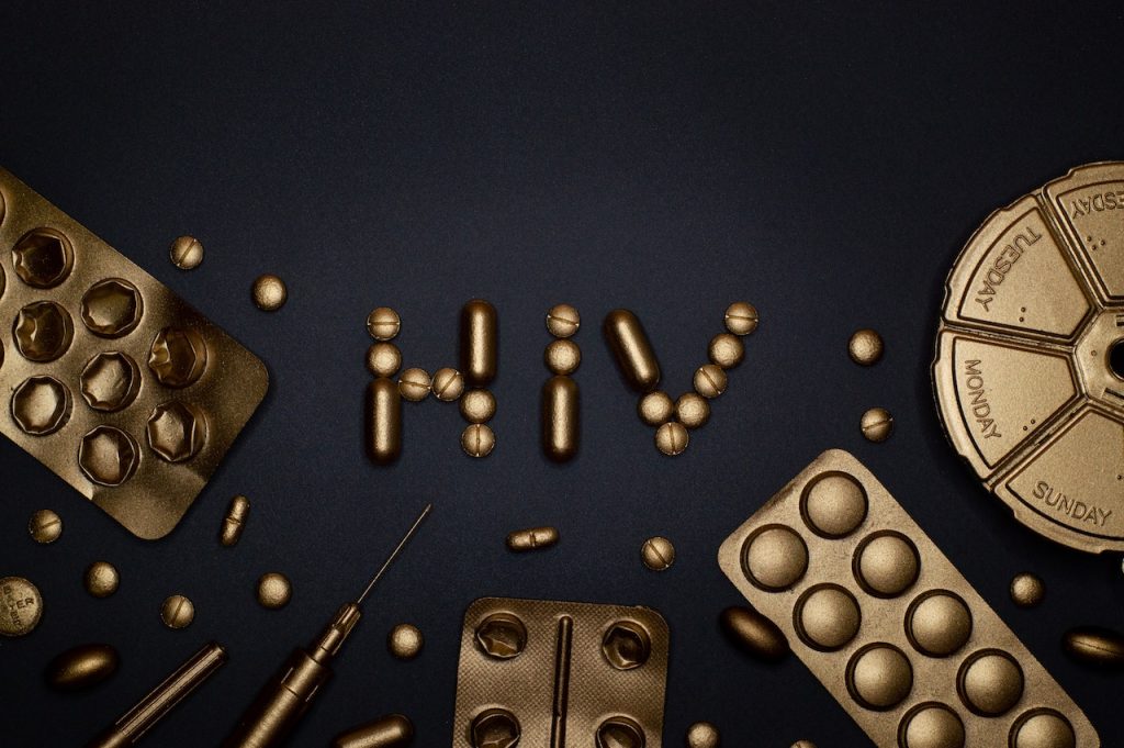
8 Million People Living with HIV in SA, According to Latest Estimates

Photo by Miguel Á. Padriñán

By Marcus Low

The number of people living with HIV in South Africa has for the first time reached the eight million mark. Of these, around 6.2 million are on treatment, according to new estimates.

The number of people living with HIV in South Africa continues to rise, surpassing eight million in 2024. This is according to just-released estimates from Thembisa, the leading mathematical model of HIV and TB in South Africa. The eight million amounts to 12.8% of the population.

The continued rise is due to the fact that there are more people becoming newly infected with HIV than there are people with HIV who are dying. The increasing numbers are thus a reflection of the fact that antiretroviral medicines are keeping people alive who would otherwise have died.

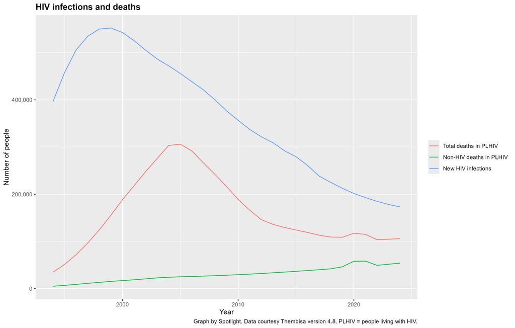
There was an estimated 178 000 new HIV infections in 2023/2024 (mid-2023 to mid-2024). Over the same period, around 105 000 people with HIV passed away – 53 000 due to HIV-related causes and 52 000 for reasons not related to HIV.

The estimates of new HIV infections are slightly higher than in last year’s Thembisa publications. According to Dr Leigh Johnson, of the University of Cape Town and the key developer of the Thembisa model, this is mainly due to the model factoring in new evidence that condom usage is declining.

78% treatment coverage

Of the eight million people living with HIV, around 6.2 million, or 78%, were taking antiretroviral treatment in 2024. Around one in five people living with the virus were thus not on treatment. Treatment is recommended for everyone living with HIV.

On the UNAIDS 95-95-95 targets, also endorsed in South Africa’s National Strategic Plan for HIV, TB and STIs 2023 – 2028, the middle target, helping people start and stay on treatment, continues to be the main area of underperformance. Around 95% of people living with HIV in South Africa knew their status in 2024, around 81.5% of these were on antiretroviral treatment, and of those on treatment, around 92% had viral suppression. (Note that the 78% treatment coverage figure is the product of multiplying the performance on the first two 95 targets.)

There continues to be stark gender disparities in South Africa’s HIV epidemic. On the one hand, there are many more women living with HIV than men – 5.2 million compared to 2.6 million as of mid-2024. On the other hand, slightly more men died of HIV-related causes than women in 2023/2024 – 27 100 men compared to 24 200 women.

Worrying trends

One ongoing area of concern is that many people only start treatment once their immune systems have been severely compromised. In 2023/2024, around 54 000 adults started treatment for the first time with CD4 counts below 200 cells/mm3. A CD4 count above 500 cells/mm3 is generally considered to be healthy. CD4 cells are a type of white blood cell that is vital to the functioning of the immune system. People who start treatment with low CD4 counts tend to have worse long-term outcomes.

The latest Thembisa outputs also contain worrying findings on the extent to which people drop in and out of care. In 2023/2024, an estimated 714 000 people restarted antiretroviral treatment after previously having stopped for at least a month – of these, around 326 000 had CD4 counts below 200 cells/mm3.

Finally, on a more positive note, the latest Thembisa outputs continue to show a rise in life expectancy in South Africa. As shown in the above graph, life expectancy declined severely round the turn of the century, largely due to people dying of AIDS, but then increased over time as antiretroviral therapy started keeping people living with HIV alive. The blip in 2020 and 2021 is due to the COVID-19 pandemic.

Note: This article is based on outputs from Thembisa version 4.8 – published in late March 2025. We have quoted 2023/2024 figures since they are based on more data, and thus more reliable than the estimates for 2024/2025. We have rounded some numbers to make the text more accessible. Graphs were made using the R package ggplot2. Spotlight will soon publish an #InTheSpotlight special briefing in which we will unpack the Thembisa 4.8 outputs in more detail.

Republished from Spotlight under a Creative Commons licence.

Read the original article.

Radiopharmaceuticals Being Tested for Brain Tumours in Children

Credit: National Cancer Institute

Neuroblastoma is a rare disease that affects children, often before the age of two. Some are born with the disease. Paediatric surgeon Jakob Stenman is investigating whether targeted radioactive drugs can slow down the disease in those with the most severe form.

Neuroblastoma is a complicated disease, with the most aggressive variant called high-risk neuroblastoma. Children with this disease are treated very intensively. They may undergo surgery, chemotherapy, high-dose chemotherapy with stem cell transplantation, radiotherapy and antibody treatment. Treatment often lasts up to a year and a half.

Despite this, the survival rate is around 60%, according to the Swedish Childhood Cancer Foundation.

“Some relapse in their disease, and we currently lack curative treatment for them,” says Jakob Stenman, a researcher at the Department of Women’s and Children’s Health at Karolinska Institutet.

It is these children, those who have relapsed, that he is treating in a study with targeted radioactive drugs. These are molecules that attach to the surface of cancer cells. These molecules have an appendage: the radioactive substance lutetium-177. The drug first moves through the bloodstream but then attaches to the cancer cells. The emitted radiation damages the cancer cells but unfortunately also the neighbouring healthy cells.

“We have treated ten children so far. Unfortunately, the disease has not disappeared in any of these cases, but it seems to be slowing down, and some benefit more than others from the treatment. When it comes to side effects, the children have tolerated the treatment well,” says Jakob Stenman.

The hope is to be able to prevent relapse

He reports that the interest has been great from clinics in other countries where these children are treated. Hospitals from Lithuania, the Netherlands, the United Kingdom and are now involved.

In neuroblastoma, cancer cells often look very different, even in the same patient. In some metastases, there may be many cells with a surface where the drug attaches, while in other metastases there may be fewer such cells. This means that the targeted drug attaches to fewer cells in some of the metastases. As a result, the local radiation dose is too low in these metastases, which can then continue to grow and spread further.

Jakob Stenman therefore believes that the treatment could be more effective if the radioactive substance used is even more potent, which in this context means that it emits even more energy (ie, radiation). If it then attaches to fewer cells in a metastasis, it might still be able to eliminate all the cancer cells there. But it must act even more locally to protect other tissues from the higher radiation dose.

The researchers have identified several substances they believe could work in this way. These include actinium-225, astatine-211 or lead-212. The effects and side effects of actinium-225 are now being investigated in cell studies and animal experiments. The goal is to start a clinical trial with actinium in three to five years.

“If what we believe turns out to be true, we hope to be able to prevent relapse and thereby enable a cure for a larger proportion of children who have developed high-risk neuroblastoma,” says Jakob Stenman.

Text: Annika Lund for Medicinsk Vetenskap nr 4 2024 

Source: Karolinska Institutet

Persistent Parasites are Not Totally Protected from Immune Response

Source: Wikimedia CC0

Most humans have long-lived infections in various tissues, including in the nervous system, that typically do not result in disease. The microbes associated with these infections, such as Toxoplasma gondii, enter a latent stage during which they quietly hide in cells, playing the long game to evade capture and ensure their own survival. But a lack of natural models to study these quiescent stages has led to gaps in scientists’ understanding of how latency contributes to pathogen persistence and whether these stages can be targeted by the immune system.

Now, a team led by University of Pennsylvania School of Veterinary Medicine researchers shows that the immune system indeed recognises the latent stage of the parasite Toxoplasma gondii, which causes toxoplasmosis. The work, published in Nature Microbiology, challenges some common assumptions about how the immune system deals with infections in the brain. Senior author Christopher A. Hunter, professor at PennU Vet, says this knowledge supports the idea that Toxoplasma gondii cysts can be targeted and perhaps even cleared, and the findings have implications for other infections and potential future therapies. The paper also demonstrates how cysts promote the mutual survival of the parasite and host.

In its latent stage, Toxoplasma gondii forms long-lived cysts in neurons in the brain, which helps the parasite evade the host’s immune response. In this study, the researchers found that certain T cells can target neurons containing cysts, thereby promoting parasite control. But there’s a tradeoff: They also found that when cysts are not formed, there is an even higher parasite burden and increased damage to the brain. The study is published in Nature Microbiology.

“There’s this balance of the pathogen needing to take hold in the host but not expand so much that it’s detrimental to the host, because if the host dies, the pathogen may not survive,” says author Lindsey A. Shallberg, who at the time of the research was a doctoral student in Hunter’s lab.

Toxoplasma gondii causes toxoplasmosis, an infection that is asymptomatic for most healthy people but poses a greater risk for those who are immunocompromised or pregnant. It is caused by eating contaminated, poorly cooked meat and by exposure to infected cat faeces, as felines are the only animal in which the parasite can sexually reproduce.

Co-author Julia N. Eberhard, an immunology doctoral student, points to two findings that run counter to preexisting literature and common notions among immunologists. She says scientists long thought that Toxoplasma gondii cysts could hide out in neurons to prevent immune recognition, but this study showed that “neurons aren’t this complete refuge for pathogens.”

This image shows Toxoplasma gondii (red) and a neuron (green) in a mouse brain.
(Image: Courtesy of Anita Koshy)

Eberhard says another commonly held belief was that the parasite needs to form cysts to be able to persist, but in looking at a parasite strain that couldn’t convert to the cyst stage, the researchers found that the immune system did not clear the parasite. They could still identify parasites in mice six months later, which Eberhard found very surprising.

Mathematical modelling independently confirmed experimental findings and indicated that immune pressure on the latent stage of Toxoplasma gondii could explain the observed rise and fall in cyst numbers. This was done by Aaron Winn, a doctoral student in the Department of Physics and Astronomy.

Shallberg says this paper came about because co-author Sebastian Lourido, an associate professor of biology at MIT, had identified the key molecular mechanism that allows the parasite to become latent and wanted to know what would happen if the parasite could not form cysts. In addition, co-author Anita Koshy, a neurologist and scientist at the University of Arizona, had evidence that some neurons could rid themselves of this infection. 

While Toxoplasma gondii is a relevant microorganism to study in and of itself, it is also useful in furthering scientists’ understanding of nervous system infections with latent stages in humans that don’t have mouse models, such as cytomegalovirus. “What makes it special is the fact that it’s a tractable model that we can use in the lab and then apply what we’ve learned to other infections,” Shallberg says.

Looking ahead, Hunter says that his laboratory continues to investigate whether T cells directly recognise the neurons and to study the T cell response in more detail.

Source: University of Pennsylvania

Three Quarters of People Who Have Taken Antidepressants Say They Were Helpful

Photo by Danilo Alvesd on Unsplash

About 75% of a sample of nearly 20 000 people who have taken selective serotonin reuptake inhibitors (SSRIs) report they found them helpful, according to new research from the Institute of Psychiatry, Psychology & Neuroscience (IoPPN) at King’s College London.

Published in Psychological Medicine, the study explored different factors that could explain why SSRIs work for some people with major depressive disorder, but not others.

Researchers analysed data from UK Biobank on 19 516 participants who had tried at least one SSRI, such as citalopram, fluoxetine, paroxetine or sertraline, for at least two weeks. Participants reported whether the SSRI helped them “feel better” using a single item questionnaire with possible responses “yes, at least a little”, “no”, “do not know”, or “prefer not to answer”. This is the first detailed analysis of this large-scale study which assesses SSRIs using self-reported experiences rather than clinician-reported remission from symptoms.

Overall, 74.9% felt SSRIs helped them feel better. 18.8% said the prescribed drug was not helpful.

Using a range of data collected by UK Biobank, the study analysed what factors might influence whether people found SSRIs helpful.

It found that sociodemographic factors such as age, gender and household income were linked to differences in how people perceived the effectiveness of SSRIs. Those participants who were older, male, had lower incomes, and reported alcohol or illicit drug use were more likely to say that they did not find antidepressants helpful.

Participants who had experienced no mood improvement even when positive events occurred or whose worst episode of depression lasted more than two years, were also less likely to report that SSRIs were helpful. Lastly those who had a greater genetic risk for depression, calculated using polygenic risk scores, were less likely to report that SSRIs were helpful.

The use of antidepressants, and the rate at which they are prescribed in the UK, has been the source of much debate both in the public and media. While antidepressants don’t work for every user, this research provides reassuring evidence that many people report that this common type of medication is helping them manage what can be a severe illness.

Dr Michelle Kamp, Postdoctoral Research Associate at King’s IoPPN and first author on the study

We know that not all people respond to antidepressants prescribed, but most studies have focussed on clinician’s perspectives of response. Using participant reports, we found a strong support for antidepressants, with three quarters of people saying the drugs had helped them. The factors that make people more likely to respond to antidepressants mirror findings in clinical trials which use measures reported by clinicians. This suggests that patient-focussed responses can capture valuable insights into the effectiveness of antidepressants.

Professor Cathryn Lewis, Professor of Genetic Epidemiology & Statistics at King’s IoPPN and senior author on the study

Professor Andrew McIntosh, Professor of Biological Psychiatry at the University of Edinburgh’s Centre for Clinical Brain Sciences and co-investigator on the study, said: “The findings from this large study show that nearly three-quarters of people in UK Biobank who were treated with antidepressants found them helpful. There is already excellent evidence from clinical trials that antidepressants work for people with depression. However those studies focus on addressing only whether they are more effective than placebos, and not why they are more effective in some people than others. We must now focus on developing a better understanding of how antidepressants work and how we can predict which people are most likely to benefit from these treatments.”

The study offers key insights into antidepressant response, however the sample may not fully represent the general population and reliance on retrospective self-reports can lead to inaccurate recollection.

Source: King’s College London

Shorter, Higher-dose Radiotherapy for Prostate Cancer Delivers Promising Results

Photo by Jo McNamara

A new large-scale study co-led by UCLA Health Jonsson Comprehensive Cancer Center investigators provides the strongest evidence yet that a shorter, standard-dose course radiation treatment is just as effective as conventional radiotherapy for early-stage prostate cancer, without compromising the safety of patients.

The shorter approach, known as isodose moderately hypofractionated radiotherapy (MHFRT), delivers slightly higher doses of radiation per session, allowing the total treatment duration to be over four to five weeks instead of seven to eight weeks.

According to the study, patients who received this type of MHFRT had the same cancer control rates as those who received conventional radiotherapy. Additionally, the risk of long-term side effects affecting the bladder and intestines was no higher with MHFRT, confirming its safety. 

The findings were published in the Lancet Oncology.

“We believe these data strongly support that isodose MHFRT should become the preferred standard of care MHFRT regimen for prostate cancer,” said Dr Amar Kishan, executive vice chair of radiation oncology at the David Geffen School of Medicine at UCLA and co-first author of the study. “More broadly, there appears to be little reason to consider conventional radiotherapy over MHFRT for the types of patients enrolled in these trials given these results.”

While MHFRT is now the most commonly used radiotherapy regimen for prostate cancer, concerns remain about whether delivering a higher daily dose increases the risk of urinary and bowel issues, such urinary incontinence, chronic diarrhoea and rectal bleeding.

MHFRT: isodose versus dose-escalated

To better understand whether there might be an increased risk of toxicity with the delivery of a higher dose per day of radiation, Kishan and the team of researchers examined data from more than 5800 patients across seven randomised clinical trials comparing standard therapy with two different MHFRT approaches: isodose MHFRT, which maintains the total radiation dose at a level similar to standard therapy, and dose-escalated MHFRT, which increases the total dose in hopes of enhancing tumour control.

The analysis found patients who received isodose MHFRT (60Gy in 20 fractions) had similar cancer control and side effects compared to those receiving conventional radiation therapy, with no significant difference in the five-year progression-free survival (77.0% for MHFRT vs 75.6% for conventional).

Patients who received higher dose-escalated MHFRT did not improve cancer control when compared to those receiving standard doses, with five-year progression-free survival rates being identical to conventional therapy (82.7% in both groups). Patient-reported outcomes also showed significantly higher gastrointestinal side effects (7.2% vs 4.9%), particularly bowel issues. 

While dose-escalated MHFRT was expected to improve outcomes, the data showed no additional benefit in cancer control and a higher risk of gastrointestinal side effects, noted Kishan. This underscores the advantage of isodose MHFRT, which provides the same effectiveness as conventional therapy without increasing toxicity.

“These findings reinforce isodose MHFRT as the standard of care, offering the same cancer control as conventional treatment but with fewer side effects than dose-escalated MHFRT,” said Kishan, who is also a researcher in the UCLA Health Jonsson Comprehensive Cancer Center. “Patients can safely opt for a shorter treatment schedule without compromising their outcomes, ensuring they receive effective care with fewer visits and minimal added risk. Less time in treatment can still mean the best possible results.”

Source: University of California – Los Angeles Health Sciences

An Arthritis Drug Might Unlock Lasting Relief from Epilepsy and Seizures

Source: Pixabay

A drug typically prescribed for arthritis halts brain-damaging seizures in mice that have a condition like epilepsy, according to researchers at the University of Wisconsin–Madison. The drug, called tofacitinib, also restores short-term and working memory lost to epilepsy in the mice and reduces inflammation in the brain caused by the disease.

If the drug proves viable for human patients, it would be the first to provide lasting relief from seizures even after they stopped taking it.

“It ticks all the boxes of everything we’ve been looking for,” says Avtar Roopra, a neuroscience professor in the UW–Madison School of Medicine and Public Health and senior author of the study, which appears in Science Translational Medicine.

Epilepsy is one of the most common neurological diseases, afflicting more than 50 million people around the world. While there are many known causes, the disease often appears after an injury to the brain, like a physical impact or a stroke.

Some days, months or even years after the injury, the brain loses the ability to calm its own activity. Normally balanced electrical activity through the brain goes haywire.

“The system revs up until all the neurons are firing all the time, synchronously,” says Roopra. “That’s a seizure that can cause massive cell death.”

And the seizures repeat, often at random intervals, forever. Some drugs have been useful in addressing seizure symptoms, protecting patients from some of the rampant inflammation and memory loss, but one-third of epilepsy patients do not respond to any known drugs, according to Olivia Hoffman, lead author of the study and a postdoctoral researcher in Roopra’s lab. The only way to stop the most damaging seizures has been to remove a piece of the brain where disruptive activity starts.

On their way to identifying tofacitinib’s potential in epilepsy, Hoffman and co-authors used relatively new data science methods to sift through the way thousands of genes were expressed in millions of cells in the brains of mice with and without epilepsy. They found a protein called STAT3, key to a cell signaling pathway called JAK, at the centre of activity in the seizure-affected mouse brains.

“When we did a similar analysis of data from brain tissue removed from humans with epilepsy, we found that was also driven by STAT3,” Hoffman says.

Meanwhile, Hoffman had unearthed a study of tens of thousands of arthritis patients in Taiwan aimed at describing other diseases associated with arthritis. It turns out, epilepsy was much more common among those arthritis patients than people without arthritis — but surprisingly less common than normal for the arthritis patients who had been taking anti-inflammatory drugs for more than five-and-a-half years.

“If you’ve had rheumatoid arthritis for that long, your doctor has probably put you on what’s called a JAK-inhibitor, a drug that’s targeting this signaling pathway we’re thinking is really important in epilepsy,” Hoffman says.

The UW researchers ran a trial with their mice, dosing them with the JAK-inhibitor tofacitinib following the administration of a brain-damaging drug that puts them on the road to repeated seizures. Nothing happened. The mice still developed epilepsy like human patients.

Remember, though, that epilepsy doesn’t often present right after a brain-damaging event. It can take years. In the lab mice, there’s usually a lull of weeks of relatively normal time between the brain damage and what the researchers call “reignition” of seizures. If it’s not really epilepsy until reignition, what if they tried the drug then? They devised a 10-day course of tofacitinib to start when the mouse brains fell out of their lull and back into the chaos of seizures.

“Honestly, I didn’t think it was going to work,” Hoffman says. “But we believe that initial event sort of primes this pathway in the brain for trouble. And when we stepped in at that reignition point, the animals responded.”

The drug worked better than they could have imagined. After treatment, the mice stayed seizure-free for two months, according to the paper. Collaborators at Tufts University and Emory University tried the drug with their own mouse models of slightly different versions of epilepsy and got the same, seizure-free results.

Roopra’s lab has since followed mice that were seizure-free for four and five months. And their working memory returned.

“These animals are having many seizures a day. They cannot navigate mazes. Behaviourally, they are bereft. They can’t behave like normal mice, just like humans who have chronic epilepsy have deficits in learning and memory and problems with everyday tasks,” Roopra says. “We gave them that drug, and the seizures disappear. But their cognition also comes back online, which is astounding. The drug appears to be working on multiple brain systems simultaneously to bring everything under control, as compared to other drugs, which only try to force one component back into control.”

Because tofacitinib is already FDA-approved as safe for human use for arthritis, the path from animal studies to human trials may be shorter than it would be for a brand-new drug. The next steps toward human patients largely await NIH review of new studies, which have been paused indefinitely amid changes at the agency.

For now, the researchers are focused on trying to identify which types of brain cells are shifted back to healthy behavior by tofacitinib and on animal studies of even more of the many types of epilepsy. Hoffman and Roopra have also filed for a patent on the use of the drug in epilepsy.

Source: University of Wisconsin-Madison

Immune Cell Networks Found to be Driving Idiopathic Pulmonary Fibrosis

Photo by Robina Weermeijer on Unsplash

Rutgers Health researchers have discovered that networks of misplaced immune cells drive an aggressive lung disease, potentially opening a path to new treatments for a condition that kills 80% of patients within a decade.

Idiopathic pulmonary fibrosis (IPF) scars lung tissue and makes breathing increasingly difficult until patients can’t get enough oxygen. Available drugs provide minimal benefit. Lung transplantation works for some patients, but transplants have a 50% five-year mortality rate.

This study in the European Respiratory Journal used advanced spatial mapping techniques to compare healthy lung tissues and tissues from patients with fatal IPF. The researchers discovered that disease-scarred lung tissue abounds in plasma cells – specialised immune cells that typically reside in bone marrow and produce antibodies.

“What we found most striking in this study is that all the fibrotic regions of IPF patients’ lungs are covered by antibody-producing plasma cells,” Qi Yang, an associate paediatrics professor at Rutgers and a senior author of the study. “In normal lungs, there are almost no plasma cells. But in IPF patients, the lungs are full of them.”

The researchers identified previously unknown cellular networks orchestrating this abnormal immune response. They discovered novel mural cells wrapping around blood vessels and producing signal proteins that organize immune responses. They also found unique fibroblasts secreting a protein that attracts plasma cells to damaged areas.

“This particular type of fibroblast has never been described before,” said Reynold Panettieri, director of the Rutgers Institute for Translational Medicine and Science and a senior author of the study. “People have shown that fibroblasts are the cell types responsible for scarring – in the skin, the lungs and the brain – but this particular type of fibroblast seems unique to the lung.”

Having found the plasma cells in lung tissue taken from people who died of IPF, the team began using live mice to see if reducing plasma in the lungs slowed disease formation. This work demonstrated that blocking signaling pathways reduced plasma cell accumulation and alleviated lung scarring. Targeting these same signaling pathways may thus prove an effective disease treatment in humans, the researchers said.

The research is particularly promising because drugs targeting plasma cells already exist. Medications used to treat multiple myeloma, a plasma cell cancer, could potentially be repurposed to treat IPF.

“If the plasma cells are really making the bad antibodies, I assume we may have to get rid of them,” said Yang, a member of the Institute for Translational Medicine and Science. “Otherwise, patients will keep making these antibodies that drive the disease.”

Previous studies have shown that IPF patients have heightened antibody responses and elevated lung antibody levels. The new research explains the origin of these antibodies and reveals how abnormal antibody-producing cells accumulate in the lungs.

The researchers said the antibodies may drive tissue damage through several mechanisms. Their data suggest that antibody-antigen complexes stimulate the production of transforming growth factor-beta from pulmonary macrophages, thus promoting fibrosis.

“Now that we have a target, a cell, a unique cell that Dr Yang has identified and phenotyped, we’re optimistic that we could affect that cell and not other fibroblasts that are important in normal injury repair response,” Panettieri said.

For patients with IPF, the findings offer hope of new treatments for a debilitating condition with limited therapeutic options. The disease typically affects men over 60 years of age, with most patients dying within five years of diagnosis.

The next steps for the research team include determining whether the plasma cells are producing autoantibodies against healthy tissues and further investigating how fibroblasts and mural cells develop their abnormal properties in IPF.

“Our research suggests that IPF might have a strong autoimmune link,” Yang said.

Source: Rutgers University

Cold Plunges Fire up Repair Functions in Cells

Photo by Yaroslav Shuraev

Investigating the effects of trendy ice baths, scientists at the University of Ottawa have made an interesting discovery on the beneficial way they affect cell functions, and have published their findings in Advanced Biology.

A new study conducted at the Human and Environmental Physiology Research lab (HEPRU) at the University of Ottawa has unveiled significant findings on the effects of cold water acclimation on autophagic (the cells’ recycling system, which promotes cellular health) and apoptotic (the programmed cell death that gets rid of damaged cells) responses in young males. The research highlights the potential for cold exposure to enhance cellular resilience against stress.

The study, conducted by Kelli King, postdoctoral fellow, and Glen Kenny, Full Professor at uOttawa’s School of Human Kinetics and Director of HEPRU, involved ten healthy young males who underwent cold-water immersion at 14°C for one hour across seven consecutive days. Blood samples were collected to analyse the participants’ cellular responses before and after the acclimation period.

“Our findings indicate that repeated cold exposure significantly improves autophagic function, a critical cellular protective mechanism,” says Professor Kenny. “This enhancement allows cells to better manage stress and could have important implications for health and longevity.”

The research revealed that while autophagy was initially dysfunctional after high-intensity cold stress, consistent exposure over a week led to increased autophagic activity and decreased cellular damage signals.

“By the end of the acclimation, we noted a marked improvement in the participants’ cellular cold tolerance,” explains King, the study’s first author. “This suggests that cold acclimation may help the body effectively cope with extreme environmental conditions.”

The implications of this study extend beyond athletic performance. Cold water immersion has gained popularity for its potential health benefits, and this research provides some scientific backing for its efficacy. The findings suggest that proper autophagic activity could not only extend cellular longevity but also prevent the onset of various diseases.

As the use of cold exposure becomes increasingly mainstream, understanding its effects on cellular mechanisms is vital. Professor Kenny emphasises, “This work underscores the importance of acclimation protocols in enhancing human health, especially in contexts where individuals are exposed to extreme temperatures.”

“We were amazed to see how quickly the body adapted,” notes King. “Cold exposure might help prevent diseases and potentially even slow down aging at a cellular level. It’s like a tune-up for your body’s microscopic machinery.”

These results apply to young males and more research is needed to see if it would also apply to other cohorts.

Source: University of Ottawa

How Antibiotics in Infancy may Increase Diabetes Risk

Gut Microbiome. Credit Darryl Leja National Human Genome Research Institute National Institutes Of Health

Exposure to antibiotics during a key developmental window in infancy can stunt the growth of insulin-producing cells in the pancreas and may boost risk of diabetes later in life, new research in mice suggests. The study, published this month in the journal Science, also pinpoints specific microorganisms that may help those critical cells proliferate in early life.

The findings are the latest to shine a light on the importance of the human infant microbiome—the constellation of bacteria and fungi living on and in us during our first few years. The research could lead to new approaches for addressing a host of metabolic diseases.

“We hope our study provides more awareness for how important the infant microbiome actually is for shaping development,” said first author Jennifer Hill, assistant professor in molecular, cellular and developmental biology at CU’s BioFrontiers Institute. “This work also provides important new evidence that microbe-based approaches could someday be used to not only prevent but also reverse diabetes.”

Something in the environment

More than 2 million U.S. adults live with Type 1 diabetes. The disease typically emerges in childhood, and genetics play a strong role. But scientists have found that, while identical twins share DNA that predisposes them to Type 1 diabetes, only one twin usually gets the disease.

“This tells you that there’s something about their environmental experiences that is changing their susceptibility,” said Hill.

For years, she has looked to microbes for answers.

Previous studies show that children who are breastfed or born vaginally, which can both promote a healthy infant microbiome, are less likely to develop Type 1 diabetes than others. Some research also shows that giving babies antibiotics early can inadvertently kill good bugs with bad and boost diabetes risk.

The lingering questions: What microbes are these infants missing out on?

“Our study identifies a critical window in early life when specific microbes are necessary to promote pancreatic cell development,” said Hill.

A key window of opportunity

She explained that human babies are born with a small amount of pancreatic “beta cells,” the only cells in the body that produce insulin. But some time in a baby’s first year, a once-in-a-lifetime surge in beta cell growth occurs.

“If, for whatever reason, we don’t undergo this event of expansion and proliferation, that can be a cause of diabetes,” Hill said.

She conducted the current study as a postdoctoral researcher at the University of Utah with senior author June Round, a professor of pathology.

They found that when they gave broad-spectrum antibiotics to mice during a specific window (the human equivalent of about 7 to 12 months of life), the mice developed fewer insulin producing cells, higher blood sugar levels, lower insulin levels and generally worse metabolic function in adulthood.

“This, to me, was shocking and a bit scary,” said Round. “It showed how important the microbiota is during this very short early period of development.”

Lessons in baby poop

In other experiments, the scientists gave specific microbes to mice, and found that several they increased their production of beta cells and boosted insulin levels in the blood. The most powerful was a fungus called Candida dubliniensis.

The team used faecal samples from The Environmental Determinants of Diabetes in the Young (TEDDY) study to make what Hill calls “poop slushies” and fed them to the mice.

When the researchers inoculated newborn mice with poop from healthy infants between 7 to 12 months in age, their beta cells began to grow. Poop from infants of other ages did not do the same. Notably, Candida dublineinsis was abundant in human babies only during this time period.

“This suggests that humans also have a narrow window of colonisation by these beta cell promoting microbes,” said Hill.

When male mice that were genetically predisposed to Type 1 diabetes were colonised with the fungus in infancy, they developed diabetes less than 15% of the time. Males that didn’t receive the fungus got diabetes 90% of the time.

Even more promising, when researchers gave the fungus to adult mice whose insulin-producing cells had been killed off, those cells regenerated.

Too early for treatments

Hill stresses that she is not “anti-antibiotics.” But she does imagine a day when doctors could give microbe-based drugs or supplements alongside antibiotics to replace the metabolism-supporting bugs they inadvertently kill.

Poop slushies (faecal microbiota transplants) have already been used experimentally to try to improve metabolic profiles of people with Type 2 diabetes, which can also damage pancreatic beta cells.

But such approaches can come with real risk, since many microbes that are beneficial in childhood can cause harm in adults. Instead, she hopes that scientists can someday harness the specific mechanisms the microbes use to develop novel treatments for healing a damaged pancreas—reversing diabetes.

She recently helped establish a state-of-the-art “germ-free” facility for studying the infant microbiome at CU Boulder. There, animals can be bred and raised entirely without microbes, and by re-introducing them one by one scientists can learn they work.

“Historically we have interpreted germs as something we want to avoid, but we probably have way more beneficial microbes than pathogens,” she said. “By harnessing their power, we can do a lot to benefit human health.”

Source: University of Colorado at Boulder

Social Skills not as Relevant for Autism Diagnosis than Thought

Photo by Peter Burdon on Unsplash

People with autism are typically diagnosed by clinical observation and assessment. To deconstruct the clinical decision process, which is often subjective and difficult to describe, researchers used a large language model (LLM) to synthesize the behaviours and observations that are most indicative of an autism diagnosis. Their results, publishing in the Cell Press journal Cell, show that repetitive behaviours, special interests, and perception-based behaviours are most associated with an autism diagnosis.

These findings have potential to improve diagnostic guidelines for autism by decreasing the focus on social factors – which the established guidelines in the DSM-5 focus on but the model did not classify among the most relevant in diagnosing autism.

“Our goal was not to suggest that we could replace clinicians with AI tools for diagnosis,” says senior author Danilo Bzdok of the Mila Québec Artificial Intelligence Institute and McGill University in Montreal. “Rather, we sought to quantitatively define exactly what aspects of observed behaviour or patient history a clinician uses to reach a final diagnostic determination. In doing so, we hope to empower clinicians to work with diagnostic instruments that are more in line with their empirical realities.”

The scientists leveraged a transformer language model, which was pre-trained on about 489 million unique sentences. They then fine-tuned the LLM to predict the diagnostic outcome from a collection of more than 4000 reports written by clinicians working with patients considered for autism diagnosis. The reports, which were often used by multiple clinicians, included accounts of observed behaviour and relevant patient history but did not include a suggested diagnostic outcome.

The team developed a bespoke LLM module that pinpointed specific sentences in the reports that were most relevant to a correct diagnosis prediction. They then extracted the numerical representation of these highly autism-relevant sentences and compared them directly with the established diagnostic criteria enumerated in the DSM-5.

“Modern LLMs, with their advanced natural language processing capabilities, are natively suited to this textual analysis,” Bzdok says. “The key challenge we faced was in designing sentence-level interpretability tools to pinpoint the exact sentences, expressed by the healthcare professional themselves, that were most essential to a correct diagnosis prediction by the LLM.”

The researchers were surprised by how clearly the LLM was able to distinguish between the most diagnostically relevant criteria. For example, their framework flagged that repetitive behaviours, special interests, and perception-based behaviour were the criteria most relevant to autism. While these criteria are used in clinical settings, current criteria focus more on deficits in social interplay and lack of communication skills.

The authors note that there are limitations to this study, including a lack of geographical diversity. Additionally, the researchers did not analyse their results based on demographic variables, with the goal of making the conclusions more broadly applicable.

The team expects their framework will be helpful to researchers and medical professionals working with a range of psychiatric, mental health, and neurodevelopmental disorders in which clinical judgement forms the bulk of the diagnostic decision-making process.

“We expect this paper to be highly relevant to the broader autism community,” Bzdok says. “We hope that our paper motivates conversations about grounding diagnostic standards in more empirically derived criteria. We also hope it will establish common threads that link seemingly diverse clinical presentations of autism together.”

Source: ScienceDaily