Tag: neurodegenerative diseases

Breathing Low-oxygen Air Slows Parkinson’s Progression in Mice

Photo by Mike Markov on Unsplash

Researchers from the Broad Institute and Mass General Brigham have shown that a low-oxygen environment – similar to the thin air found at Mount Everest base camp – can protect the brain and restore movement in mice with Parkinson’s-like disease.

The new research, in Nature Neuroscience, suggests that cellular dysfunction in Parkinson’s leads to the accumulation of excess oxygen molecules in the brain, which then fuel neurodegeneration – and that reducing oxygen intake could help prevent or even reverse Parkinson’s symptoms.

“The fact that we actually saw some reversal of neurological damage is really exciting,” said co-senior author Vamsi Mootha, an institute member at the Broad, professor of systems biology and medicine at Harvard Medical School, a Howard Hughes Medical Institute investigator in the Department of Molecular Biology at Massachusetts General Hospital (MGH), a founding member of the Mass General Brigham healthcare system. “It tells us that there is a window during which some neurons are dysfunctional but not yet dead – and that we can restore their function if we intervene early enough.”

“The results raise the possibility of an entirely new paradigm for addressing Parkinson’s disease,” added co-senior author Fumito Ichinose, the William T. G. Morton professor of anesthesia at Harvard Medical School and MGH.

The researchers caution that it’s too soon to translate these results directly to new treatments for patients. They emphasize that unsupervised breathing of low-oxygen air, especially intermittently such as only at night, can be dangerous and may even worsen the disease. But they’re optimistic their findings could help spur the development of new drugs that mimic the effects of low oxygen.

The study builds on a decade of research from Mootha and others into hypoxia – the condition of having lower than normal oxygen levels in the body or tissues – and its unexpected ability to protect against mitochondrial disorders.

“We first saw that low oxygen could alleviate brain-related symptoms in some rare diseases where mitochondria are affected, such as Leigh syndrome and Friedreich’s ataxia,” said Mootha, who leads the Friedreich’s Ataxia Accelerator at Broad. “That raised the question: Could the same be true in more common neurodegenerative diseases like Parkinson’s?”

Eizo Marutani, an instructor of anesthesia at MGH and Harvard Medical School, is the first author of the new paper. 

A long-standing link

Parkinson’s disease, which affects more than 10 million people worldwide, causes the progressive loss of neurons in the brain, leading to tremors and slowed movements. Neurons affected by Parkinson’s also gradually accumulate toxic protein clumps called Lewy bodies. Some biochemical evidence has suggested that these clumps interfere with the function of mitochondria, that Mootha knew were altered in other diseases that could be treated with hypoxia.

Moreover, anecdotally, people with Parkinson’s seem to fare better at high altitudes. And long-term smokers – who have elevated levels of carbon monoxide, leading to less oxygen in tissues – also appear to have a lower risk of developing Parkinson’s.

“Based on this evidence, we became very interested in the effect of hypoxia on Parkinson’s disease,” said Ichinose.

Mootha and Ichinose turned to a well-established mouse model of Parkinson’s in which animals are injected with clumps of the α-synuclein proteins that seed the formation of Lewy bodies. The mice were then split into two groups: one breathing normal air (21% oxygen) and the other continuously housed in chambers with 11% oxygen – comparable to living at an altitude of about 4800m. 

A new paradigm for Parkinson’s

The results were striking. Three months after receiving α-synuclein protein injections, the mice breathing normal air had high levels of Lewy bodies, dead neurons, and severe movement problems. Mice that had breathed low-oxygen air from the start didn’t lose any neurons and showed no signs of movement problems, despite developing abundant Lewy bodies.

The findings show that hypoxia wasn’t stopping the formation of Lewy bodies but was protecting neurons from the damaging effects of these protein clumps – potentially suggesting a new mode of treating Parkinson’s without targeting α-synuclein or Lewy bodies, Ichinose said. 

What’s more, when hypoxia was introduced six weeks after the injection, when symptoms were already appearing, it still worked. The mice’s motor skills rebounded, their anxiety-like behaviors faded, and the loss of neurons in the brain stopped.

To further explore the underlying mechanism, the team analyzed brain cells of the mice and discovered that mice with Parkinson’s symptoms had much higher levels of oxygen in some parts of the brain than control mice and those that had breathed low-oxygen air. This excess oxygen, the researchers said, likely results from mitochondrial dysfunction. Damaged mitochondria can’t use oxygen efficiently, so it builds up to damaging levels. 

“Too much oxygen in the brain turns out to be toxic,” said Mootha. “By reducing the overall oxygen supply, we’re cutting off the fuel for that damage.”

Hypoxia in a pill

More work is needed before the findings can be directly used to treat Parkinson’s. In the meantime, Mootha and his team are developing “hypoxia in a pill” drugs that mimic the effects of low oxygen to potentially treat mitochondrial disorders, and they think a similar approach might work for some forms of neurodegeneration.

While not all neurodegenerative models respond to hypoxia, the approach has now shown success in mouse models of Parkinson’s, Leigh syndrome, Friedreich’s ataxia, and accelerated aging.

“It may not be a treatment for all types of neurodegeneration,” said Mootha, “but it’s a powerful concept – one that might shift how we think about treating some of these diseases.”

Source: Broad Institute

Hibernation ‘Superpowers’ May Be Hidden in Human DNA

Photo by Sangharsh Lohakare on Unsplash

Animals that hibernate are incredibly resilient. They can spend months without food or water, muscles refusing to atrophy, body temperature dropping to near freezing as their metabolism and brain activity slow to a crawl. When they emerge from hibernation, they recover from dangerous health changes similar to those seen in type 2 diabetes, Alzheimer’s disease, and stroke.

New genetic research suggests that hibernating animals’ superpowers could lie hidden in human DNA – with clues on how to unlock them, perhaps one day leading to treatments that could reverse neurodegeneration and diabetes.

Two studies describing the results are published in Science.

The genetics of metabolism and obesity

A gene cluster called the “fat mass and obesity (FTO) locus” plays an important role in hibernators’ abilities, the researchers found. Intriguingly, humans have these genes too. “What’s striking about this region is that it is the strongest genetic risk factor for human obesity,” says Chris Gregg, PhD, professor in neurobiology and human genetics at University of Utah Health and senior author on the studies. But hibernators seem able to use genes in the FTO locus in new ways to their advantage.

The team identified hibernator-specific DNA regions that are near the FTO locus and that regulate the activity of neighbouring genes, tuning them up or down. The researchers speculate that adjusting the activity of neighbouring genes, including those in or near the FTO locus, allows hibernators to pack on the pounds before settling in for the winter, then slowly use their fat reserves for energy throughout hibernation.
 
Indeed, the hibernator-specific regulatory regions outside of the FTO locus seem crucial for tweaking metabolism. When the researchers mutated those hibernator-specific regions in mice, they saw changes in the mice’s weight and metabolism. Some mutations sped up or slowed down weight gain under specific dietary conditions; others affected the ability to recover body temperature after a hibernation-like state or tuned overall metabolic rate up or down. 

Intriguingly, the hibernator-specific DNA regions the researchers identified weren’t genes themselves. Instead, the regions were DNA sequences that contact nearby genes and turn their expression up or down, like an orchestra conductor fine-tuning the volume of many musicians. This means that mutating a single hibernator-specific region has wide-ranging effects extending far beyond the FTO locus, explains Susan Steinwand, research scientist in neurobiology at U of U Health and first author on one of the studies.  “When you knock out one of these elements – this one tiny, seemingly insignificant DNA region – the activity of hundreds of genes changes,” she says. “It’s pretty amazing.”
 
Understanding hibernators’ metabolic flexibility could lead to better treatments for human metabolic disorders like type 2 diabetes, the researchers say. “If we could regulate our genes a bit more like hibernators, maybe we could overcome type 2 diabetes the same way that a hibernator returns from hibernation back to a normal metabolic state,” says Elliott Ferris, MS, bioinformatician at U of U Health and first author on the other study.

Uncovering the regulation of hibernation

Finding the genetic regions that may enable hibernation is a problem akin to excavating needles from a massive DNA haystack. To narrow down the regions involved, the researchers used multiple independent whole-genome technologies to ask which regions might be relevant for hibernation. Then, they started looking for overlap between the results from each technique.

First, they looked for sequences of DNA that most mammals share but that had recently changed in hibernators. “If a region doesn’t change much from species to species for over 100 million years but then changes rapidly and dramatically in two hibernating mammals, then we think it points us to something that is important for hibernation, specifically,” Ferris says.

To understand the biological processes that underlie hibernation, the researchers tested for and identified genes that turn up or down during fasting in mice, which triggers metabolic changes similar to hibernation. Next, they found the genes that act as central coordinators, or “hubs,” of these fasting-induced changes to gene activity.

Many of the DNA regions that had recently changed in hibernators also appeared to interact with these central coordinating hub genes. Because of this, the researchers expect that the evolution of hibernation requires specific changes to the controls of the hub genes. These controls comprise a shortlist of DNA elements that are avenues for future investigation.

Awakening human potential

Most of the hibernator-associated changes in the genome appeared to “break” the function of specific pieces of DNA, rather than confer a new function. This hints that hibernators may have lost constraints that would otherwise prevent extreme flexibility in the ability to control metabolism. In other words, it’s possible that the human “thermostat” is locked to a narrow range of continuous energy consumption. For hibernators, that lock may be gone.

Hibernators can reverse neurodegeneration, avoid muscle atrophy, stay healthy despite massive weight fluctuations, and show improved aging and longevity. The researchers think their findings show that humans may already have the needed genetic code to have similar hibernator-like superpowers—if we can bypass some of our metabolic switches. 

“Humans already have the genetic framework,” Steinwand says. “We just need to identify the control switches for these hibernator traits.” By learning how, researchers could help confer similar resilience to humans.

Source: University of Utah Health

MRI and Ultrasound Combo Opens Blood-brain Barrier

In a mouse model study of MRI-guided focused ultrasound-induced blood-brain barrier (BBB) opening at MRI field strengths ranging from ­approximately 0 T (outside the magnetic field) to 4.7 T, the static magnetic field dampened the detected microbubble cavitation signal and decreased the BBB opening volume. Credit: Washington University School of Medicine in St. Louis

Using a combination of ultrasound, MRI field strength and microbubbles can open the blood-brain barrier (BBB) and allow therapeutic drugs to reach the diseased brain location with MRI guidance. 

Using the physical phenomenon of cavitation, it is a promising technique that has been shown safe in patients with various brain diseases, such as Alzheimer’s diseases, Parkinson’s disease, ALS, and glioblastoma.
While MRI has been commonly used for treatment guidance and assessment in preclinical research and clinical studies, until now, researchers did not know the impact that MRI scanner’s magnetic field had on the BBB opening size and drug delivery efficiency.

Hong Chen, associate professor of biomedical engineering at Washington University in St. Louis, and her lab have found for the first time that the magnetic field of the MRI scanner decreased the BBB opening volume by 3.3-fold to 11.7-fold, depending on the strength of the magnetic field, in a mouse model. The findings were in Radiology.

Prof Chen conducted the study on four groups of mice. After they were injected microbubbles, three groups received focused-ultrasound sonication at different strengths of the magnetic field: 1.5 T (teslas), 3 T and 4.7 T, and one group was never exposed to the field. 

The researchers found that the microbubble cavitation activity, or the growing, shrinking and collapse of the microbubbles, decreased by 2.1 decibels at 1.5 T; 2.9 decibels at 3 T; and 3 decibels at 4.7 T, compared with those that had received the dose outside of the magnetic field. Additionally, the magnetic field decreased the BBB opening volume by 3.3-fold at 1.5 T; 4.4-fold at 3 T; and 11.7-fold at 4.7 T. No tissue damage from the procedure was seen.

Following focused-ultrasound sonication, the team injected a model drug, Evans blue dye, to investigate whether the magnetic field affected drug delivery across the BBB. The images showed that the fluorescence intensity of the Evans blue was lower in mice that received the treatment in one of the three strengths of magnetic fields compared with mice treated outside the magnetic field. The Evans blue trans-BBB delivery was decreased by 1.4-fold at1.5 T, 1.6-fold at 3.0 T and 1.9-fold at 4.7 T when compared with those treated outside of the magnetic field.

“The dampening effect of the magnetic field on the microbubble is likely caused by the loss of bubble kinetic energy due to the Lorentz force acting on the moving charged lipid molecules on the microbubble shell and dipolar water molecules surrounding the microbubbles,” said Yaoheng (Mack) Yang, a doctoral student in Prof Chen’s lab and the lead author of the study.

“Findings from this study suggest that the impact of the magnetic field needs to be considered in the clinical applications of focused ultrasound in brain drug delivery,” Prof Chen said.

In addition to brain drug delivery, cavitation is also used in several other therapeutic techniques, such as histotripsy, the use of cavitation to mechanically destroy regions of tissue, and sonothrombolysis, a therapy used after acute ischaemic stroke. The magnetic field’s damping effect on cavitation is expected to affect the treatment outcomes of other cavitation-mediated techniques when MRI-guided focused-ultrasound systems are used.

Source: Washington University in St. Louis

Journal information: Yang, Y., et al. (2021) Static Magnetic Fields Dampen Focused Ultrasound–mediated Blood-Brain Barrier Opening. Radiology. doi.org/10.1148/radiol.2021204441

New Treatment Candidate May Reverse Neurodegenerative Decline

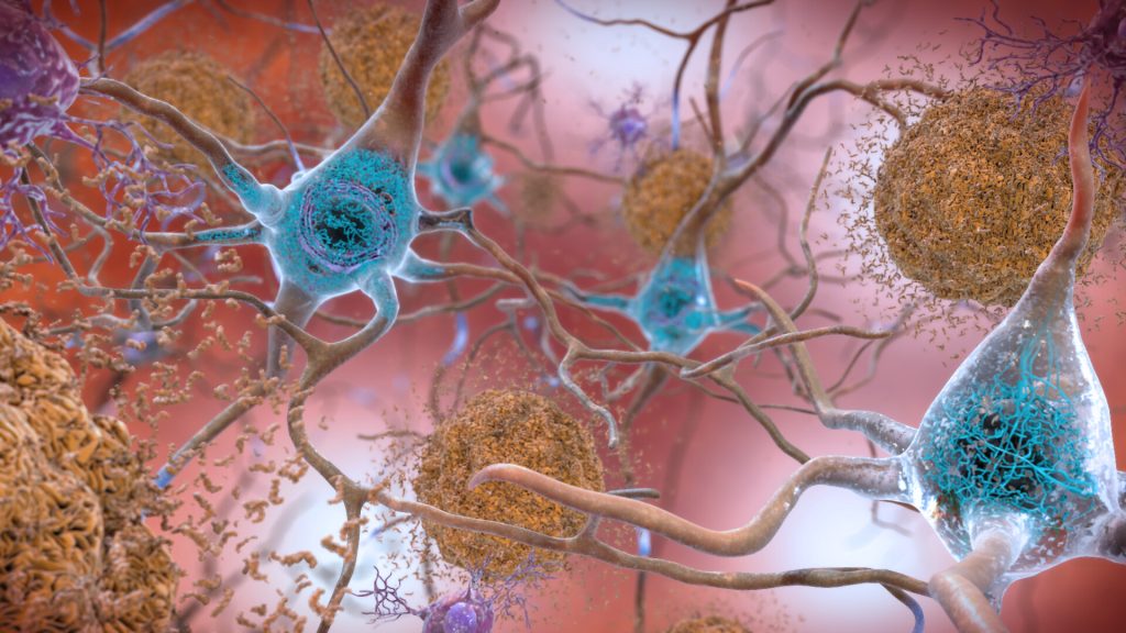
In the Alzheimer’s affected brain, abnormal levels of the beta-amyloid protein clump together to form plaques (seen in brown) that collect between neurons and disrupt cell function. Abnormal collections of the tau protein accumulate and form tangles (seen in blue) within neurons, harming synaptic communication between nerve cells.
Credit: National Institute on Aging, NIH

Researchers  at Tohoku University in Japan have identified a new treatment candidate that seems to not only halt but partially reverse neurodegenerative symptoms in mouse models of dementia and Alzheimer’s disease.

Kohji Fukunaga, professor emeritus in Tohoku University’s Graduate School of Pharmaceutical Sciences and paper author, said: “There are currently no disease-modifying therapeutics for neurodegenerative disorders such as Alzheimer’s disease, Lewy body dementia, Huntington disease and frontotemporal dementia in the world. We discovered the novel, disease-modifying therapeutic candidate SAK3, which, in our studies, rescued neurons in most protein-misfolding, neurodegenerative diseases.”

In previous work, the team found that the SAK3 molecule – the base structure of which is found in the enhancement of T-type Ca2+ channel activity – apparently improved memory and learning in a mouse model of Alzheimer’s disease.

SAK3 enhances the function of a cell membrane channel thereby promoting neuronal activity in the brain. Typically, SAK3 promotes neurotransmitter releases of acetylcholine and dopamine — neurotransmitters which are lowered in Alzheimer’s disease and Lewy body dementia. The Ca2+ channel enhancement is thought to trigger a change from resting to active in neuronal activity. When the Ca2+ channel is dysregulated in the brain, less acetylcholine and dopamine is released. Cognitive confusion and uncoordinated motor function arises from this dysregulated system.

SAK3 binds directly  to the subunit of this channel, enhancing neurotransmission and so improving cognitive deficits. The researchers found that the same process also seemed to work in a mouse model of Lewy body dementia, which is characterised by a buildup of proteins known as Lewy bodies.

“Even after the onset of cognitive impairment, SAK3 administration significantly prevented the progression of neurodegenerative behaviors in both motor dysfunction and cognition,” Prof Fukunaga said.

In comparison, Aduhelm, the Alzheimer’s drug recently approved by the US Food and Drug Administration, reduces the number of amyloid plaques in the brain, but whether the amyloid reduction actually prevents further cognitive or motor decline in patients is not yet known. According to Prof Fukunaga, SAK3 helps destroy amyloid plaque – at least in mice.

SAK3 also helps destroy misfolded alpha-synuclein, which normally helps regulate neurotransmitter transmission in the brain. The misfolded protein can aggregate, contributing to what researchers suspect may be an underlying cause of neurodegenerative symptoms. This aggregation can also cause loss of dopamine neurons, which are associated with learning and memory.

“We found that chronic administration of SAK3 significantly inhibited the accumulation of alpha-synuclein in the mice,” Prof Fukunaga said, noting that the mice received a daily oral dose of SAK3.

According to Prof Fukunaga, SAK3 enhances the activity of the system that identifies and destroys misfolded proteins. In neurodegenerative diseases, this system is often dysfunctional, leaving misfolded proteins to wreak havoc in the cell’s machinery.

“SAK3 is the first compound targeting this regulatory activity in neurodegenerative disorders,” Fukunaga said. “SAK3 administration promotes the destruction of misfolded proteins, meaning the therapeutic has the potential to solve the problems of diverse protein misfolding diseases such as Parkinson’s disease, Lewy body dementia and Huntington disease, in addition to Alzheimer’s disease.”

The team published their results in the International Journal of Molecular Sciences. This treatment candidate has been declared safe by Japan’s governing board, and the researchers are planning to start human clinical trials in the next year.

Source: Tohoku University

Journal information: Xu, J., et al. (2021) T-Type Ca2+ Enhancer SAK3 Activates CaMKII and Proteasome Activities in Lewy Body Dementia Mice Model. International Journal of Molecular Sciencesdoi.org/10.3390/ijms22126185.

Are We Wrong About Amyloid Plaques in Alzheimer’s?

A recent study sheds new light on the disease and the highly debated aducanumab, a new drug recently approved by the FDA that treats the amyloid plaques.

Led by the University of Cincinnati and conducted in collaboration with the Karolinska Institute in Sweden, the study claims that the treatment of Alzheimer’s disease might lie in normalising levels of a brain protein called amyloid-beta peptide. This protein is needed in its original, soluble form to keep the brain healthy, but it sometimes hardens into ‘brain stones’ or clumps, called amyloid plaques.

“It’s not the plaques that are causing impaired cognition,” said senior author Alberto Espay, professor of neurology at UC. “Amyloid plaques are a consequence, not a cause,” of Alzheimer’s disease, stated Prof Espay, who is also a member of the UC Gardner Neuroscience Institute.

Since its discovery, scientists have focused on treatments to eliminate the plaques. But the UC team, he said, viewed it differently: Cognitive impairment could be due to a decline in soluble amyloid-beta peptide instead of the corresponding accumulation of amyloid plaques. 
To test their hypothesis, they analyzed the brain scans and spinal fluid from 600 individuals enrolled in the Alzheimer’s Disease Neuroimaging Initiative study, who all had amyloid plaques. From there, they compared the amount of plaques and levels of the peptide in the individuals with normal cognition to those with cognitive impairment. They found that individuals with high levels of the peptide were cognitively normal, despite the numbers of plaques in their brains.

They also found that higher levels of soluble amyloid-beta peptide were associated with a larger hippocampus, the area of the brain most important for memory.

According to the authors, as we age most people develop amyloid plaques, but few people develop dementia. In fact, by the age of 85, 60% of people will have these plaques, but only 10% develop dementia.

“The key discovery from our analysis is that Alzheimer’s disease symptoms seem dependent on the depletion of the normal protein, which is in a soluble state, instead of when it aggregates into plaques,” said co-author Kariem Ezzat from the Karolinska Institute.

The most relevant future therapeutic approach for the Alzheimer’s program would then be to restore these brain soluble proteins to their normal levels, said Prof Espay.

The research team is now working to test their findings in animal models. If successful, future treatments may be very different from those tried over the last two decades. Treatment, says Espay, may consist of increasing the soluble version of the protein in a manner that keeps the brain healthy while preventing the protein from hardening into plaques.  

Source: University of Cincinnati 

Journal information: Andrea Sturchio et al, High cerebrospinal amyloid-β 42 is associated with normal cognition in individuals with brain amyloidosis, EClinicalMedicine (2021). DOI: 10.1016/j.eclinm.2021.100988

New Imaging Technique Picks up Earliest Stages of Neurological Disorders

A new imaging technique has the potential to detect neurological disorders such as Alzheimer’s disease at their earliest stages. 

The imaging methodology, called super-resolution, combines position emission tomography (PET) with an external motion tracking device to create highly detailed images of the brain. PET scanning, which is mostly used for oncology, where the activity of radioactive tracers introduced into the body is measured. Higher activity corresponds to greater uptake of that particular tracer.

In normal brain PET imaging, image quality is often limited by unwanted movements of the patient during scanning. In this study, researchers utilised super-resolution to make use of the normally unwanted head motion of subjects to enhance the resolution in brain PET.

Moving phantom and non-human primate experiments were performed on a PET scanner in conjunction with an external motion tracking device that continuously measured head movement with extremely high precision. Static reference PET acquisitions with no induced movement were also performed. After combining data from the imaging devices, researchers obtained PET images with higher resolution than the standard static reference scans.

Yanis Chemli, MSc, PhD candidate, Gordon Center for Medical Imaging, said: “This work shows that one can obtain PET images with a resolution that outperforms the scanner’s resolution by making use, counterintuitively perhaps, of usually undesired patient motion. Our technique not only compensates for the negative effects of head motion on PET image quality, but it also leverages the increased sampling information associated with imaging of moving targets to enhance the effective PET resolution.”

Though this super-resolution technique has only been tested in preclinical studies, researchers are preparing to try it with human subjects. Looking to the future, Chemli noted the important impact that super-resolution may have on brain disorders, specifically Alzheimer’s disease. “Alzheimer’s disease is characterized by the presence of tangles composed of tau protein. These tangles start accumulating very early on in Alzheimer’s disease–sometimes decades before symptoms–in very small regions of the brain. The better we can image these small structures in the brain, the earlier we may be able to diagnose and, perhaps in the future, treat Alzheimer’s disease,” he noted.

Source: Society of Nuclear Medicine and Molecular Imaging

Lifestyle Changes Shown to Reduce Risk of Dementia

Photo by Ketut Subiyanto from Pexels

After almost two decades, a new drug for Alzheimer’s disease has been approved in the US. However, some experts say it doesn’t really work — only treating amyloid plaques which are thought to cause the disease — and worry that it may cost a lot.

The amount of attention around this news reflects the importance of preventing dementia, with its devastating toll on families and patients. But millions of adults could lower their chances of needing such a drug by taking preventative measures.

That’s why a national panel of experts including the University of Michigan’s Deborah Levine, MD, MPH, recently published a guide for primary care providers on this topic as an official Scientific Statement from the American Heart Association.

People dread Alzheimer’s disease, she said. Helping people understand that they can prevent or slow future dementia by taking specific steps now could motivate them to increase their healthy behaviours for a positive effect.

The first step is to recognise that dementia risk is higher among people with seven major modifiable risk factors.

These are: depression, hypertension, physical inactivity, diabetes, obesity, hyperlipidaemia, poor diet, smoking, social isolation, excessive alcohol use, sleep disorders and hearing loss. Addressing each of these factors can, to varying extents, help reduce the risk of developing dementia, a fact backed by decades of research.

The second step is using medication, lifestyle change and other interventions to help patients reduce their dementia risk.

“Dementia is not inevitable,” said Dr Levine, a primary care provider at the University of Michigan Health, part of Michigan Medicine. “Evidence is growing that people can better maintain brain health and prevent dementia by following healthy behaviours and controlling vascular risk factors.”

These strategies can help preserve cognitive function and lower risk for heart attacks and strokes, said Dr Levine, who heads the Cognitive Health Services Research Program and sees patients at the Frankel Cardiovascular Center.

“We need to address the significant disparities that lead women, Black, Hispanic and less-educated Americans to have a much higher risk of dementia,” said Levine, a member of the U-M Institute for Healthcare Policy and Innovation.

She added that it’s never too late in life to start working on cognitive risk factor control.

“We have no treatments that will halt dementia – so it’s important to protect your brain health.”

Source: University of Michigan

Molecule Found to Play a Key Role in Brain Rejuvenation

Image source: Pixabay

A new study shows that a molecule could play a key role in support cells in the brain, allowing them to repair and properly communicate.

Studies have shown that new brain cells continually formed in response to injury, physical exercise, and mental stimulation. Glial cells, and in particular oligodendrocyte progenitors, are highly responsive to external signals and injuries. They can detect changes in the nervous system and form new myelin, which forms a sheath around nerves, providing metabolic support and accurate transmission of electrical signals. However, less myelin is formed with age, and this progressive decline has been linked to the age-related cognitive and motor deficits observed in older people. Impaired myelin formation also has been reported in older individuals with neurodegenerative diseases such as Multiple Sclerosis or Alzheimer’s and identified as one of the causes of their progressive clinical deterioration.

A new study from the Neuroscience Initiative team at the Advanced Science Research Center at The Graduate Center, CUNY (CUNY ASRC) has identified a molecule called ten-eleven-translocation 1 (TET1) as a necessary component of myelin repair. shows that TET1 modifies the DNA in specific glial cells in adult brains so they can form new myelin in response to injury. The study was published in Nature Communications.

“We designed experiments to identify molecules that could affect brain rejuvenation,” said lead author Sarah Moyon, PhD, a research assistant professor with the CUNY ASRC Neuroscience Initiative. “We found that TET1 levels progressively decline in older mice, and with that, DNA can no longer be properly modified to guarantee the formation of functional myelin.”

The authors are currently exploring whether raising levels of TET1 in older mice could rejuvenate the oligodendroglial cells, restoring their regenerative functions.

Combining whole-genome sequencing bioinformatics, the authors showed that the DNA modifications induced by TET1 in young adult mice were essential to promote healthy communication among central nervous system cells and for ensuring proper function. The authors also showed that young adult mice with a genetic modification of TET1 in the myelin-forming glial cells could not produce functional myelin, and so behaved like older mice.

“This newly identified age-related decline in TET1 may account for the inability of older individuals to form new myelin,” said Patrizia Casaccia, founding director of the CUNY ASRC Neuroscience Initiative, a professor of Biology and Biochemistry at The Graduate Center, CUNY, and the study’s primary investigator. “I believe that studying the effect of aging in glial cells in normal conditions and in individuals with neurodegenerative diseases will ultimately help us design better therapeutic strategies to slow the progression of devastating diseases like multiple sclerosis and Alzheimer’s.”

The findings could also hold important implications for molecular rejuvenation of ageing brains in healthy individuals, the researchers noted. Future studies aimed at increasing TET1 levels in older mice are underway to define whether the molecule could restore new myelin formation and favour proper neuro-glial communication. The long-term goal of the team is to promote recovery of cognitive and motor functions in older people and in patients with neurodegenerative diseases.

Source: Advanced Science Research Center

A Neurologist Confronts His Alzheimer’s Disease

Image by valelopardo from Pixabay

Neurologist Daniel Gibbs, MD, PhD, related his experiences of having been diagnosed with Alzheimer’s disease and taking part in clinical trials of possible treatments for it.

“I’m fascinated by this disease that, for my entire career as a scientist and a neurologist, I could only observe from the outside,” Dr Gibbs wrote in his new book, A Tattoo on my Brain: A Neurologist’s Personal Battle against Alzheimer’s Disease. “Now I’ve got a front-row seat — or rather, I’m in the ring with the tiger.”

Dr Gibbs stumbled upon his diagnosis accidentally, when he and his wife tested their DNA to learn about their ancestry that he discovered he had two copies of the APOE4 allele, the most common genetic risk factor for Alzheimer’s disease.

Because he had an early diagnosis, Dr Gibbs has volunteered to participate in several Alzheimer’s clinical trials in recent years, including one for aducanumab, the controversial Alzheimer’s treatment the FDA is expected to decide upon in June.

During a trial of aducanumab, he developed a serious amyloid-related imaging abnormality (ARIA) involving both brain oedema and intracerebral haemorrhage, which he recovered from. Dr Gibbs went on to co-author a case report about the clinical course and treatment of his complication. In the wake of much controversy, aducanumab has today received FDA approval.

MedPage Today interviewed Dr Gibbs on his experiences and perspectives since his Alzheimer’s diagnosis.

Dr Gibbs said that “as a patient and as a neurologist” it is a coping mechanism which gives hime “a huge advantage” to be able to look at the disease through his two “masks”. “Looking at it from the neurologist scientist’s point of view is a lot less threatening and is intellectually very satisfying. I enjoy reading and writing about it,” he said.

Regarding his future, he said: “One of the messages I try to get across in the book is that you need to plan for the future while you are still cognitively intact, and make very clearly known what you want done when you’re unable to give instructions about your care. I’ve done that. My family knows, my doctor knows: I don’t want anything done if I can’t participate in making decisions.” 

Dr Gibbs said he was excited to volunteer for the aducanumab study partly because of the way aducanumab was discovered; a reverse-engineered antibody found in cognitively normal aged people. Another reason was the more aggressive nature of the trial. He explained the meaning of “tattoo on my brain” alluded to in the title of his book, an adverse effect of the experimental drug.

“For me, a ‘tattoo on my brain’ has two forms. In the ARIA — the amyloid-related imaging abnormality complication I had from aducanumab — there was both leakage of fluid causing swelling in my brain and leakage of blood, microhaemorrhages. Those went away, as did the swelling in my brain, but they left behind this haemosiderin, this iron-containing pigment which is not dissimilar to tattoo ink, if you will.

“I haven’t had a recent MRI scan, but at least the last one I looked at a year or two ago still showed those little dots of hemosiderin. In a literal sense, that is the tattoo on my brain. In the figurative sense, the tattoo is a symbol of a kind of coming out of the closet and showing something that you’re not ashamed of.” 

The book, he said, is about people with early disease and the children of people with Alzheimer’s disease because they’re at risk. The aim is to “loosen up the conversation” so that interventions such as lifestyle changes can take place.

He suspects that the first disease-modifying drugs will be effective in early stages, which are going to be really hard studies to do. Recruiting participants without cognitive impairment but the pathology of  of Alzheimer’s disease is extremely difficult.
Finally, he offered some advice on dealing with Alzheimer’s.

“What I would recommend is for everybody to start doing things that are good for them. A heart-healthy diet is good for you in so many ways. It’s hard to say that’s not a good idea, although we’re a country of hamburger-loving people. And exercise — I don’t know how you overcome that bar of convincing people if you want to be a healthy 70- or 80-year-old, you have to exercise and get a good diet. And good sleep.”

Source:MedPage Today

Precise Ultrasound Heating of Neurons Could Treat Neurological Disorders

Image source: Fakurian Design on Unsplash

A multidisciplinary team at Washington University in St. Louis has developed a new brain stimulation technique using focused ultrasound that is able to turn specific types of neurons in the brain on and off and precisely control motor activity without surgical device implantation.

Being able to turn neurons on and off can treat certain neurological disorders such as Parkinson’s disease and epilepsy. Used for over six decades, deep brain stimulation techniques have had some treatment success in neurological disorders, but those require surgical device implantation. 

The team, led by Hong Chen, assistant professor of biomedical engineering in the McKelvey School of Engineering and of radiation oncology at the School of Medicine, is the first to provide direct evidence showing noninvasive activation of specific neuron types in mammalian brains by combining an ultrasound-induced heating effect and genetics, which they have named sonothermogenetics. It is also the first work to show that the ultrasound- genetics combination can robustly control behaviour by stimulating a specific target deep in the brain.

The results of the three years of research were published online in Brain Stimulation

“Our work provided evidence that sonothermogenetics evokes behavioural responses in freely moving mice while targeting a deep brain site,” Chen said. “Sonothermogenetics has the potential to transform our approaches for neuroscience research and uncover new methods to understand and treat human brain disorders.”

Chen and colleagues delivered a viral construct containing TRPV1 ion channels to genetically-selected neurons in a mouse model. Then, they delivered small pulses of heat generated by low-intensity focused ultrasound to the selected neurons in the brain via a wearable device. The heat, only a few degrees warmer than body temperature, activated the TRPV1 ion channel, which then acted as a switch to turn the neurons on or off.

“We can move the ultrasound device worn on the head of free-moving mice around to target different locations in the whole brain,” said Yaoheng Yang, first author of the paper and a graduate student in biomedical engineering. “Because it is noninvasive, this technique has the potential to be scaled up to large animals and potentially humans in the future.”

Building on prior research from his lab, professor of biomedical engineering Jianmin Cui and his team found for the first time that ion channel activity can be influenced by ultrasound alone, possibly leading to new and noninvasive ways to control the activity of specific cells. They discovered that focused ultrasound modulated the currents flowing through the ion channels on average by up to 23%, depending on channel and stimulus intensity. Following this work, researchers found close to 10 ion channels with this capability, but all of them are mechanosensitive, not thermosensitive.

The work also builds on the concept of optogenetics, the combination of the targeted expression of light-sensitive ion channels and the precise delivery of light to stimulate neurons deep in the brain. While optogenetics has increased discovery of new neural circuits, it has limited penetration depth due to light scattering, requiring surgical implantation of optical fibres to reach deeper into the brain.

Sonothermogenetics has the promise to target any location in the mouse brain with millimetre-scale resolution without causing any damage to the brain, Chen said. She and her team are further refining the technique and validating their work.

Source: Sci Tech Daily

Journal information: Yaoheng Yang et al, Sonothermogenetics for noninvasive and cell-type specific deep brain neuromodulation, Brain Stimulation (2021). DOI: 10.1016/j.brs.2021.04.021