Looking to nature for answers to complex questions can reveal new and unprecedented results that can even affect cells on molecular levels. For instance, human cells oxidise glucose to produce ATP (adenosine triphosphate), an energy source necessary for life.
Cancer cells produce ATP through glycolysis, which does not utilise oxygen even under conditions where oxygen is present, and convert glucose into pyruvic acid and lactic acid. This method of producing ATP, known as the Warburg effect, is considered inefficient, thus raising questions as to why cancer cells choose this energy pathway to fuel their proliferation and survival.
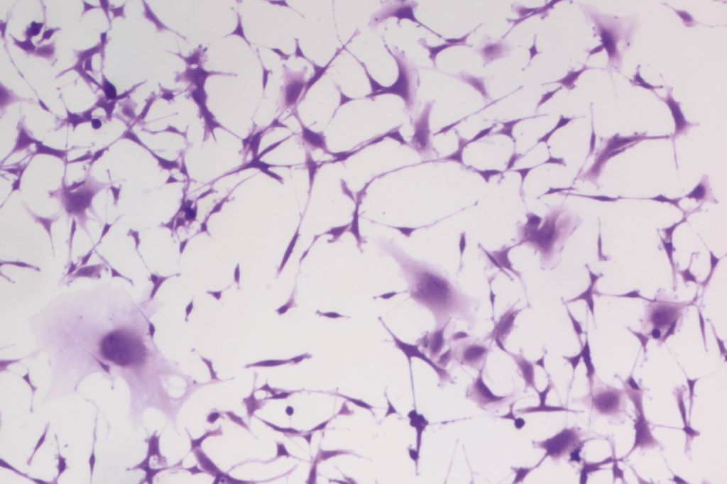
In search for this energy catalyst, Associate Professor Akiko Kojima-Yuasa’s team at Osaka Metropolitan University’s Graduate School of Human Life and Ecology analysed the cinnamic acid ester ethyl p-methoxycinnamate, a main component of kencur ginger, and its mechanism of action. In previous research, the team discovered that ethyl p-methoxycinnamate has inhibitory effects on cancer cells. Furthering their study, the acid ester was administered to Ehrlich ascites tumour cells to assess which component of the cancer cells’ energy pathway was being affected.
Results revealed that the acid ester inhibits ATP production by disrupting de novo fatty acid synthesis and lipid metabolism, rather than through glycolysis as commonly theorised. Further, the researchers discovered acid ester-induced inhibition triggered increased glycolysis, which acted as a possible survival mechanism in the cells. This adaptability was theorised to be attributed to ethyl p-methoxycinnamate’s inability to induce cell death.
“These findings not only provide new insights that supplement and expand the theory of the Warburg effect, which can be considered the starting point of cancer metabolism research, but are also expected to lead to the discovery of new therapeutic targets and the development of new treatment methods,” stated Professor Kojima-Yuasa.
Genetic mutations and cell maturity as key factors in acute myeloid leukaemia drug resistance
Photo by Tima Miroshnichenko on Pexels
An international study led by the University of Colorado Cancer Center has uncovered why a widely used treatment for acute myeloid leukaemia (AML) doesn’t work for everyone. The findings could help doctors better match patients with the therapies most likely to work for them.
Researchers analysed data from 678 AML patients, the largest group studied to date for this treatment, and found that both gene mutations and the maturity of leukaemia cells affect how patients respond to a drug combination of venetoclax and hypomethylating agents (HMA).
“Venetoclax-based therapies are now the most common treatment for newly diagnosed AML,” said Daniel Pollyea, MD, MS, professor of medicine at University of Colorado. “But not all patients respond the same way. Our goal was to figure out why and give doctors better tools to predict outcomes at the start.”
Mutations and maturity of leukaemia cells
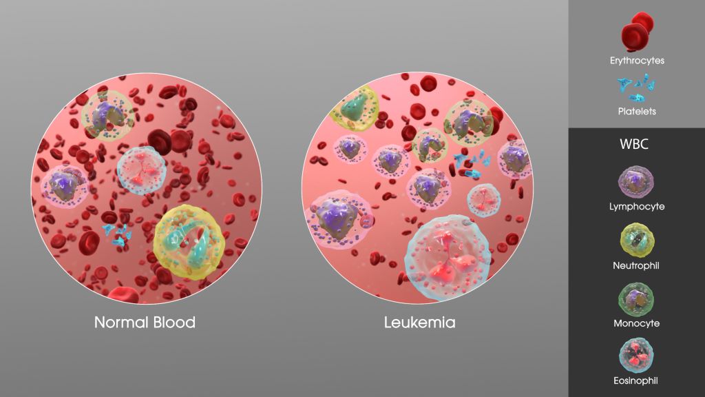
AML is a fast-growing cancer of the blood and bone marrow, most often seen in older adults. Many patients can’t tolerate traditional chemotherapy, so doctors treat them with venetoclax plus HMA. This combination has improved survival for many, but some patients still relapse or don’t respond.
The study found that patients with a certain type of AML, called “monocytic,” had worse outcomes especially if they did not have a helpful gene mutation known as NPM1. These patients were also more likely to carry other mutations, such as KRAS, that are linked to drug resistance.
“Patients with monocytic AML and no NPM1 mutation were nearly twice as likely to die from the disease,” said Pollyea. “So, it’s not just about the gene mutations. It’s also about how developed or mature the cancer cells are when treatment begins.”
Previous research often focused only on either genetic mutations or cell type. Pollyea’s team looked at both, giving them a clearer understanding of how these two factors work together to influence treatment response.
Designing therapies that shut down cancer cell escape routes
“We learned that some cancer cells basically find a back door to evade the treatment,” said Pollyea. “By identifying how and why that happens, we can begin designing therapies that shut down those escape routes.”
This is a powerful new way to classify AML patients by risk, enabling doctors to better predict who is likely to respond to venetoclax and who might need another approach.
“This is a major step toward personalised medicine in AML,” said Pollyea. “We’re moving closer to a world where we can look at a patient’s leukaemia on day one and know which therapy gives them the best chance and ultimately improve survival rates.”
Pollyea and his team are working to expand the study with even more patient data and hope to design a clinical trial that uses this model to guide treatment decisions.
Selective serotonin reuptake inhibitors (SSRIs) could help the immune system fight cancer, according to recent UCLA research. The study, published in Cell, found that SSRIs significantly enhanced the ability of T cells to fight cancer and suppressed tumour growth across a range of cancer types in both mouse and human tumour models.
“It turns out SSRIs don’t just make our brains happier; they also make our T cells happier – even while they’re fighting tumours,” said Lili Yang, PhD, senior author of the new study. “These drugs have been widely and safely used to treat depression for decades, so repurposing them for cancer would be a lot easier than developing an entirely new therapy.”
According to the CDC, one out of eight adults in the US takes an antidepressant, and SSRIs are the most commonly prescribed. These drugs increase levels of serotonin the brain’s “happiness hormone” by blocking the activity of a protein called serotonin transporter, or SERT.
While serotonin is best known for the role it plays in the brain, it’s also a critical player in processes that occur throughout the body, including digestion, metabolism and immune activity.
Dr Yang and her team first began investigating serotonin’s role in fighting cancer after noticing that immune cells isolated from tumours had higher levels of serotonin-regulating molecules. At first, they focused on MAO-A, an enzyme that breaks down serotonin and other neurotransmitters, including norepinephrine and dopamine.
In 2021, they reported that T cells produce MAO-A when they recognise tumours, which makes it harder for them to fight cancer. They found that treating mice with melanoma and colon cancer using MAO inhibitors, also called MAOIs – the first class of antidepressant drugs to be invented – helped T cells attack tumours more effectively.
However, because MAOIs have safety concerns, including serious side effects and interactions with certain foods and medications, the team turned its attention to a different serotonin-regulating molecule: SERT.
“Unlike MAO-A, which breaks down multiple neurotransmitters, SERT has one job – to transport serotonin,” explained Bo Li, PhD, first author of the study and a senior research scientist in the Yang lab. “SERT made for an especially attractive target because the drugs that act on it – SSRIs – are widely used with minimal side effects.”
The researchers tested SSRIs in mouse and human tumour models representing melanoma, breast, prostate, colon and bladder cancer. They found that SSRI treatment reduced average tumour size by over 50% and made the cancer-fighting T cells, known as killer T cells, more effective at killing cancer cells.
“SSRIs made the killer T cells happier in the otherwise oppressive tumour environment by increasing their access to serotonin signals, reinvigorating them to fight and kill cancer cells,” said Dr Yang, who is also a professor of microbiology, immunology and molecular genetics and a member of the UCLA Health Jonsson Comprehensive Cancer Center.
How SSRIs could boost the effectiveness of cancer therapies
The team also investigated whether combining SSRIs with existing cancer therapies could improve treatment outcomes. They tested a combination of an SSRI and anti-PD-1 antibody – a common immune checkpoint blockade (ICB) therapy – in mouse models of melanoma and colon cancer. ICB therapies block immune checkpoint molecules that normally suppress immune cell activity, allowing T cells to attack tumours more effectively.
The results were striking: the combination significantly reduced tumour size in all treated mice and even achieved complete remission in some cases.
“Immune checkpoint blockades are effective in fewer than 25% of patients,” said James Elsten-Brown, a graduate student in the Yang lab and co-author of the study. “If a safe, widely available drug like an SSRI could make these therapies more effective, it would be hugely impactful.”
To confirm these findings, the team will investigate whether real-world cancer patients taking SSRIs have better outcomes, especially those receiving ICB therapies. About 20% of cancer patients are already taking the medication, Dr Yang said.
Dr Yang added that using existing FDA-approved drugs could speed up the process of bringing new cancer treatments to patients, making this research especially promising.
“Studies estimate the bench-to-bedside pipeline for new cancer therapies costs an average of $1.5 billion,” she said. “When you compare this to the estimated $300 million cost to repurpose FDA-approved drugs, it’s clear why this approach has so much potential.”
New research published in the Journal of Magnetic Resonance Imaging has uncovered changes in brain connectivity during chemotherapy in patients with breast cancer.
In the study of 55 patients with breast cancer and 38 controls without cancer, investigators conducted functional magnetic resonance imaging scans of participants’ brains over several months.
Scans from patients revealed changes in brain connectivity, particularly in the frontal-limbic system (involved in executive functions) and the cerebellar cortex (linked to memory) throughout the course of treatment. These changes got worse and spread more as chemotherapy continued.
“The findings suggest that chemotherapy can quickly disrupt brain function in breast cancer patients, potentially contributing to cognitive issues,” the authors wrote.
A new large-scale study co-led by UCLA Health Jonsson Comprehensive Cancer Center investigators provides the strongest evidence yet that a shorter, standard-dose course radiation treatment is just as effective as conventional radiotherapy for early-stage prostate cancer, without compromising the safety of patients.
The shorter approach, known as isodose moderately hypofractionated radiotherapy (MHFRT), delivers slightly higher doses of radiation per session, allowing the total treatment duration to be over four to five weeks instead of seven to eight weeks.
According to the study, patients who received this type of MHFRT had the same cancer control rates as those who received conventional radiotherapy. Additionally, the risk of long-term side effects affecting the bladder and intestines was no higher with MHFRT, confirming its safety.
“We believe these data strongly support that isodose MHFRT should become the preferred standard of care MHFRT regimen for prostate cancer,” said Dr Amar Kishan, executive vice chair of radiation oncology at the David Geffen School of Medicine at UCLA and co-first author of the study. “More broadly, there appears to be little reason to consider conventional radiotherapy over MHFRT for the types of patients enrolled in these trials given these results.”
While MHFRT is now the most commonly used radiotherapy regimen for prostate cancer, concerns remain about whether delivering a higher daily dose increases the risk of urinary and bowel issues, such urinary incontinence, chronic diarrhoea and rectal bleeding.
MHFRT: isodose versus dose-escalated
To better understand whether there might be an increased risk of toxicity with the delivery of a higher dose per day of radiation, Kishan and the team of researchers examined data from more than 5800 patients across seven randomised clinical trials comparing standard therapy with two different MHFRT approaches: isodose MHFRT, which maintains the total radiation dose at a level similar to standard therapy, and dose-escalated MHFRT, which increases the total dose in hopes of enhancing tumour control.
The analysis found patients who received isodose MHFRT (60Gy in 20 fractions) had similar cancer control and side effects compared to those receiving conventional radiation therapy, with no significant difference in the five-year progression-free survival (77.0% for MHFRT vs 75.6% for conventional).
Patients who received higher dose-escalated MHFRT did not improve cancer control when compared to those receiving standard doses, with five-year progression-free survival rates being identical to conventional therapy (82.7% in both groups). Patient-reported outcomes also showed significantly higher gastrointestinal side effects (7.2% vs 4.9%), particularly bowel issues.
While dose-escalated MHFRT was expected to improve outcomes, the data showed no additional benefit in cancer control and a higher risk of gastrointestinal side effects, noted Kishan. This underscores the advantage of isodose MHFRT, which provides the same effectiveness as conventional therapy without increasing toxicity.
“These findings reinforce isodose MHFRT as the standard of care, offering the same cancer control as conventional treatment but with fewer side effects than dose-escalated MHFRT,” said Kishan, who is also a researcher in the UCLA Health Jonsson Comprehensive Cancer Center. “Patients can safely opt for a shorter treatment schedule without compromising their outcomes, ensuring they receive effective care with fewer visits and minimal added risk. Less time in treatment can still mean the best possible results.”
Developed by the University of South Australia in collaboration with the Medical School at Stanford University, this world first formulation uniquely combines limonene (a citrus essential oil) with a lipid-based drug delivery system to treat dry mouth (xerostomia), a common side effect of radiotherapy.
The new formula demonstrated 180-fold better solubility than pure limonene in lab experiments and boosted relative bioavailability by over 4000% compared to pure limonene in pre-clinical trials.
Dry mouth is the most reported side effect following radiotherapy for the treatment of head and neck cancer, affecting up to 70% of patients due to salivary gland damage. It can lead to difficulty speaking and swallowing, significantly reducing quality of life.
Limonene has protective effects on saliva production during radiotherapy, but its poor solubility means high doses are needed to take effect, and these cause indigestion, abdominal discomfort and unpleasant ‘citrus burps’.
Lead researcher, Professor Clive Prestidge says UniSA’s new limonene-lipid combination creates a ‘super-solubilising’ treatment that reduces dry mouth at lower dose and without uncomfortable side effects.
“The therapeutic benefits of limonene are well known. It’s used as an anti-inflammatory, antioxidant, and mood-enhancing agent, and can also improve digestion and gut function. But despite its widespread use, its volatility and poor solubility have limited its development as an oral therapy,” Prof Prestidge says.
“As limonene is an oil, it forms a film on the top of the stomach contents, causing significant stomach pain and discomfort.
“Our novel formulation combines limonene with healthy fats and oils – called lipids – to create a super-solubilising compound that the body can easily absorb with reduced uncomfortable side effects.
“This increases the dispersion of limonene in the stomach, boosts absorption, and controls biodistribution – all while increasing a patient’s saliva production and reducing dry mouth.”
Co-researcher Dr Leah Wright says the formulation has the potential to significantly improve the quality of life for cancer patients and others suffering dry mouth conditions.
“Cancer patients undergoing radiotherapy and other medical treatments regularly experience dry mouth, which not only prevents them from comfortably swallowing, but can also have other negative and potentially life-threatening outcomes,” Dr Wright says.
“While limonene can be ingested directly, it’s not well tolerated, especially by those with dry mouth. Plus, its poor absorption prevents it from effectively reaching the salivary glands – the target site.
“This inventive and highly impactful limonene-lipid formulation could provide a simple, effective oral solution for dry mouth, offering cancer patients long-lasting relief and comfort, improved oral health, and a higher quality of life during a difficult time.”
Clinical trials for the new formula are ongoing, with next steps to be announced soon.
Researchers have uncovered a stealth survival strategy that melanoma cells use to evade targeted therapy, offering a promising new approach to improving treatment outcomes.
The study, published in Cell Systems and conducted by researchers at the Institute for Systems Biology (ISB) and Massachusetts Institute of Technology (MIT) identifies a non-genetic, reversible adaptation mechanism that allows melanoma cells to survive treatment with BRAF inhibitors. By identifying and blocking this early response, researchers proposed a combination therapy that could delay resistance and enhance the effectiveness of existing treatments.
Cracking the Code of Melanoma’s Drug Escape
Melanoma, the deadliest form of skin cancer, is often driven by mutations in the BRAF gene, which fuels uncontrolled tumor growth. While BRAF inhibitors (such as vemurafenib) initially halt tumor growth, many tumors quickly adapt and survive treatment, leading to therapy failure.
Unlike traditional resistance driven by genetic mutations, this study uncovers an early, dynamic adaptation process that occurs within hours to days of drug treatment – long before genetic resistance takes hold. Surprisingly, this process does not rely on reactivating the BRAF-ERK pathway, which is the usual resistance mechanism.
Using cutting-edge mass spectrometry-based phosphoproteomics and deep transcriptomics analyses, researchers mapped the molecular shifts in melanoma cells over minutes, hours, and days of BRAF inhibitor treatment.
“We found that while the BRAF-ERK signaling pathway was quickly and durably suppressed, cancer cells did not rely on reactivating ERK to survive. Instead, they triggered an alternative SRC family kinase (SFK) signaling pathway, which promoted cell survival and eventual recovery,” said Chunmei Liu, PhD, a bioinformatics scientist at ISB and co-first author of the paper.
Turning a Weakness Into a Target
A key discovery in this study came when researchers linked SFK activation to reactive oxygen species (ROS), a cellular stress response that builds up under BRAF inhibition. As ROS levels surged, SFK activity spiked, helping melanoma cells withstand treatment. However, this adaptation was reversible – when treatment was removed, cells returned to their original state.
Recognizing this Achilles’ heel, the team tested a combination approach: pairing BRAF inhibitors with the SFK inhibitor dasatinib.
“By adding dasatinib, we blocked this adaptive escape mechanism, significantly reducing melanoma cell survival and stabilising tumours in animal models,” said ISB Associate Professor Wei Wei, PhD, co-corresponding author.
Importantly, SFK inhibition alone had little effect on melanoma cells, highlighting the need for a strategic combination therapy to suppress melanoma adaptation before resistance fully develops.
“This approach has the potential to prolong the effectiveness of BRAF inhibitors and improve patient outcomes,” said ISB President and Professor Jim Heath, PhD, co-corresponding author.
Looking Ahead: A Path to the Clinic
Beyond uncovering a key mechanism of drug adaptation, this research underscores the importance of early intervention to prevent it from happening. It also highlights ROS accumulation and SFK activation as potential biomarkers for identifying patients who may benefit from this combination therapy.
Further preclinical studies and clinical trials will be necessary to validate this combination therapy strategy and determine its potential for broader clinical use.
Breast cancer cells. Image by National Cancer Institute
Research by UMass Chan Medical School scientists poses a new explanation for how PARP inhibitor drugs attack and destroy BRCA1 and BRCA2 tumour cells. Published in Nature Cancer, this study illustrates how a small DNA nick – a break in one strand of the DNA – can expand into a large single-stranded DNA gap, killing BRCA mutant cancer cells, including drug-resistant breast cancer cells. These findings identify a novel vulnerability that may be a potential target for new therapeutics.
Mutations in BRCA1 and BRCA2, tumour suppressor genes that play a crucial role in DNA repair, substantially increase the likelihood of cancer. These cancers are, however, quite sensitive to anticancer drugs such as poly (ADP-ribose) polymerase inhibitors (PARPi). When successful, these cancer treatments cause enough DNA damage to trigger cancer cell death. However, the array of different damages potentially induced by these drugs makes it difficult to pinpoint the exact cause of cell death. Additionally, PARPi resistance does occur, complicating treatment and leading to recurrent cancer.
“The conventional thinking has been that single-stranded DNA breaks from PARPi ultimately generated DNA double-strand breaks, and that was what was killing the BRCA mutant cancer cells,” said Sharon Cantor, PhD, professor of molecular, cell and cancer biology. “Yet, there wasn’t much in the literature that experimentally confirmed this belief. We decided to go back to the beginning and use genome engineering tools to see how these cells dealt with single-strand nicks to their DNA.”
Using CRISPR technology, Cantor and Jenna M. Whalen, PhD, a postdoctoral researcher in the Cantor lab, introduced small, single strand breaks into several breast cancer cell lines, such as those with the BRCA1 and BRCA2 mutation, as well BRCA-proficient cells. They found that cells with BRCA1 or BRCA2 deficiency were uniquely sensitive to nicks. They also found that breast cancer cells that lose components of the complex that protects DNA from unnecessary DNA end cuts become resistant to chemotherapy drugs such as PARP inhibitors. However, restoring double strand DNA repair functions in breast cancer cells did not save the cells from dying, thus demonstrating that these repair functions are not critical for breast cancer cell survival. Instead, the cells become even more sensitive to single strand nicks, which then accumulate and form large gaps.
“Our findings reveal that it is the resection of a nick into a single-stranded DNA gap that drives this cellular lethality,” said Whalen. “This highlights a distinct mechanism of cytotoxicity, where excessive resection, rather than failed DNA repair by homologous recombination, underpins the vulnerability of BRCA-deficient cells to nick-induced damage.”
The findings suggest that PARPi may also work by generating nicks in BRCA1 and BRCA2 cancer cells, exploiting their inability to effectively process these lesions. For cancers that have developed PARPi-resistance, nick-inducing therapies provide a promising mechanism to bypass resistance and selectively target resection-dependent vulnerabilities.
“Importantly, our findings suggest a path forward for treating PARPi-resistant cells that regained homologous recombination repair: to kill these cells, nicks could be induced such as through ionizing radiation,” said Cantor. “By targeting nicks in this way, therapies could effectively exploit the persistent vulnerabilities of these resistant cancer cells.”
A new study led by researchers from Moffitt Cancer Center, in collaboration with investigators from the University of Michigan, shows that artificial intelligence (AI) can help doctors make better decisions when treating cancer. However, it also highlights challenges in how doctors and AI work together. The study, published in Nature Communications, focused on AI-assisted radiotherapy for non-small cell lung cancer and hepatocellular carcinoma.
Radiotherapy is a common treatment for cancer that uses high-energy radiation to kill or shrink tumors. The study looked at a treatment approach known as knowledge-based response-adaptive radiotherapy (KBR-ART). This method uses AI to optimize treatment outcomes by suggesting treatment adjustments based on how well the patient responds to the therapy.
The study found that when doctors used AI to help decide the best treatment plan, they made more consistent choices, reducing differences between doctors’ decisions. However, the technology didn’t always change doctors’ minds. In some cases, doctors disagreed with the AI suggested and made treatment decisions based on their experience and patient needs.
Doctors were asked to make treatment decisions for cancer patients, first without any technological assistance, and then with the help of AI. The AI system developed by the researchers uses patient data like medical imaging and test results to recommend changes in radiation doses. While some doctors found the suggestions helpful, others preferred to rely on their own judgment.
“While AI offers insights based on complex data, the human touch remains crucial in cancer care,” said Moffitt’s Issam El Naqa, PhD. “Every patient is unique, and doctors must make decisions based on both AI recommendations and their own clinical judgment.”
The researchers noted that while AI can be a helpful tool, doctors need to trust it for it to work well. Their study found that doctors were more likely to follow AI suggestions when they felt confident in its recommendations. “Our research shows that AI can be a powerful tool for doctors,” said Dipesh Niraula, PhD, an applied research scientist in Moffitt’s Machine Learning Department. “But it’s important to recognise that AI works best when it’s used as a support, not a replacement, for human expertise. Doctors bring their expertise and experience to the table, while AI provides data-driven insights. Together, they can make better treatment plans, but it requires trust and clear communication.”
The study’s authors hope that their findings can lead to better integration of AI tools and collaborative relationships that doctors can use to make more personalised treatment decisions for cancer patients. They also plan to further investigate how AI can support doctors in other medical fields.
A fascinating new study, published in the Journal of Clinical Investigation, has revealed an unexpected potential benefit of severe COVID infection: it may help shrink cancer.
This surprising finding, based on research conducted in mice, opens up new possibilities for cancer treatment and sheds light on the complex interactions between the immune system and cancer cells – but it certainly doesn’t mean people should actively try to catch COVID.
The data outlining the importance of the immune system in cancer is considerable and many drugs target the immune system, unlocking its potential, an important focus of my own research.
The study here focused on a type of white blood cell called monocytes. These immune cells play a crucial role in the body’s defence against infections and other threats. However, in cancer patients, monocytes can sometimes be hijacked by tumour cells and transformed into cancer-friendly cells that protect the tumour from the immune system.
What the researchers discovered was that severe COVID infection causes the body to produce a special type of monocyte with unique anti-cancer properties. These “induced” monocytes are specifically trained to target the virus, but they also retain the ability to fight cancer cells.
To understand how this works, we need to look at the genetic material of the virus that causes COVID. The researchers found that these induced monocytes have a special receptor that binds well to a specific sequence of COVID RNA. Ankit Bharat, one of the scientists involved in this work from Northwestern University in Chicago explained this relationship using a lock-and-key analogy: “If the monocyte was a lock, and the COVID RNA was a key, then COVID RNA is the perfect fit.”
Remarkable
To test their theory, the research team conducted experiments on mice with various types of advanced (stage 4) cancers, including melanoma, lung, breast and colon cancer. They gave the mice a drug that mimicked the immune response to a severe COVID infection, inducing the production of these special monocytes. The results were remarkable. The tumours in the mice began to shrink across all four types of cancer studied.
Unlike regular monocytes, which can be converted by tumours into protective cells, these induced monocytes retained their cancer-fighting properties. They were able to migrate to the tumour sites – a feat that most immune cells cannot accomplish – and, once there, they activated natural killer cells. These killer cells then attacked the cancer cells, causing the tumours to shrink.
This mechanism is particularly exciting because it offers a new approach to fighting cancer that doesn’t rely on T cells, which are the focus of many current immunotherapy treatments.
While immunotherapy has shown promise, it only works in about 20% to 40% of cases, often failing when the body can’t produce enough functioning T cells. Indeed it’s thought that the reliance on T cell immunity is a major limitation of current immunotherapy approaches.
This new mechanism, by contrast, offers a way to selectively kill tumours that is independent of T cells, potentially providing a solution for patients who don’t respond to traditional immunotherapy.
It’s important to note that this study was conducted in mice, and clinical trials will be necessary to determine if the same effect occurs in humans.
Maybe aspects of this mechanism could work in humans and against other types of cancer as well, as it disrupts a common pathway that most cancers use to spread throughout the body.
While COVID vaccines are unlikely to trigger this mechanism (as they don’t use the full RNA sequence as the virus), this research opens up possibilities for developing new drugs and vaccines that could stimulate the production of these cancer-fighting monocytes.
Few would have imagined that there’d be an upside to COVID. Photo by Kelly Sikkema on Unsplash
Trained immunity
The implications of this study extend beyond COVID and cancer. It shows how our immune system can be trained by one type of threat to become more effective against another. This concept, known as “trained immunity”, is an exciting area of research that could lead to new approaches for treating a wide range of diseases.
However, it’s crucial again to emphasise that this doesn’t mean people should seek out COVID infection as a way to fight cancer, and this is especially dangerous as I have described. Severe COVID can be life-threatening and has many serious long-term health consequences.
Instead, this research provides valuable insights that could lead to the development of safer, more targeted treatments in the future. As we continue to grapple with the aftermath of the COVID pandemic, new infections and long COVID, studies like this remind us of the importance of basic scientific research.
Even in the face of a global health crisis, researchers are finding ways to advance our understanding of human biology and disease. This work not only helps us combat the immediate threat of COVID, but also paves the way for breakthroughs in treating other serious conditions such as cancer.
While there’s still much work to be done before these findings can be translated into treatments for human patients, this study represents an exciting step forward in our understanding of the complex relationship between viruses, the immune system and cancer. It offers hope for new therapeutic approaches and underscores the often unexpected ways in which scientific discoveries can lead to medical breakthroughs.