Cancer is one of the fastest-growing health challenges in South Africa, with over 100 000 new cases diagnosed annually, according to the National Cancer Registry. While most conversations focus on the physical and emotional impact, the financial strain of the disease often goes unspoken.
According to the South African Medical Journal, treatment costs for cancer can exceed R1 million per year, particularly when advanced therapies and prolonged care are required. This leaves many families facing difficult decisions that extend far beyond the hospital ward. With medical aid often falling short and with only 16% of South Africans covered by medical schemes, as reported by the Council for Medical Schemes the financial burden of cancer can be as devastating as the diagnosis itself.
“Medical aid alone often isn’t enough to cover the full cost of treatment, especially when it comes to critical illnesses like cancer,” says Matthew Green, Product Portfolio Manager at FNB Life. “We’ve seen firsthand how having the right insurance can make a real difference.”
The true impact of cancer is often measured in rands and cents: savings depleted, debt accumulated, and households forced to sacrifice essentials to pay for treatment. Myths about affordability and a lack of awareness mean that too many people enter this battle unprepared. The result is a financial shock that can be as devastating as the diagnosis itself. Beyond the direct medical expenses, families often face a range of additional costs from transport to and from treatment centres, specialised nutrition, home modifications, and caregiving support, to lost income due to time off work. Critical illness cover is designed to help bridge these financial gaps, providing a lump-sum payout that can be used not only for treatment, but also for these broader, often overlooked expenses that impact the entire household.
“Against this backdrop, insurers are under growing pressure to offer support that reflects the lived realities of ordinary South Africans. FNB Insure is among those stepping in to help close the gap – focusing on making financial protection more accessible, flexible, and relevant to everyday needs,” says Green.
Rather than positioning insurance cover as a luxury, the emphasis is on practical tools that help households navigate the rising costs of treatment and the economic strain that often follows a serious diagnosis. Whether it’s support during hospital stays, assistance with unexpected medical shortfalls, or a payout that enables immediate action after a diagnosis, the goal is to empower customers to focus on recovery – not financial survival.
This is evident from our customer feedback, where individuals have shared how timely access to cover helped them act quickly, avoid financial delays, and prioritise their health during some of life’s most difficult moments. “And its stories like this underline the importance of early financial planning and the role of accessible insurance in giving families space to focus on recovery rather than financial survival,” says Green.
With October marking Breast Cancer Awareness Month and November bringing the spotlight on men’s health through initiatives like Movember, FNB Insure is adding its voice to the broader call for awareness. “The message is clear: cancer doesn’t only affect health; it reshapes every aspect of life. Building resilience means preparing not just medically, but financially too,” concludes Green.
Study of 2,800 patients suggests moving beyond chronological cut-offs in cancer care
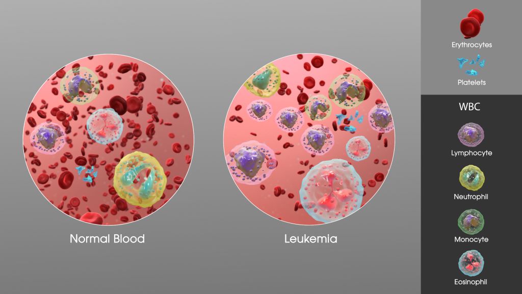
SAG Leukaemia. Credit: Scientific Animations CC0
An international study conducted by the Alliance for Clinical Trials in Oncology and the Acute Myeloid Leukemia Cooperative Group reveals that age-based classifications in the treatment of acute myeloid leukaemia (AML) may be outdated and overly simplistic.
AML is a fast-growing cancer of the blood and bone marrow that disproportionately affects older adults. Historically, age has been a key factor in determining treatment intensity, eligibility for clinical trials, and access to targeted therapies. However, this new research suggests that age alone is not a reliable indicator of disease biology or prognosis.
“Our findings support a more flexible, biology-driven approach to AML treatment and trial design. Age alone should not be a gatekeeper to potentially life-saving therapies,” said Alliance researcher and lead author Ann-Kathrin Eisfeld, MD, associate professor of Internal Medicine and director of the Clara D. Bloomfield Center for Leukemia Outcomes Research at The Ohio State University. “Our results suggest reconsidering age-based eligibility criteria for treatments. By focusing on molecular and genetic profiles rather than chronological age, clinicians may better tailor treatments to individual patients, improving outcomes and expanding access to novel therapies.”
Published in Leukemia, the study analysed data from 2823 adult AML patients treated in the setting of large cooperative group frontline trials across the United States (CALGB/Alliance) and Germany (AMLCG), uncovering nuanced age-related trends in genetic mutations and survival outcomes that challenge current clinical practices. This research is the first large-scale, cross continental study to analyse the mutational patterns and outcomes among patients of all age groups with AML.
The analysis included patients treated with frontline cytarabine-based chemotherapy between 1986 and 2017. Molecular profiling was conducted using targeted sequencing platforms, and survival outcomes were assessed using the 2022 European LeukemiaNet (ELN) genetic-risk classification.
The study found no clear age threshold that could biologically or prognostically separate patients into distinct groups. Instead, genetic mutations and survival outcomes varied continuously across the age spectrum. This challenges the long-standing practice of using arbitrary age cut-offs, such as 60 or 65 years, to guide treatment decisions.
Survival outcomes also declined steadily with age, even among patients classified as having favourable genetic risk. For example, patients aged 18 to 24 with favourable-risk AML had a five-year overall survival rate of 73%, while those aged 75 and older had a survival rate of just 21%. This trend was consistent across all risk categories, indicating that age negatively impacts prognosis regardless of genetic classification.
“This research arrives at a critical moment in oncology, as precision medicine continues to transform cancer care,” added Dr Eisfeld. “Most targeted treatment options are still only available for patients above a certain age threshold that is dictated by corresponding inclusion criteria of pivotal clinical trials, even though patients outside of that age range might equally benefit from these often less toxic treatments.”
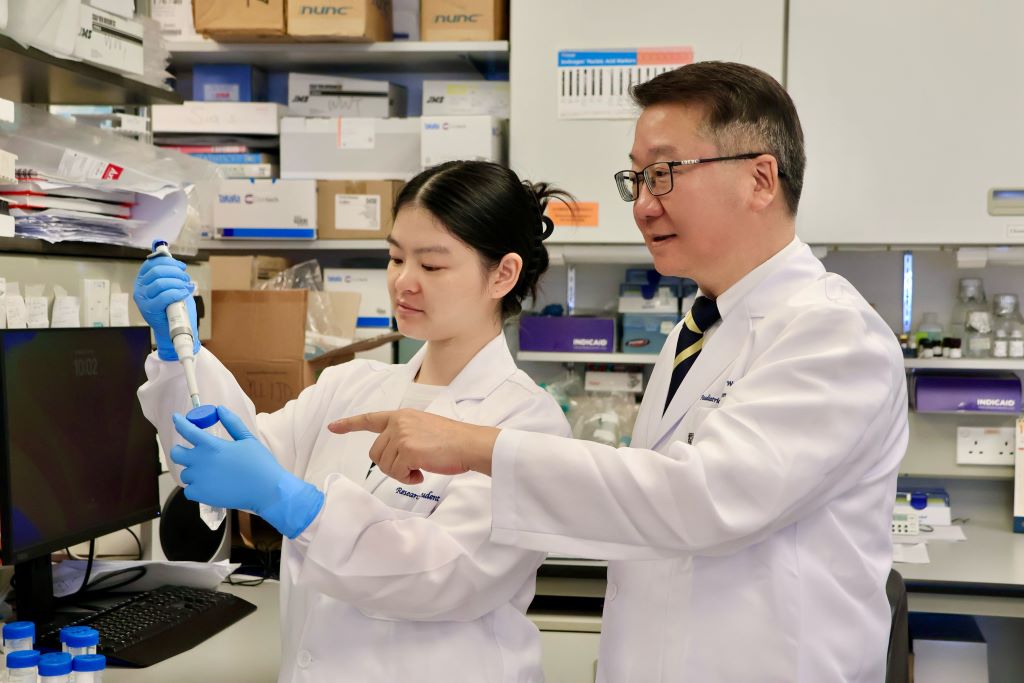
A research team at the LKS Faculty of Medicine of the University of Hong Kong (HKUMed) discovered that certain dietary fatty acids can supercharge the human immune system’s ability to fight cancer. The team found that a healthy fatty acid found in olive oil and nuts, called oleic acid (OA), enhances the power of immune γδ-T cells, specialised cells known for their cancer-fighting properties.
Conversely, they found that another fatty acid, called palmitic acid (PA), commonly found in palm oil and fatty meats, diminishes the ability of these immune cells to attack tumours. This groundbreaking study, published in the academic journal Signal Transduction and Targeted Therapy, offers an innovative approach using dietary OA supplementation to strengthen the antitumour immunity of γδ-T cells.
Dietary fatty acids and cancer immunotherapy
Dietary fatty acids are essential for health, helping with growth and body functions. They may also play a role in cancer prevention and treatment, but understanding how they affect cancer is challenging because of the complexity of people’s diets and the lack of detailed studies. Recently, scientists have learned that fatty acids can influence the immune system, especially in how it fights cancer. Specialised immune cells, called γδ-T cells, are particularly good at attacking tumours. These cells, once activated, have helped some lung and liver cancer patients live longer. However, this therapy is not effective for all patients, partly because the variation of the metabolic status, such as fatty acid metabolism, can influence its efficacy in the patients.
Oleic acid may improve cancer treatment outcomes
The research team identified a correlation between PA and OA levels and the efficacy of cancer therapies. ‘Our research suggests that dietary fatty acid supplementation, particularly with foods rich in OA, such as olive oil and avocados, could enhance γδ-T cell immunosurveillance, leading to more effective cancer treatments,’ said Professor Tu Wenwei from the Department of Paediatrics and Adolescent Medicine, School of Clinical Medicine, HKUMed, who led the study.
The team also discovered that another fatty acid, called PA, can weaken these immune cells and how OA can counteract this. ‘The results indicate that cancer patients should avoid PA and consider OA supplementation in their diets to improve clinical outcomes of γδ-T cell-based cancer therapies,’ added Professor Tu.
Significant impact from simple dietary changes
Professor Tu said, ‘This study is the first to show that the fatty acids we eat can directly affect how well our immune cells fight cancer.’ It reveals how PA can harm these cells and how OA helps them through a specific process involving a protein called IFNγ. By analysing blood samples, the researchers confirmed that the levels of these fatty acids are linked to the outcome of cancer immunotherapy.
‘For cancer patients, this discovery suggests simple changes, like eating more foods rich in OA (such as olive oil, avocados and nuts) and cutting back on PA (found in processed foods, palm oil and fatty meats), could improve the effectiveness of cancer treatments. The study also points to novel strategies, like combining dietary changes with specific drugs to further boost the immune system,’ added Professor Tu.
This study demonstrates that personalised nutrition may serve as an effective strategy to enhance immune function and support cancer treatment. It also suggests that new drugs targeting the processes affected by these fatty acids could enhance the power of γδ-T cell therapies. By integrating nutritional interventions with immunotherapy, this discovery could help more cancer patients achieve better outcomes.
Researchers at Children’s Hospital of Philadelphia (CHOP) found that combining a specialised diet with an approved medication interrupts the growth of high-risk neuroblastoma, a deadly paediatric cancer, by reprogramming tumour behaviour. The findings were published in the journal Nature.
Neuroblastoma originates from primitive cells meant to form nerve tissues but that remain “undifferentiated,” indicating cancer cells that haven’t specialized, often suggesting a more aggressive and unfavourable prognosis. These tumours rely on a steady supply of chemicals called polyamines that are essential for rapid cell growth and tumour progression. A medicine called difluoromethylornithine (DFMO) was approved by the Food and Drug Administration (FDA) to treat children with high-risk neuroblastoma, as DFMO blocks polyamine production. However, researchers sought to improve the effectiveness of the drug by using it at high doses and combining it with a diet that is depleted of the nutrients used by the body to make polyamines (arginine). This two-step approach was anticipated to lower polyamines substantially more than low dose DFMO alone.
“Our findings show that this treatment reduced polyamines in tumours to roughly 10% of their usual levels. This reduction greatly slowed tumour growth, and in many cases, completely eliminated the tumours,” said Michael D. Hogarty, MD, a lead author and an Attending Physician in the Division of Oncology at Children’s Hospital of Philadelphia. “Notably, the treatment altered the way the tumour cells make proteins, making it harder for them to grow and easier for them to mature, or differentiate.”
Hogarty and his team used a preclinical model to mimic MYCN-driven neuroblastoma, directly addressing the strong association between extra MYCN gene copies and aggressive neuroblastoma with poor prognosis. Animal models with tumours were divided into groups: one fed a normal diet and the other lacking amino acids for polyamine production. Each group either received DFMO in their drinking water or did not. The special diet or DFMO alone partially lowered polyamines and extended survival, but the combination had the most significant impact on tumours due to the profound polyamine depletion it caused.
The researchers plan to conduct additional preclinical studies, followed hopefully by clinical trials in children to determine the safety and efficacy of targeting this specific metabolic dependency of neuroblastoma cells. By complementing existing treatments, they hope to substantially improve patient outcomes, and because the therapy targets polyamines it may be effective in many other types of cancer that have frequent MYC gene activation.
Restorative programme helps post-cancer treatment patients regain hair, confidence, and quality of life after facing cancer
Photo by Natasha Brazil on Unsplash
The Cancer Association of South Africa (CANSA) has partnered with internationally renowned hair restoration clinic Alvi Armani South Africa, with head offices in Beverly Hills Los Angeles, to launch an initiative offering complimentary consultations and assessments to those recovering or recovered from cancer.
For many, completing cancer treatment is an experience that brings immense relief. However, it doesn’t always mark the end of the emotional journey. While chemotherapy and radiation often save lives, they can leave lasting reminders – and hair loss is among the most visible.
Cindy Pretorius, a cancer survivor who was diagnosed with basal cell carcinoma, an invasive skin cancer knows firsthand how the impact of the disease affects not just self-confidence but self-worth. After the cancer was removed, the surgery left lasting and visible scarring on her hairline. A hairline that was subsequently treated and restored through a minimally invasive hair transplant at Alvi Armani South Africa. “The team at Alvi Armani restored not only my hairline, but also my confidence,” said Pretorius.
Launching in August 2025, the initiative will offer CANSA-affiliated patients in recovery access to complimentary, in-depth, and personalised consultations. This may include scalp density and mapping assessments, as well as checks for lingering treatment effects. Where needed, survivors will receive advice and support with restorative hair treatments or transplants at Alvi Armani South Africa – offering significant financial relief and a renewed sense of hope.
“This isn’t about vanity. It’s about healing the whole person,” notes Dr Kashmal Kalan, Medical Director of Alvi Armani South Africa. “Unfortunately, even when cancer treatments end, the physical and emotional recovery continues. Many individuals in remission are confronted with reminders every time they look in the mirror and see someone who still looks like a patient, often making it difficult to reconnect with the person they were before cancer.”
For those recovering from cancer, the devastation of hair loss can continue to weigh heavily on their mental well-being. Studies show that persistent thinning, patchiness, or recession after treatment can fuel anxiety, depression, and social withdrawal. Even when remission is achieved, hair regrowth can be slow, and this gap between survival and self-image can take a heavy toll.
“Hair plays an important role in how we express identity; by restoring it, we help people feel like themselves again – more confident to re-enter public life, apply for jobs, or socialise without feeling marked by illness,” he explains.
In cases where hair loss is permanent, transplants using Alvi Armani’s minimally invasive Vitruvian or Maximus follicular unit extraction (FUE) technique may also be performed. Recognised as global leaders in hair transplant procedures, Alvi Armani’s network – spanning Beverly Hills, Salt Lake City, Phoenix, San Diego, Buenos Aires, Montevideo, and Johannesburg – all use state of the art protocols, ensuring that South African patients receive the same world-class standard of care they would get at any other Alvi Armani clinic globally.
“People who’ve overcome cancer deserve more than just a life saved. They deserve the chance to live it fully, with confidence and joy. We’re extremely proud to walk this journey with them, and to help them reclaim their full sense of self.”
Alvi Armani are committing extensive financial and medical resources to support the initiative. A patient referral and screening process is in place to ensure clinical suitability, but any CANSA-affiliated person in remission may apply directly and will be guided accordingly.
CANSA and Alvi Armani will also collaborate at national events such as CANSA Relay For Life, and the CANSA High Tea, where participants will receive expert advice on scalp health, treatment options, and realistic expectations around regrowth.
“When you’ve fought so hard to stay alive, the last thing you want is to be reminded daily of what you lost. This partnership is ultimately about giving people that final piece of the puzzle back, so they can look in the mirror and not only see what they’ve overcome, but truly see themselves again,” concludes Dr Kalan.
“At CANSA, we understand that the cancer experience doesn’t end with treatment – healing also means restoring dignity, self-confidence, and quality of life. Our partnership with Alvi Armani South Africa reflects our commitment to holistic survivorship care. By offering complimentary consultations and access to world-class restorative hair solutions, we’re helping survivors reclaim not only their appearance but also their sense of self,” says Makoma Raolane, CANSA’s Sustainability Manager.
Individuals affected by cancer who are interested in the initiative can contact Alvi Armani South Africa directly, referencing their affiliation with CANSA, to schedule a complimentary consultation.
Credit: Darryl Leja National Human Genome Research Institute National Institutes Of Health
Most men who are treated for prostate cancer according to modern guidelines have good survival rates and the majority of these men will die of causes other than prostate cancer. This is revealed in a new study from Uppsala University published in the Journal of the National Comprehensive Cancer Network.
“We were surprised by how much life expectancy affected the prognosis. This shows the importance of a thorough assessment of the general health of a man with newly diagnosed prostate cancer. The patient’s life expectancy has a substantial impact on the choice of appropriate treatment strategy,” says Marcus Westerberg, researcher at the Department of Surgical Sciences at Uppsala University, who led the study.
In prostate cancer, the disease progression often takes decades and the risk of dying from prostate cancer therefore depends on both the characteristics of the cancer and life expectancy based on the man’s age and other diseases at the time of diagnosis. Recommendations in guidelines and care programmes are therefore also based on both cancer characteristics and life expectancy. This means that the recommended initial treatment can range from active monitoring for low-risk cancer to combinations of local and systemic treatment for high-risk cancer.
High average age at disease onset
As the average age at diagnosis of prostate cancer is often high and the cancer often progresses very slowly, it is particularly important to know the long-term risk of death from prostate cancer in order to choose the best treatment for patients. Previously, not much has been known about this.
“We wanted to fill that knowledge gap, so we looked at outcomes up to 30 years after the men were diagnosed. In all cases, we had information about the characteristics of the cancer, treatment and the patient’s life expectancy based on age and comorbidity,” says Westerberg.
The researchers used data from the Prostate Cancer Database Sweden (PCBase), which contains information from the National Prostate Cancer Register (NPCR) and other health data registers. They focused on men who had received the recommended treatment for prostate cancer that had not spread in the body. Using statistical modelling, the researchers estimated the lifetime risk of dying from prostate cancer and other causes.
11 per cent risk of dying of cancer
For men with low-risk cancer and short life expectancy (less than 10 years), the risk of dying from prostate cancer was 11% and the risk of dying from other causes was 89% within 30 years of diagnosis.
For men with high-risk cancer (eg stage T3, PSA 30ng/mL and Gleason score 8) and long life expectancy (over 15 years), the risk of dying from prostate cancer was 34% and the risk of dying from other causes was 55% within 30 years of diagnosis.
“We hope that our results will be used to provide a realistic picture of the prognosis for men with prostate cancer. Our study shows that most men who receive the recommended treatment have a good prognosis,” Westerberg concludes.
Life expectancy was based on age and comorbidity. Examples of low-risk cancers are stage T1, PSA 5ng/mL and Gleason score 6. Examples of high-risk cancers are stage T3, PSA 30ng/mL and Gleason score 8.
Scientists at The Wistar Institute have discovered that a class of FDA-approved cancer drugs known as PARP1 inhibitors can effectively combat Epstein-Barr virus (EBV)-driven lymphomas. The findings, published in the Journal of Medical Virology, demonstrate that these drugs, which work by blocking the activity of the PARP1 enzyme, can halt tumour growth by interfering with the EBV’s ability to activate key cancer-promoting genes.
“We’ve uncovered a completely different mechanism for how PARP inhibitors work in EBV-positive cancers,” said Italo Tempera, PhD, associate professor at Wistar’s Ellen and Ronald Caplan Cancer Center and senior author of the study. “Instead of preventing DNA damage from repairing itself in the tumours, like these drugs do in other cancers, they essentially cut off the virus’s ability to hijack cellular machinery to drive cancer growth. This opens up exciting possibilities for repurposing existing FDA-approved drugs to treat EBV-associated cancers.”
EBV infects over 90% of the global population. While most people with the virus remain symptom-free, immunocompromised individuals such as people with HIV and transplant recipients have an increased risk of EBV causing several types of cancer, including various lymphomas and carcinomas. Despite the virus’s clear role in driving these malignancies, no specific therapies currently target EBV-driven cancer.
In search of such a therapy, Tempera and his research team focused on PARP1, a cellular protein that is known primarily for its role in DNA repair. In cancer treatment, PARP inhibitors typically work by preventing cancer cells from repairing their DNA, causing them to die. However, Tempera’s team had previously discovered that PARP1 plays a very different role in EBV infection: It helps control which genes are accessible and active, essentially acting as a master regulator of gene expression.
“Think of PARP1 as a key that opens up DNA to make certain genes readable,” explained Tempera. “EBV uses this key to unlock cancer-promoting genes. When we block PARP1, we’re essentially taking away the key so the virus can’t get in and use our DNA for its own purposes.”
Using a mouse model of EBV-driven lymphoma, the researchers treated the animals with BMN 673 (talazoparib/talzenna), a PARP inhibitor that has already been approved for breast cancer treatment. Compared to controls, the treated mice showed an 80% reduction in tumour growth, and the cancer’s ability to spread to other organs was significantly reduced. Further, when the team analysed the tumours, they found no increase in DNA damage in the treated animals – the hallmark of how PARP inhibitors typically work. Instead, they discovered that PARP1 inhibition disrupted a critical partnership between the viral protein EBNA2 and the cellular oncogene MYC.
“EBNA2 is like the conductor of an orchestra, directing cellular genes to play a cancer symphony,” said Tempera. “It specifically turns on MYC, which is one of the most important cancer-promoting genes. When we inhibit PARP1, EBNA2 can’t effectively activate MYC anymore, and the whole cancer program falls apart.”
The findings have significant therapeutic implications. Because PARP inhibitors are already FDA-approved and their safety profiles are well established, the path to clinical application could be accelerated compared to developing entirely new drugs.
The research also suggests this approach might work beyond EBV-associated lymphomas. The team is now investigating whether PARP inhibitors could be effective against other EBV-driven cancers, including nasopharyngeal and gastric carcinomas. Additionally, given EBV’s suspected role in autoimmune diseases, the researchers are exploring whether PARP1’s regulation of viral gene expression might contribute to these conditions.
“This work really showcases the power of understanding fundamental viral biology,” said Tempera. “We’re taking insights from basic virology research and translating them into potential therapies. With further development, this approach could provide new hope for patients with EBV-associated cancers who currently have limited treatment options.”
Pretoria, 15 July 2025 – The South African Health Products Regulatory Authority (SAHPRA) was notified of the Lancet Global Health 2025; 13: e1250, an investigational study and its findings on substandard anti-cancer medications in Sub-Saharan African countries, including Ethiopia, Kenya, Malawi, and Cameroon. This study did not include South Africa. The seven (7) medicines/dosage forms mentioned in the study are cisplatin, oxaliplatin, methotrexate, doxorubicin, cyclophosphamide, ifosfamide, and leucovorin. The specific brands mentioned/shown in the article are neither registered nor marketed in South Africa.
SAHPRA, in terms of the Medicines and Related Substances Act 101 of 1965, as amended, and its General Regulations, requires medicines marketed in the country to meet prescribed requirements and adhere to set standards. Every batch of medicine produced must undergo testing to ensure that the integrity of the product is consistent with approved specifications before the release for sale, and imported medicines must additionally comply with the Guideline for Post-Importation Testing.
SAHPRA commenced internal processes to verify whether any of the South African-registered cancer medicines with the mentioned Active Pharmaceutical Ingredients (API) might have been affected or implicated. The cancer products registered and marketed in South Africa were not implicated/affected by the investigational study and its findings on substandard anti-cancer medicines. SAHPRA conducts risk-based post-market surveillance (PMS), sampling, and testing on high-risk medical products.
SAHPRA is satisfied that the marketed and registered cancer medicines meet the appropriate specifications; therefore, no substandard cancer medicines were detected.
“SAHPRA is committed to the three pillars of quality, safety, and efficacy. I am satisfied that our rigorous regulatory processes have borne fruit and that all patients, especially cancer patients, can rest assured that their health and well-being are not compromised,” indicated SAHPRA CEO, Dr Boitumelo Semete-Makokotlela.
Serious quality defects were found in a significant number of cancer medications from sub-Saharan Africa, according to new research from the University of Notre Dame.
For the study published in The Lancet Global Health, researchers collected different cancer medications from Cameroon, Ethiopia, Kenya and Malawi and evaluated whether each drug met regulatory standards. Researchers considered a variety of factors, including appearance, packaging, labelling and, most importantly, the assay value.
The assay value is the quantity of active pharmaceutical ingredient (API) found in each drug. To meet safety standards, most products should be within a range of 90 to 110% of the right amount of API. Researchers measured the API content of each product and compared that number to what was designated on the medication packaging.
“It is important that cancer medications contain the right amount of the active ingredients so the patient gets the correct dose,” said Marya Lieberman, professor of chemistry and biochemistry at Notre Dame and lead author of the study. “If the patient’s dose is too small, the cancer can survive and spread to other locations. If the patient’s dose is too high, they can be harmed by toxic side effects from the medicine.”
One in six cancer medications tested was found to contain the incorrect quantity of API, with tested medications having APIs ranging from 28 to 120%. The study evaluated 251 samples of cancer medications collected from major hospitals and private markets in all four countries.
The study, funded by the National Cancer Institute of the National Institutes of Health, is among the first to evaluate cancer drug quality in sub-Saharan Africa. Currently, sub-Saharan Africa has no pharmaceutical regulatory laboratories carrying out chemical analyses for cancer drugs according to the standards required for regulatory purposes.
Yet, the need for cancer drugs is growing.
“We found bad-quality cancer medications in all of the countries, in all of the hospital pharmacies and in the private markets,” said Lieberman, an affiliate of Notre Dame’s Eck Institute for Global Health and Harper Cancer Research Institute. “We learned that visual inspection, which is the main method for detecting bad-quality cancer drugs in sub-Saharan Africa today, only found one in 10 of the bad products.”
In their study, the researchers explained how a combination of high demand for cancer medications, lack of regulatory capacity, and poor manufacturing, distribution and storage practices likely created a problematic environment throughout sub-Saharan Africa. They also argue that given these factors and the global supply chain for pharmaceuticals, substandard cancer medications are likely present in other low and middle-income countries as well.
Lieberman and her team identified several strategies that could help the global community address poor-quality cancer medications:
Provide inexpensive technologies at the point of care to screen for bad-quality cancer medicines and create policies for how to respond to products that fail screening tests.
Help regulatory agencies in low and middle-income countries get safety equipment and training so they can analyze the quality of cancer medicines in their markets, conduct root-cause investigations when products fail testing, take quick regulatory actions enabled by lab data and share data about bad-quality products.
Perform cost-benefit analyses of interventions that tackle common problems (such as medications being out of stock, unsafe shipping, storage or dispensing practices, and lack of availability or affordability of medications) to help policymakers and funders get the most impact on patient outcomes from their available resources.
Work with care providers to develop site-specific response policies and messaging for patients and engage regulators, donors and other resources.
Lieberman and her lab are developing a user-friendly technology called the chemoPAD for screening cancer medications. This low-cost paper device could potentially help hospitals, pharmacies and health care professionals in low and middle-income countries monitor drug quality without restricting a patient’s access to the medication.
“This is all part of a bigger project aimed at developing the ChemoPAD as a point-of-care testing device that we can use, something that’s more accurate in detecting poor-quality products than just visual inspection,” Lieberman said.
“There are lots of medicines where the regulators don’t have enough resources to verify the quality, and some manufacturers take advantage of that to cut corners. There are also problems with distribution systems, so even if a product is good quality when it leaves the manufacturer, it may be degraded during shipping or storage. These products flow into low and middle-income countries, and they get used on patients. I want to change that.”
A cancer patient receiving care at a public health facility in Gauteng. (Photo: Rosetta Msimango/Spotlight)
By Chris Bateman
Experts say cancer patients in the public sector in South Africa are dying for avoidable reasons like dysfunctional referral systems and a lack of medical imaging and treatment. We look at efforts to get the country’s battle with cancer back on track.
Many people with cancer in Gauteng have not been able to access the treatment and care they require in recent years. Though activists and the provincial government are at odds about what should, or should have been, done about it, nobody is denying that there is a problem.
At the same time, there have also been issues at a national level, with South Africa’s key cancer strategy having lapsed. The National Cancer Strategic Framework for South Africa 2017 – 2022 was previously extended to also cover 2023. Medical Brief recently reported that a new strategy is on the verge of being signed by the Director-General of Health.
The committee meant to advise the minister on cancer has also lapsed. Dr Busisiwe Ndlovu, the top government official in charge of non-communicable diseases (NCDs), said that the term of the Ministerial Advisory Committee on Cancer expired in early 2024, and new members were pending the approval of Health Minister Dr Aaron Motsoaledi. She was speaking at the KwaZulu-Natal leg of a cancer research and innovation strategy workshop in May. These consultative meetings are taking place across the country’s provinces. It aims to shape a national research and innovation strategy based on the World Health Organization’s cancer control pillars: prevention, early detection and diagnosis, treatment, and palliative care and survivorship.
The scale of the problem
While researchers anticipate that rates of infectious diseases like HIV and tuberculosis in South Africa will decline in the coming decade or two, rates of NCDs, including diabetes and cancers, are expected to increase. According to the WHO, an estimated one in five people will develop some form of cancer in their lifetimes. Increases in developing countries are expected to be particularly steep.
According to a StatsSA report published in 2023, and based on National Cancer registry (NCR) numbers and StatsSA’s mortality data, cancer-related deaths in the country increased by 29% from 2008 to 2018. They reported that 85 000 people were diagnosed with cancer in 2019 and that 44 000 died of cancers in 2018. Experts previously told Spotlight that the estimate of cancer cases may be an undercount of as much as 40%.
The most common cancers in men were prostate, colorectal, and lung – around one in four cancer diagnoses in men were for prostate cancer. Bronchus and lung cancer accounted for just under 19% of cancer-related deaths in men, while prostate cancer accounted for around 17%.
Among women, the most diagnosed cancers were breast cancer at around 23% of diagnoses and cervical cancer at around 16% of diagnoses. Cervical cancer accounted for just under 18% of all cancer deaths in women and breast cancer for 17%.
The NCR recorded 87 853 new laboratory-confirmed cancer cases in 2023, although this figure likely underestimated the true burden as it excluded clinically or radiologically diagnosed cancers, Dr Judith Mwansa-Kambafwile, senior epidemiologist with the NCR told attendees at the Durban workshop.
In a paper published in the South African Journal of Oncology in 2022, researchers calculate that cancer incidence (new cases per year) in South Africa could double from around 62 000 in 2019 to 121 000 in 2030. This is due to two factors: firstly, South Africa’s population is aging and cancers generally become more common as people age. And secondly, the risk of cancers is generally increasing for people of all ages. The researchers focused on only the five most common types of cancer, but an NCR report shows a very wide variety of cancers are being diagnosed in the country.
Since not all cancers are diagnosed, the real numbers are likely substantially higher than reported. There is also no single repository of all cancer diagnoses in the country – for the above quoted article researchers used both data from Discovery Health Medical Scheme and from the NCR.
The data gap
Cancer statistics in South Africa has been largely based on pathology results, which is to say blood or biopsies that were tested in the lab. Other types of diagnoses, such as those based on symptoms and scans have not always been counted systematically. One recent initiative aimed at addressing this data gap is a patient-led registry that feeds information into the NCR.
Mwansa-Kambafwile, explained that the NGO, Living with Cancer, was driving the patient-led registry, aimed at cross referencing and supplementing patient records with her NCR’s own patient database. Leaflets in oncologists’ reception rooms encouraged patients to upload their pathology/histology test results onto the Living with Cancer website via a standard online National Department of Health form. A national shopping mall campaign in May was aimed at boosting awareness.
“Living with Cancer had a Memorandum of Understanding with us and in addition, links cancer survivors with the same type of cancer to one another in support groups online where they can share experiences and knowledge,” she added.
Dr Mazvita Muchengeti, who heads up work on the NCR at the National Health Laboratory Services which is part of the National Institute for Communicable Diseases (NICD), previously told Spotlight that cancer was made a reportable disease under the National Health Act in 2011. While compulsory reporting has improved data on cancer cases, she added: “There is an increase in the number of reported cancers; this does not necessarily translate to an increase in cancer, we are just counting cancer cases better because reporting is now compulsory.”
Another new strategy
In light of the country’s cancer burden, a group of organisations is leading the development of a new National Cancer Research and Innovation Strategy. This collective includes the Nuclear Medicine Research Infrastructure at the University of Pretoria, the South African Medical Research Council, and the Department of Science, Technology and Innovation, in partnership with the National Department of Health.
They are hosting provincial workshops to help understand the current state of cancer research in South Africa, identify key challenges, set national priorities, and develop a strong, future-focused strategy. These workshops are part of a broader plan to make sure the strategy is inclusive, based on evidence, and meets the country’s needs.
This research and innovation strategy differs from the health department’s National Cancer Strategic Framework, which guides provinces as to what the cancer priorities are.
‘Integrated cancer care approach’
At the Durban workshop, Ndlovu, emphasised the need for an integrated cancer care approach across all levels of the healthcare system. She noted the importance of streamlined referral pathways and urgent attention to waiting times, care packages, registry improvements, and financing. The expired national cancer strategy required urgent evaluation and revision, Ndlovu added.
A clear pattern emerging from these workshops is one whereby cancers are often diagnosed too late, and patients frequently struggle to access timely, appropriate care.
Also at the Durban workshop, Professor Jeannette Parkes, Head of Radiation Oncology at Groote Schuur Hospital and the University of Cape Town, outlined the many systemic barriers to early detection. These included socio-cultural factors, urban-rural divides, and broken referral systems.
“We have a massive issue with accessing imaging services, biopsy support, pathology services, and their costs,” she said.
Parkes, who is also President of the College of Radiation Oncology of South Africa and clinical director of the Access to Care Cape Town programme, said early cancer detection was better in the private sector because patients could access and afford the necessary systems and diagnostic technology. The remaining 85% of the population depended on the public sector, in particular overburdened primary healthcare clinics but also on all levels of care.
“There’s a bias towards urban versus rural areas and too often a failure to refer. The referral pathway is problematic and differs from province to province and in various settings. We have a massive issue with regards to accessing imaging services, while biopsy support and pathology services and their costs are also a big issue,” she told the workshop.
Late diagnosis
At the Johannesburg meeting, late diagnosis was singled out as a particular problem when it comes to cervical cancer. Dr Mary Kawonga, public health specialist with the Gauteng Department of Health and Wits School of Public Health, said that 16% of women screened at Charlotte Maxeke Academic Hospital’s drainage district had pre-cancerous lesions, underlining the lack of preventative care. “Patients often only begin treatment on their sixth visit,” she said, citing the failure of diagnostic tools, referral inefficiencies, and poor implementation of available technologies.
Dr Mariza Vorster, Head of Nuclear Medicine at the University of KwaZulu-Natal and Inkosi Albert Luthuli Academic Hospital, said that insufficient specialists and excessive patient loads result in unacceptable turnaround times for diagnosis.
Clinicians often get blamed for delays, but as Dr Sheynaz Bassa, Head of Radiation Oncology at Steve Biko Academic Hospital, pointed out, many patients wait weeks or months to afford transport to care facilities. “By the time they get to us, they’re already in crisis mode,” she said. “Peripheral clinics and hospitals must improve referral systems before we can make real progress.”
Salomé Meyer, Director of Cancer Alliance, alleged that survivorship care is almost entirely absent in both the public and private sectors. “Supportive and palliative care often ends when treatment stops. Survivors are left without co-ordinated care,” she said.
Apart from improving screening and referral systems, other recommendations emerging from the workshops included better coordination between clinicians and the NCR, leveraging mobile technology like the health department’s Mom Connect app to reduce clinic visits and fast-track referrals. Greater community involvement in setting research priorities, using mobile clinics to conduct cancer screening in rural areas, and increasing awareness for breast self-examination. More research into the genetic factors relating to cancers in South Africa was also argued for.
Call for new cancer institute
Meyer has been leading a call for South Africa to establish a National Cancer Institute (NCI).
“An NCI would develop clear guidelines on treatment protocols, workforce allocation, and facility requirements,” she said. With South Africa transitioning toward a National Health Insurance system, Meyer said an NCI would help plan resource allocation based on cancer projections, enabling smarter investments in infrastructure, technology, and staffing.
The lapsed National Cancer Strategic Framework lacked province-specific detail, leaving provinces to adapt guidelines as they saw fit, often leading to fragmented service delivery, she added. Meyer said decentralisation was essential. “We can no longer restrict cancer treatment to tertiary hospitals. Many district and regional facilities could provide diagnostics and some treatments if properly resourced,” she said.
A reset of South Africa’s disease monitoring and research infrastructure has been on the cards for some time. The NICD was set to be replaced by the new National Public Health Institute of South Africa (NAPHISA) after the NAPHISA Act became law in 2020. Five years later, NAPHISA has not yet been established. On the face of it, NAPHISA would be a natural home for an entity like the proposed NCI were it to be created.