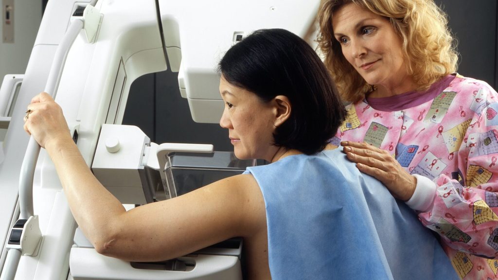
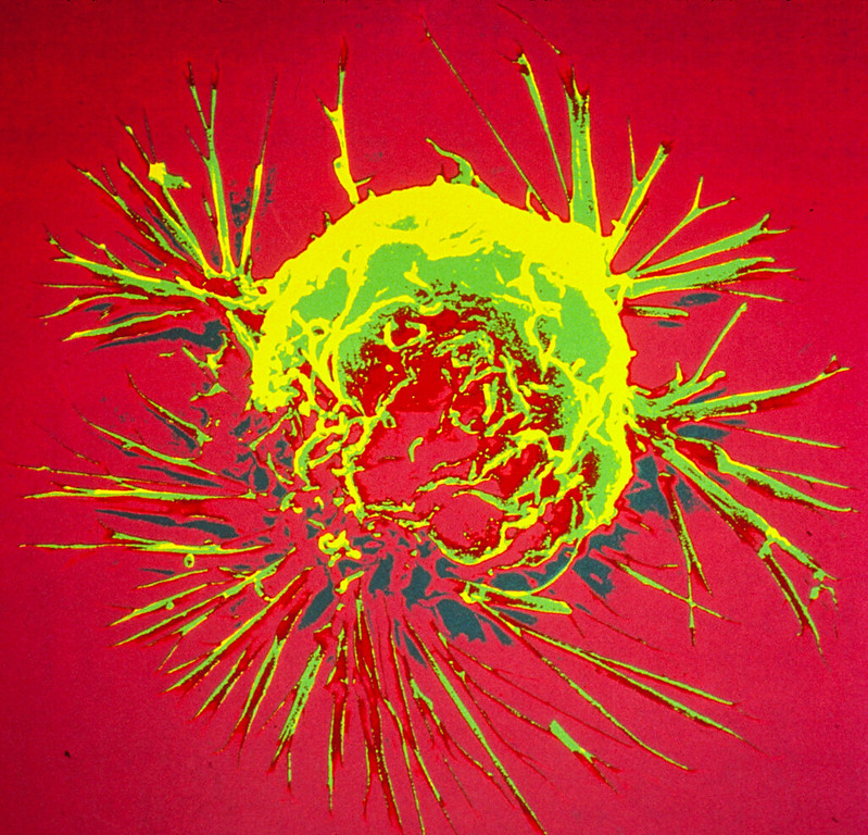
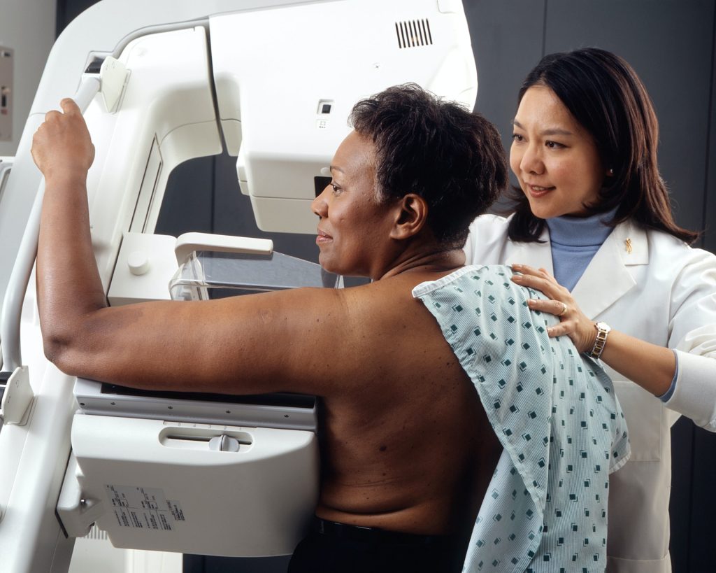
Tag: breast cancer

Lymph Node Transfer Reduces Lymphoedema After Breast Cancer Surgery

Photo by Michelle Leman on Pexels

A multicentre study led from Finland has shown that lymph node transfer is a viable treatment for the swelling in the affected limb, a condition known as lymphoedema, after breast cancer surgery. Unfortunately, a drug to improve the outcomes of the transfer treatment was not shown to be effective. 

“I am becoming increasingly convinced that lymphoedema is not just a lymphatic problem, but is connected to an immunological factor,” says Plastic Surgeon and InFLAMES Flagship Researcher Pauliina Hartiala from the University of Turku in Finland.

The study by Hartiala and collaborators was published in the journal Plastic and Reconstructive Surgery.

Around one in four women with breast cancer undergo an axillary lymph node removal surgery. The surgery is performed if tests show that the cancer has spread from the breast tissue to the lymph nodes and is often followed by radiotherapy.

After the treatment, around 20–40% of women develop lymphoedema, a lymphatic drainage disorder in the affected arm. In 2022, about 2.3 million women were diagnosed with breast cancer worldwide. In men, the disease is rare.

Swelling can start years after treatment

“Lymphedema usually starts about six months after cancer surgery, but can also occur with a delay of several years after the cancer has been treated,” says Pauliina Hartiala.

In lymphedema, fluid accumulates in the tissue at first, but over time fat and firm connective tissue also begin to accumulate in the arm. Eventually, the upper limb becomes thick and clumsy. An elastic compression sleeve is used to try to control the problem by applying pressure to prevent the limb from swelling. However, the swelling can become so severe that the arm clearly interferes with everyday life, both at work and at leisure.

Lymphedema can be treated with surgical options including liposuction, lymphatic bypass procedure, or lymph node transfer. In the transfer surgery, the patient’s lymph nodes are transferred from the groin area to the armpit, or axilla, to replace the removed lymph nodes. The procedure involves extensive scar removal from the armpit.

Lymph node transfer is often performed at the same time as the breast operated on for cancer is reconstructed with a tissue flap taken from the patient’s abdomen. 

A lymph node transfer involves removing lymph nodes from the groin area and transferring them into the armpit. Image: Pauliina Hartiala

Additional benefits were expected from a growth factor

Pauliina Hartiala was one of the leaders of a multicentre study in Turku, Finland, focused on investigating whether the outcome of lymph node transfer could be improved by a growth factor, a drug called Lymfactin, that promotes growth and repair of lymphatic vessels. The study was carried out in five research centres in Finland and Sweden and builds on extensive basic research led by Finnish Professors Kari AlitaloSeppo Ylä-Herttuala and Anne Saarikko. Lymfactin is a research product from the Finnish pharmaceutical company Herantis Pharma.

The study tested whether the growth factor could improve lymph node flap function compared to a lymph node transfer conducted without the drug. The study involved 39 women. Of these, 20 underwent a transfer procedure where the tissue flap was injected with the lymphatic growth factor before it was transferred. For the second group, the transfer was carried out by adding only saline (placebo) to the tissue flap.

“Even though the drug therapy had worked well in combination with lymph node transfer in the animal model, it did not provide sufficient additional benefit to surgery in humans,” says Pauliina Hartiala.

Although Lymfactin did not work as expected in humans, Pauliina Hartiala is pleased with the other results of the study. In both study groups, the excess arm volume reduced during follow-up. In addition, the patients treated with Lymfactin had a significantly greater reduction in skin interstitial fluid than the placebo group.

“We are the first to show, with a double-blind study, that lymph node transfer is a viable treatment for some patients with lymphoedema after breast cancer surgery. One of the results of our study was the fact that the operation significantly improved women’s quality of life, which is an important finding.”

Pauliina Hartiala works as a Plastic Surgeon at Turku University Hospital, alongside her research work. She now believes that besides a lymphatic problem, lymphedema is linked to an immunological factor. It may be one or more of the immune cells that are involved in the accumulation of connective tissue and fat in the lymphoedema.   

“If this is the case, further research will allow us to investigate whether regulating the functions of this cell population could reduce fat accumulation in the limb,” concludes Hartiala.

Source: University of Turku

Coping with the Fear of Breast Cancer Recurrence

Photo by Michelle Leman on Pexels

Breast cancer is the world’s most prevalent cancer. Although earlier detection and targeted treatment have resulted in high survival rates, many breast cancer survivors experience fear of cancer recurrence. For some survivors this fear is occasional, for others it is persistent and often debilitating.

A new study of breast cancer survivors has found this psychosocial challenge impacts almost every important domain of their lives – the emotional, behavioural, cognitive, relational and professional. A larger number of domains was affected, and they were affected more frequently in those with greater fear of recurrence.

“Study participants were reportedly disease free and trying to rebuild their lives during their post-treatment survivorship,” said senior author Shelley Johns, PsyD, a researcher-clinician with the Regenstrief Institute, the Indiana University School of Medicine and the IU Melvin and Bren Simon Comprehensive Cancer Center. “Our findings provide clarity about how breast cancer survivors are impacted by fear of recurrence and insight into how they cope with this understandable fear.”

The study was published in Supportive Care in Cancer.

The impact of fear of recurrence ranged from mildly to severely disruptive. Women experiencing mild fear reported sporadic occurrences. Those with significant fear described it as persistent and/or easily triggered across multiple life domains.

Disturbed sleep prior to mammograms was reported by survivors with mild fear, while frequent need to absent themselves from social activities, get into bed and pull the blanket over their eyes to avoid thinking about cancer was an example of severe, also known as clinical, fear of recurrence. Approximately 74 percent of study participants were experiencing clinical fear of recurrence.

347 women completed the study’s open-ended survey:

  • Many reported feelings of stress, irritability and sadness.
  • Some said fear of recurrence frequently interrupted their train of thought, for example interfering with their job when their disease popped into their mind.
  • Survivors who thought that they were more worried than they should be compared to other breast cancer survivors reported feelings of embarrassment.
  • Some indicated it was too hard to be around their family because they were constantly wondering how many more Christmases and birthdays they were going to have with their children.

The paper’s title includes the phrase, “out of a dark place,” a direct quote from a breast cancer survivor who said that she joined the study to support “getting out of a dark place.”

Other survivors noted the specific impact of fear of cancer recurrence on daily life:

  • “It motivates me to maintain healthy habits. Such as eating five servings of fruits and vegetables, working out and drinking less alcohol. It also motivates me to maintain mental health and physical health.”
  • “Whenever I feel any kind of pain or discomfort in the area where I had cancer it concerns me and I feel anxious and irritable.”
  • “Cancer is all around us. Everything is a trigger. Anniversaries, other family/friends’ diagnosis, commercials about drugs, social media, etc. …it’s a daily thought or a daily emotion.”
  • “Sit for hours doing nothing, do not turn on TV, sleepless, find hours pass by and I am in the same place just thinking, do not participate in activities, get lost driving because I’m deep in thought, compulsive online shopping, collecting things.”

Survivors offered specifics on their coping mechanisms:

  • “Just trying to be positive, eat healthy, take my meds, get enough sleep, exercise three times a week, and hope for the best.”
  • “I try to avoid things that make me think about recurrence. For example, unfollowing social media accounts, fast forwarding or leaving the room when commercials about cancer medications are on.”
  • “I try not to focus on it. I also speak with family members who have lived with cancer longer than myself.”
  • “Prayer, meditation, staying in the moment, and focusing on making the best of each day.”

While many survivors cited avoidance of thoughts and feelings as their primary coping behaviour, Dr Johns, a health services researcher and clinical health psychologist, observes that research is needed to probe the function of various coping behaviours’ to determine if they are helpful.

In a question seldom posed to participants in a clinical trial, when asked what they hoped to gain by participating in the study, the majority indicated that they sought senses of purpose, belonging, control and connection with others.

The paper concludes, “Fear of cancer recurrence is one of the most common psychological challenges for cancer survivors. Understanding affected life domains, coping strategies employed prior to intervention, and reasons for seeking guidance can inform the development and implementation of evidence-based interventions to effectively address fear of cancer recurrence among persons living with breast cancer.”

Source: Regenstrief Institute

Inhibitor Drugs to Treat Aggressive Breast Cancer Identified

Colourised scanning electron micrograph of a breast cancer cell. Credit: NIH

A US study seeking more effective treatment for deadly metaplastic breast cancer has identified two inhibitor drugs with the potential to interrupt disease progression.

Houston Methodist and a team of researchers from across the country examined the biology of metaplastic breast cancer, comparing it to non-metaplastic triple negative breast cancer. They discovered metaplastic breast cancers typically exhibit two unique signaling pathways in their cell interaction. Researchers were able to disrupt these pathways using a class of inhibitors typically used to treat advanced cancers – phosphoinositide 3 kinase inhibitor (P13K) – in combination with a nitric oxide inhibitor (NOS) typically used to treat septic shock, cardiovascular disease and other conditions. When introduced to the cell, these drugs disrupted these pathways, making the treatment more effective.

A rare and aggressive form of disease, metaplastic breast cancer typically grows faster and is more likely to metastasise than other breast cancers. It is also more likely to recur after successful initial treatment. Patients with metaplastic breast cancer will often receive the same treatment as a patient with triple negative breast cancer, another aggressive and deadly form of the disease. However, metaplastic breast cancer often does not respond well.

The findings are published in Nature Communications The study’s corresponding author is Dr Jenny Chang, the executive vice president, president and CEO, and chief academic officer at the Houston Methodist Academic Institute.

“This is a significant finding because it offers a promising therapeutic option for one of the most aggressive and difficult-to-treat subtypes of breast cancer,” said Chang. “We have the potential to improve outcomes for patients who currently face limited treatment options and poor prognoses, marking an important step forward in cancer research and therapy.”

The first author, Dr Tejaswini Reddy, hopes these findings will help develop a specific care plan for metaplastic cancer patients and improve long-term survival of the disease.

“Our findings highlight a promising therapeutic combination that could hopefully change the landscape of metaplastic breast cancer treatment. Translating this research into a National Cancer Institute-funded clinical trial is crucial to improving outcomes for patients facing this rare and aggressive disease. Moreover, this approach may have broader implications, potentially benefiting patients with other cancers with similar biology,” said Reddy. 

The findings of this preclinical study have translated into a National Cancer Institute (NCI)-funded phase 2 clinical trial to help patients with this rare and aggressive malignancy (https://clinicaltrials.gov/study/NCT05660083).

Source: Houston Methodist

New Combination Immunotherapy for Melanoma and Breast Cancer

3D structure of a melanoma cell derived by ion abrasion scanning electron microscopy. Credit: Sriram Subramaniam/ National Cancer Institute

A research team at the Medical University of Vienna led by Maria Sibilia has investigated a new combination therapy against cancer. This therapy employs systemic administration of the tissue hormone interferon-I combined with local application of Imiquimod. Promising results were seen in topically accessible tumours like melanoma and breast cancer models: the therapy led to the death of tumour cells at the treated sites and simultaneously activated the adaptive immune system to fight even distant metastases. The findings, published in Nature Cancer, could improve the treatment of superficial tumours such as melanoma and breast cancer.

In recent years, immunotherapies have had significant success in the treatment and cure of a wide range of cancers. However, for some patients, these agents are still not sufficiently effective. As part of a preclinical study, Maria Sibilia, Head of the Center for Cancer Research at the Medical University of Vienna, therefore investigated the effects of a combination immunotherapy consisting of systemic administration of the tissue hormone interferon (IFN)-I and local imiquimod therapy. Imiquimod is an active substance that activates the innate receptors TLR7/8 and used to treat basal cell carcinomas. The researchers employed various preclinical mouse tumour models of melanoma and breast cancer. What both tumours have in common is that they are accessible to local therapy and often form distant metastases.

Effective for local tumours and distant metastases

Immunotherapies use the body’s own immune system to fight cancer cells. Plasmacytoid dendritic cells (pDCs), which are activated by Imiquimod via TLR7/8, play an important role in this process. The study showed that oral imiquimod stimulates pDCs to produce the tissue hormone IFN-I. This sensitised other dendritic cells and macrophages in the tumour environment to topical imiquimod therapy, which inhibited the formation of new blood vessels via the cytokine IL12 leading to the death of tumour cells.
The combination immunotherapy not only had an effect on the treated tumours, but also on distant metastases. It reduced the formation of new metastases thus preventing tumour relapses and increasing the sensitivity of melanomas to checkpoint inhibitors.

“These findings illustrate that the combination of systemic treatment with imiquimod or IFN-I and topical therapy with imiquimod has the potential to expand treatment options for patients and improve therapy outcomes in locally accessible tumors such as melanoma or breast cancer,” emphasizes Maria Sibilia.
“Topical treatment of the primary tumor with imiquimod is essential for this combination therapy with systemic IFN-I to be effective at the treated site and also to clear distant metastases,” adds Philipp Novoszel, MedUni Vienna, one of the first authors of the study.

The results suggest that this therapeutic strategy has the potential to improve treatment outcomes in superficial and thus locally accessible tumors such as melanoma and breast cancer – on the one hand through therapy-associated cancer cell death at the locally treated tumors, but also through the induction of a T cell-induced anti-tumor immune response at distant metastases, which is further enhanced by checkpoint inhibitors.

“Our aim is to continue developing immunotherapeutic strategies in order to improve the long-term prospects for patients who are not yet responding well to these agents,” says Maria Sibilia, who is also Deputy Head of the Comprehensive Cancer Center of MedUni Vienna and University Hospital Vienna.
“As systemic interferon is a well-known cancer therapy and dendritic cells are activated in a similar way to our preclinical models, we believe that the new combination therapy can show an effect in patients,” adds Martina Sanlorenzo, dermato-oncologist at MedUni Vienna and co-first author of the study.

Publication: Nature Cancer
Systemic IFN-I combined with topical TLR7/8 agonists promotes distant tumor-suppression by c-Jun-dependent IL-12 expression in dendritic cells
Sanlorenzo M, Novoszel P, Vujic I, Gastaldi T, Hammer M, Fari O, De Sa Fernandes C, Landau AD, Göcen-Oguz BV, Holcmann M, Monshi B, Rappersberger K, Agnes Csiszar A, Sibilia M
DOI: 10.1038/s43018-024-00889-9; https://www.nature.com/articles/s43018-024-00889-9

Source: Medical University of Vienna

Routine Brain MRI Screening Urged in Asymptomatic Late Stage Breast Cancer

Photo by Anna Shvets on Pexels

A new study led by researchers at Moffitt Cancer Center shows that asymptomatic brain metastasis is more common in stage 4 breast cancer patients than previously believed. The study, published in Neuro-Oncology, suggests that doctors may need to rethink current screening guidelines for detecting brain metastasis in patients without symptoms.

Researchers examined 101 asymptomatic patients diagnosed with stage 4 breast cancer, including triple-negative, HER2-positive and hormone receptor-positive/HER2-negative breast cancer. These patients underwent MRI scans to check for brain metastasis, with a follow-up MRI six months later if the initial scan showed no signs of cancer spread.

Of the patients who completed the initial MRI, 14% had brain metastasis. The rates by subtype were:

  • 18% in triple-negative breast cancer
  • 15% in HER2-positive breast cancer
  • 10% in hormone receptor-positive/HER2-negative breast cancer

After the second MRI, the number of patients with brain metastasis grew to about 25% in each subtype. Following diagnosis, patients went on to receive early treatment for their brain metastases, including changes in systemic therapy and local therapies.

“Our study suggests that asymptomatic brain metastasis is quite common in stage 4 breast cancer,” said Kamran Ahmed, MD, associate member and section chief for Breast Radiation Oncology at Moffitt and principal investigator of the study. “Although larger studies are needed to confirm our findings, given the improvements in systemic and local therapies for breast cancer brain metastasis, the time may be appropriate to reconsider current guidelines that recommend against routine MRI surveillance in late stage breast cancer.”

Source: H. Lee Moffitt Cancer Center & Research Institute

Magnetic Fields Boost Doxorubicin Uptake in Breast Cancer Treatment

Colourised scanning electron micrograph of a breast cancer cell. Credit: NIH

Researchers at the National University of Singapore (NUS) have developed a non-invasive method to improve the effectiveness of chemotherapy while reducing its harmful side effects.

By applying brief, localised pulses of magnetic fields, the team demonstrated a significant increase in the uptake of doxorubicin (DOX), a widely used chemotherapy drug, into breast cancer cells, with minimal impact on healthy tissues. This selective uptake enables more precise targeting of cancer cells, potentially improving treatment outcomes and reducing the adverse effects often associated with chemotherapy.

The study, led by Associate Professor Alfredo Franco-Obregón at NUS, is the first to systematically show how pulsed magnetic fields enhance DOX uptake in cancer cells. The team also showed that this approach could suppress tumours at lower drug doses.

The team’s research was published in the journal Cancers. It builds on earlier work from 2022, which first revealed that certain cancer cells are more vulnerable to magnetic field therapy.

Better chemotherapy outcomes and fewer side effects

DOX is a commonly used chemotherapy drug for breast cancer. It works by binding to DNA components and disrupting cell replication and respiration, which then kills off cancer cells. Despite its efficacy, it is a non-selective drug, which means it can also damage healthy tissues, leading to side effects ranging from mild to severe, including cardiomyopathy and muscle atrophy.

To address these challenges, the NUS researchers developed a novel approach that uses brief pulses of magnetic fields to selectively increase DOX uptake into breast cancer cells. Their study revealed the role of a calcium ion channel known as TRPC1, which is often found in aggressive cancers, including breast cancer. Magnetic field exposure activates TRPC1, enhancing its ability to facilitate the entry of DOX into cancer cells.

The researchers conducted experiments comparing the effects of the magnetic field therapy on human breast cancer cells and healthy muscle cells. They found that breast cancer cells took in significantly more DOX when exposed to magnetic pulses, while normal tissues were not targeted as much. A 10-minute magnetic field exposure reduced the drug concentration needed for similar amount of cancer killing by half, particularly at low doses of the drug.

In contrast, healthy muscle cells did not show an increase in cell death in response to the combination of DOX and magnetic pulses indicating greater protection for non-cancerous tissues.

The team also demonstrated that reducing TRPC1 expression or blocking its activity eliminated this effect, which confirms the crucial role of TRPC1 channels in the process. “Importantly, when we increased the amount of TRPC1, we observed an increase in DOX uptake – this means that TRPC1 can be used as a viable therapeutic target for aggressive cancers,” said first author Mr Viresh Krishnan Sukumar, PhD candidate at NUS Centre for Cancer Research (N2CR).

“What’s promising is that this mechanism works strongest at low drug concentrations, enabling us to target cancer cells more effectively while reducing the burden of chemotherapy on healthy tissues,” Assoc Prof Franco-Obregón added.

With breast cancer remaining the leading cause of cancer-related deaths among women worldwide, the need for novel treatment strategies is urgent. “The majority of women who undergo chemotherapy experience side effects from treatment, and in some cases, doses of chemotherapy need to be reduced, or in severe cases, stopped prematurely,” said Assistant Professor Joline Lim, Principal Investigator at N2CR and Senior Consultant, Department of Haematology-Oncology, National University Cancer Institute, Singapore. “Moreover, prolonged exposure to high-dose chemotherapy can also lead to drug resistance. This targeted approach represents an excellent opportunity to potentially improve treatment outcomes while preserving patients’ quality of life.”

Advancing the frontier of precision oncology

The team’s magnetic-assisted approach addresses one of the biggest challenges of chemotherapy, namely its toxic effects on healthy tissues. By selectively enhancing drug uptake into cancer cells, this method has the potential to drastically reduce the systemic side effects often experienced by breast cancer patients. This not only improves treatment outcomes and quality of life, but also encourages earlier treatment for those hesitant about treatment side effects. The study also underscores the role of biomarkers, such as elevated TRPC1 expression, in transforming cancer care by enabling precision-driven treatment options.

Future work will focus on translating these findings into clinical practice by localising magnetic field exposure specifically to tumours in patients. This would further validate the potential to reduce systemic DOX doses while maximising localised drug delivery in cancer cells.

“Our approach will be patented and form the foundation for a startup specialising in breast cancer treatment. We are currently in discussions with potential investors in Southeast Asia and the United States to translate this technology from bench to bedside,” shared Assoc Prof Franco-Obregón. National University Cancer Institute, Singapore. “Moreover, prolonged exposure to high-dose chemotherapy can also lead to drug resistance. This targeted approach represents an excellent opportunity to potentially improve treatment outcomes while preserving patients’ quality of life.”

Source: National University of Singapore

Analysis of Repeat Mammograms Improves Cancer Prediction

Photo by National Cancer Institute on Unsplash

A new study describes an innovative method of analysing mammograms that significantly improves the accuracy of predicting the risk of breast cancer development over the following five years. Using up to three years of previous mammograms, the new method identified individuals at high risk of developing breast cancer 2.3 times more accurately than the standard method, which is based on questionnaires assessing clinical risk factors alone, such as age, race and family history of breast cancer.

The study, from Washington University School of Medicine in St. Louis, appears in JCO Clinical Cancer Informatics.

“We are seeking ways to improve early detection, since that increases the chances of successful treatment,” said senior author Graham A. Colditz, MD, DrPH, associate director, prevention and control, of Siteman Cancer Center, based at Barnes-Jewish Hospital and WashU Medicine. “This improved prediction of risk also may help research surrounding prevention, so that we can find better ways for women who fall into the high-risk category to lower their five-year risk of developing breast cancer.”

This risk-prediction method builds on past research led by Colditz and lead author Shu (Joy) Jiang, PhD, a statistician, data scientist and associate professor at WashU Medicine. The researchers showed that prior mammograms hold a wealth of information on early signs of breast cancer development that can’t be perceived even by a well-trained human eye. This information includes subtle changes over time in breast density, which is a measure of the relative amounts of fibrous versus fatty tissue in the breasts.

For the new study, the team built an algorithm based on artificial intelligence that can discern subtle differences in mammograms and help identify those women at highest risk of developing a new breast tumour over a specific timeframe. In addition to breast density, their machine-learning tool considers changes in other patterns in the images, including in texture, calcification and asymmetry within the breasts.

“Our new method is able to detect subtle changes over time in repeated mammogram images that are not visible to the eye,” said Jiang, yet these changes hold rich information that can help identify high-risk individuals.

At the moment, risk-reduction options are limited and can include drugs such as tamoxifen that lower risk but may have unwanted side effects. Most of the time, women at high risk are offered more frequent screening or the option of adding another imaging method, such as an MRI, to try to identify cancer as early as possible.

“Today, we don’t have a way to know who is likely to develop breast cancer in the future based on their mammogram images,” said co-author Debbie L. Bennett, MD, an associate professor of radiology and chief of breast imaging for the Mallinckrodt Institute of Radiology at WashU Medicine. “What’s so exciting about this research is that it indicates that it is possible to glean this information from current and prior mammograms using this algorithm. The prediction is never going to be perfect, but this study suggests the new algorithm is much better than our current methods.”

AI improves prediction of breast cancer development

The researchers trained their machine-learning algorithm on the mammograms of more than 10 000 women who received breast cancer screenings through Siteman Cancer Center from 2008–2012. These individuals were followed through 2020, and in that time 478 were diagnosed with breast cancer.

The researchers then applied their method to predict breast cancer risk in a separate set of 18 000 women who received mammograms from 2013–2020. Subsequently, 332 women were diagnosed with breast cancer during the follow-up period, which ended in 2020.

According to the new prediction model, women in the high-risk group were 21 times more likely to be diagnosed with breast cancer over the following five years than were those in the lowest-risk group. In the high-risk group, 53 out of every 1000 women screened developed breast cancer over the next five years. In contrast, in the low-risk group, 2.6 women per 1000 screened developed breast cancer over the following five years. Under the old questionnaire-based methods, only 23 women per 1000 screened were correctly classified in the high-risk group, providing evidence that the old method, in this case, missed 30 breast cancer cases that the new method found.

The mammograms were conducted at academic medical centres and community clinics, demonstrating that the accuracy of the method holds up in diverse settings. Importantly, the algorithm was built with robust representation of Black women, who are usually underrepresented in development of breast cancer risk models. The accuracy for predicting risk held up across racial groups. Of the women screened through Siteman, most were white, and 27% were Black. Of those screened through Emory, 42% were Black.

Source: Washington University School of Medicine in St. Louis

Why Does Tamoxifen Work Only for Some Patients but not Others?

Photo by Danilo Alvesd on Unsplash

A new study has shown that variation in the microbiota of the human gut impacts the pharmacokinetics of tamoxifen and thus the effectiveness of the drug. The finding, published in the journal mBio, suggests that in the future, doctors may use a simple stool test to check for certain bacteria in the gut and help predict tamoxifen’s effectiveness for them.

Tamoxifen is a selective oestrogen receptor modulator used to prevent breast cancer. It prevents breast cancer cells from being able to use oestrogen to grow.

“The key takeaway from this study is that while tamoxifen is a common and important treatment for preventing breast cancer recurrence, nearly 50% of patients don’t respond well to it,” said lead study author Yasmine Alam, a PhD candidate in the Department of Biological Chemistry, University of California Irvine. “Since tamoxifen is taken orally and passes through the gut, this difference in how patients respond may be linked to the gut microbiome – the trillions of bacteria in our intestines, which vary greatly from person to person. Our study aims to better understand how these gut bacteria influence the way tamoxifen is absorbed, broken down and recycled in the body, with the goal of improving treatment outcomes for breast cancer patients.”

In the new study, the researchers set out to define the role that gut microbes play in how tamoxifen is processed (ie, absorption, distribution, metabolism and excretion), given its significant variable efficacy across patients. The researchers provided tamoxifen to mice that had no gut microbiome and to mice with a human microbiome (introduced to the mice by a human faecal sample). They found that mice with gut bacteria had higher amounts of tamoxifen in their bloodstream. The scientists then went on to explore what part of the gut microbiome was responsible for controlling the level of drug in the bloodstream. By examining the faecal samples from people, they linked a specific enzyme in bacteria, beta-glucuronidase, as a key factor that allows the drug to enter the bloodstream.

Tamoxifen is absorbed into the bloodstream from the intestine. Tamoxifen is carried by the bloodstream to the liver, where it is converted to its cancer-fighting form. Sometimes a sugar molecule can get attached to it, which signals the body to dump the cancer-fighting form of the drug back into the intestine. This drug can only get out of the intestine by taking the sugar off the molecule – and the researchers found that beta-glucuronidase in gut bacteria can eat the sugar off the drug so it can go on to fight breast cancer.

“Specifically, we found that certain enzymes produced by gut bacteria, called β-glucuronidase, play a role in how tamoxifen is broken down. These enzymes help recycle tamoxifen back into the bloodstream, which can make the drug more effective,” Alam said. “We discovered that a particular type of bacteria, Bacteroides fragilis, was strongly linked to the ability of these enzymes to affect tamoxifen levels in the blood in a positive way. This suggests that the gut microbiome plays an important role in how tamoxifen works in the body.”

The long-term goal of the study is to pave the way for more tailored and effective therapeutic interventions in the prevention of breast cancer recurrence.

Source: American Society for Microbiology

Aerobic Exercise may Help Prevent the Brain Fog from Chemotherapy

Clinical trial reveals improved self-reported cognitive function in women with breast cancer who started an exercise program when initiating chemotherapy.

Photo by Ketut Subiyanto on Pexels

Many women who receive chemotherapy experience a decreased ability to remember, concentrate, and/or think – commonly referred to as “chemo-brain” or “brain fog” – both short- and long-term. In a recent clinical trial of women initiating chemotherapy for breast cancer, those who simultaneously started an aerobic exercise program self-reported greater improvements in cognitive function and quality of life compared with those receiving standard care. The findings are published by Wiley online in CANCER, a peer-reviewed journal of the American Cancer Society.

The study, called the Aerobic exercise and CogniTIVe functioning in women with breAsT cancEr (ACTIVATE) trial, included 57 Canadian women in Ottawa and Vancouver who were diagnosed with stage I–III breast cancer and beginning chemotherapy. All women participated in 12–24 weeks of aerobic exercise: 28 started this exercise when initiating chemotherapy and 29 started after chemotherapy completion. Cognitive function assessments were conducted before chemotherapy initiation and after chemotherapy completion (therefore, before the latter group started the exercise program).

Women who participated in the aerobic exercise program during chemotherapy self-reported better cognitive functioning and felt their mental abilities improved compared with those who received standard care without exercise. Neuropsychological testing – a performance-based method used to measure a range of mental functions – revealed similar cognitive performance in the two groups after chemotherapy completion, however.

“Our findings strengthen the case for making exercise assessment, recommendation, and referral a routine part of cancer care; this may help empower women living with and beyond cancer to actively manage both their physical and mental health during and after treatment,” said lead author Jennifer Brunet, PhD, of the University of Ottawa.

Dr Brunet noted that many women undergoing chemotherapy for breast cancer remain insufficiently active, and there are limited exercise programs tailored to their needs. “To address this, we advocate for collaboration across various sectors – academic, healthcare, fitness, and community – to develop exercise programs specifically designed for women with breast cancer,” she said. “These programs should be easy to adopt and implement widely, helping to make the benefits of exercise more accessible to all women facing the challenges of cancer treatment and recovery.”

Source: Wiley

New Research Reveals Why Breast Cancer Metastasises to Bone

Colourised scanning electron micrograph of a breast cancer cell. Credit: NIH

Researchers from Tampere University, Finland, and Izmir Institute of Technology, Turkey, have developed an in vitro cancer model to investigate why breast cancer spreads to bone. Their findings, published in PLOS One, hold promise for advancing the development of preclinical tools to predict breast cancer bone metastasis.

Breast cancer is a significant global public health challenge, with 2.3 million new cases and 700 000 deaths every year. Approximately 80% of patients with primary breast cancer can be cured, if they are diagnosed and treated promptly. However, in many cases, the cancer has already metastasised at the time of diagnosis. 

Metastatic cancer is incurable and accounts for more than 90% of cancer-related deaths. Currently, there are no reliable in vitro models to study how breast cancer spreads to secondary organs such as bone, lung, liver or brain. Now, researchers from the Precision Nanomaterials Group at Tampere University in Finland, and the Cancer Molecular Biology Lab at Izmir Institute of Technology in Turkey, have used lab-on-a-chip platforms to create a physiologically relevant metastasis model to study the factors controlling breast cancer bone metastasis. 

“Breast cancer most frequently spreads to bone, with an estimated rate of 53%, resulting in severe symptoms such as pain, pathological bone fractures, and spinal cord compressions. Our research provides a laboratory model that estimates the likelihood and mechanism of bone metastasis occurring within a living organism. This advances the understanding of molecular mechanisms in breast cancer bone metastasis and provides the groundwork for developing preclinical tools for predicting bone metastasis risk,” says Burcu Firatligil-Yildirir, postdoctoral researcher at Tampere University and the first author of the paper.

According to Nonappa, Associate Professor and leader of the Precision Nanomaterials Group at Tampere University, developing sustainable in vitro models that mimic the complexity of the native breast and bone microenvironment is a multidisciplinary challenge.

“Our work shows that physiologically relevant in vitro models can be generated by combining cancer biology, microfluidics and soft materials. The results open new possibilities for developing predictive disease, diagnostic and treatment models,” he says.

Source: Tampere University