

Tag: mammograms

Breast Cancer Screening in South Africa: Balancing Early Detection with Appropriate Care

By Dr Fatima Hoosain, specialist surgeon and Principal of Apffelstaedt, Hoosain & Associates, with a clinical focus on breast and endocrine surgery.
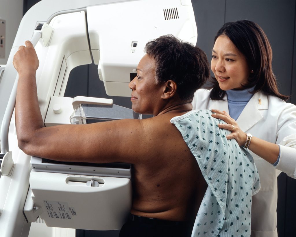
Photo by National Cancer Institute on Unsplash

When people think about breast cancer screening, the conversation often begins and ends with one message: screen more women.

As clinicians, we know it is not quite that simple.

There is no question that screening saves lives. Regular mammography reduces breast cancer mortality and gives us the opportunity to diagnose disease when it is smaller, more treatable and associated with significantly better outcomes. Few interventions in medicine demonstrate such a clear benefit.

The challenge is that good breast care is not defined simply by how many mammograms we perform. It is defined by the quality of the decisions that surround them.

This was the focus of my presentation at the recent Board of Healthcare Funders (BHF) Conference, where we explored how clinicians can balance the burden of breast cancer with evidence-based screening decisions while remaining mindful of both underdiagnosis and overdiagnosis.
Those competing risks are encountered by every clinician involved in breast care.

We all worry about the patient whose cancer is diagnosed later than it should have been. Earlier diagnosis frequently means less extensive surgery, more treatment options and, ultimately, better outcomes. The survival difference between early-stage and advanced disease is substantial, making timely diagnosis one of the most important contributors to long-term prognosis.

At the same time, screening is not without consequences.

Not every abnormality detected on imaging will become life-threatening, yet every suspicious finding understandably creates anxiety. Additional imaging, biopsies and sometimes treatment may follow. Our responsibility is therefore not simply to detect abnormalities, but to interpret them appropriately within the context of each patient’s overall clinical picture.

This is why breast screening should never be approached as a uniform process. Risk matters.

A woman with an inherited genetic mutation or a strong family history should not necessarily follow the same screening pathway as someone at average risk. Likewise, imaging should answer a clinical question. Mammography remains the cornerstone of breast screening, but dense breast tissue, patient age and individual risk factors may require supplementary investigations such as ultrasound or MRI. More imaging is not automatically better medicine. Appropriate imaging is.

These decisions have become even more complex within the South African healthcare environment. International guidelines provide an excellent evidence base, but they do not remove the practical realities we face every day. Access to imaging differs between regions. Advanced investigations may not always be readily available. Medical scheme funding, co-payments and affordability inevitably influence what is possible for many patients. These factors cannot be ignored when discussing best practice because they form part of the reality in which clinical decisions are made.

Fortunately, the treatment landscape continues to evolve.

Advances in oncoplastic surgery, targeted therapies, immunotherapy and modern radiation techniques have transformed outcomes for many patients diagnosed with breast cancer. These developments are encouraging, but they should not distract us from one fundamental principle: the earlier we diagnose clinically significant disease, the greater the opportunity to offer patients treatments that are both effective and less invasive.

Diagnosis, however, is only the beginning of the journey.

Long-term follow-up remains an essential part of breast cancer care. Ongoing surveillance, adherence to endocrine therapy where appropriate, management of treatment side effects and supporting patients through the psychological and financial impact of a cancer diagnosis all influence outcomes. Good breast care extends well beyond the operating theatre or oncology unit.

As our healthcare system continues to face increasing clinical and financial pressures, I believe we need to move beyond simplistic conversations about screening uptake alone.

The more important discussion is whether we are making consistently good clinical decisions. Are we identifying the patients who stand to benefit most? Are we investigating appropriately? Are we avoiding unnecessary intervention when the evidence suggests it is unlikely to improve outcomes?

Those are not easy questions, but they are the ones that matter.

Ultimately, breast cancer screening is not about doing more. It is about doing what is right for the patient sitting in front of us. That remains the most important clinical judgement we make.

Missing First Mammogram Raises Breast Cancer Death Risk

Photo by National Cancer Institute on Unsplash

Women who miss their first mammogram run a higher risk of being diagnosed with advanced breast cancer and dying from the disease. This is shown in a new study from Karolinska Institutet published in The BMJ.

Since the early 1990s, women in Sweden have been offered regular mammograms, which has contributed to a decrease in breast cancer mortality. Despite this, a significant proportion choose not to attend their first examination. The researchers behind the new study wanted to investigate the long-term consequences of this. 

The study is based on data from the Swedish mammography screening program and national health registries, and covers almost 433 000 women in Stockholm between 1991 and 2020, with follow-up for up to 25 years. 

The results show that 32% of all women who were invited to their first screening declined. These women were also less likely to participate in future examinations, which often led to a later diagnosis and poorer prognosis.

“Skipping the first mammogram is a strong indicator of who is at risk of late detection and higher mortality. Our results show that missing the first mammogram is not just a one-time choice, but often marks the beginning of a long-term pattern of not attending check-ups,” says the study’s first author, Ziyan Ma, a doctoral student at the Department of Medical Epidemiology and Biostatistics, Karolinska Institutet.

Were detected at a more advanced stage

When women who skipped their first screening were later diagnosed with breast cancer, the disease was more often detected at a more advanced stage. The risk of developing stage III cancer was approximately 1.5 times higher, and for stage IV, the risk was as much as 3.6 times higher compared to those who participated in the first mammogram. Over a 25-year follow-up period, almost 1 percent of those who did not participate had died of breast cancer, compared with 0.7 percent among the participants – a difference that corresponds to a 40 percent higher risk of dying from the disease. 

However, the total proportion of women who developed breast cancer was almost the same in both groups, approximately 7.7%. According to the researchers, this shows that the increased mortality is mainly due to delayed detection rather than more cases of the disease.

“Family history is a well-known, unchangeable risk factor for breast cancer. Our study shows that missing the very first screening examination carries a similar mortality risk – but unlike family history, this is a behaviour that we can change. Since over 30 percent of women skip their first screening, increased participation could save many lives. Since this group can be identified early, decades before deaths occur, healthcare providers have a chance to intervene with reminders or support to encourage participation, says the study’s last author, Kamila Czene, professor at the Department of Medical Epidemiology and Biostatistics, Karolinska Institutet

Source: Karolinska Institutet

Analysis of Repeat Mammograms Improves Cancer Prediction

Photo by National Cancer Institute on Unsplash

A new study describes an innovative method of analysing mammograms that significantly improves the accuracy of predicting the risk of breast cancer development over the following five years. Using up to three years of previous mammograms, the new method identified individuals at high risk of developing breast cancer 2.3 times more accurately than the standard method, which is based on questionnaires assessing clinical risk factors alone, such as age, race and family history of breast cancer.

The study, from Washington University School of Medicine in St. Louis, appears in JCO Clinical Cancer Informatics.

“We are seeking ways to improve early detection, since that increases the chances of successful treatment,” said senior author Graham A. Colditz, MD, DrPH, associate director, prevention and control, of Siteman Cancer Center, based at Barnes-Jewish Hospital and WashU Medicine. “This improved prediction of risk also may help research surrounding prevention, so that we can find better ways for women who fall into the high-risk category to lower their five-year risk of developing breast cancer.”

This risk-prediction method builds on past research led by Colditz and lead author Shu (Joy) Jiang, PhD, a statistician, data scientist and associate professor at WashU Medicine. The researchers showed that prior mammograms hold a wealth of information on early signs of breast cancer development that can’t be perceived even by a well-trained human eye. This information includes subtle changes over time in breast density, which is a measure of the relative amounts of fibrous versus fatty tissue in the breasts.

For the new study, the team built an algorithm based on artificial intelligence that can discern subtle differences in mammograms and help identify those women at highest risk of developing a new breast tumour over a specific timeframe. In addition to breast density, their machine-learning tool considers changes in other patterns in the images, including in texture, calcification and asymmetry within the breasts.

“Our new method is able to detect subtle changes over time in repeated mammogram images that are not visible to the eye,” said Jiang, yet these changes hold rich information that can help identify high-risk individuals.

At the moment, risk-reduction options are limited and can include drugs such as tamoxifen that lower risk but may have unwanted side effects. Most of the time, women at high risk are offered more frequent screening or the option of adding another imaging method, such as an MRI, to try to identify cancer as early as possible.

“Today, we don’t have a way to know who is likely to develop breast cancer in the future based on their mammogram images,” said co-author Debbie L. Bennett, MD, an associate professor of radiology and chief of breast imaging for the Mallinckrodt Institute of Radiology at WashU Medicine. “What’s so exciting about this research is that it indicates that it is possible to glean this information from current and prior mammograms using this algorithm. The prediction is never going to be perfect, but this study suggests the new algorithm is much better than our current methods.”

AI improves prediction of breast cancer development

The researchers trained their machine-learning algorithm on the mammograms of more than 10 000 women who received breast cancer screenings through Siteman Cancer Center from 2008–2012. These individuals were followed through 2020, and in that time 478 were diagnosed with breast cancer.

The researchers then applied their method to predict breast cancer risk in a separate set of 18 000 women who received mammograms from 2013–2020. Subsequently, 332 women were diagnosed with breast cancer during the follow-up period, which ended in 2020.

According to the new prediction model, women in the high-risk group were 21 times more likely to be diagnosed with breast cancer over the following five years than were those in the lowest-risk group. In the high-risk group, 53 out of every 1000 women screened developed breast cancer over the next five years. In contrast, in the low-risk group, 2.6 women per 1000 screened developed breast cancer over the following five years. Under the old questionnaire-based methods, only 23 women per 1000 screened were correctly classified in the high-risk group, providing evidence that the old method, in this case, missed 30 breast cancer cases that the new method found.

The mammograms were conducted at academic medical centres and community clinics, demonstrating that the accuracy of the method holds up in diverse settings. Importantly, the algorithm was built with robust representation of Black women, who are usually underrepresented in development of breast cancer risk models. The accuracy for predicting risk held up across racial groups. Of the women screened through Siteman, most were white, and 27% were Black. Of those screened through Emory, 42% were Black.

Source: Washington University School of Medicine in St. Louis