Tag: breast cancer

FDA-Approved Drug Halts EBV-Driven Lymphoma by Disrupting a Key Cancer Pathway

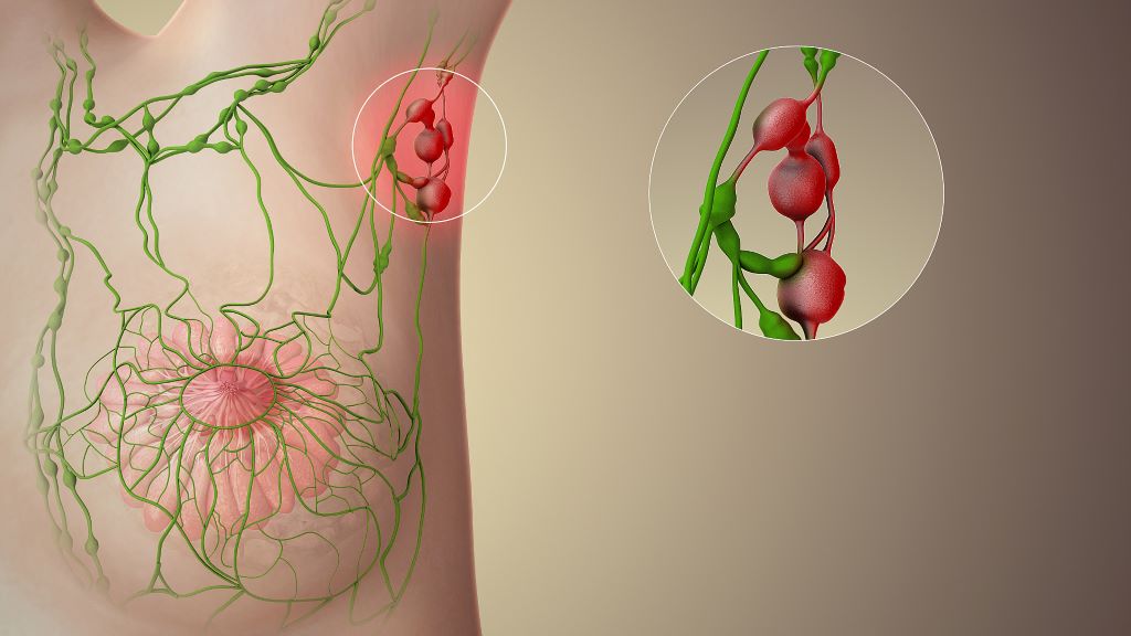
Swollen lymph nodes. Credit: Scientific Animations CC0

Scientists at The Wistar Institute have discovered that a class of FDA-approved cancer drugs known as PARP1 inhibitors can effectively combat Epstein-Barr virus (EBV)-driven lymphomas. The findings, published in the Journal of Medical Virology, demonstrate that these drugs, which work by blocking the activity of the PARP1 enzyme, can halt tumour growth by interfering with the EBV’s ability to activate key cancer-promoting genes.

“We’ve uncovered a completely different mechanism for how PARP inhibitors work in EBV-positive cancers,” said Italo Tempera, PhD, associate professor at Wistar’s Ellen and Ronald Caplan Cancer Center and senior author of the study. “Instead of preventing DNA damage from repairing itself in the tumours, like these drugs do in other cancers, they essentially cut off the virus’s ability to hijack cellular machinery to drive cancer growth. This opens up exciting possibilities for repurposing existing FDA-approved drugs to treat EBV-associated cancers.”

EBV infects over 90% of the global population. While most people with the virus remain symptom-free, immunocompromised individuals such as people with HIV and transplant recipients have an increased risk of EBV causing several types of cancer, including various lymphomas and carcinomas. Despite the virus’s clear role in driving these malignancies, no specific therapies currently target EBV-driven cancer.

In search of such a therapy, Tempera and his research team focused on PARP1, a cellular protein that is known primarily for its role in DNA repair. In cancer treatment, PARP inhibitors typically work by preventing cancer cells from repairing their DNA, causing them to die. However, Tempera’s team had previously discovered that PARP1 plays a very different role in EBV infection: It helps control which genes are accessible and active, essentially acting as a master regulator of gene expression.

“Think of PARP1 as a key that opens up DNA to make certain genes readable,” explained Tempera. “EBV uses this key to unlock cancer-promoting genes. When we block PARP1, we’re essentially taking away the key so the virus can’t get in and use our DNA for its own purposes.”

Using a mouse model of EBV-driven lymphoma, the researchers treated the animals with BMN 673 (talazoparib/talzenna), a PARP inhibitor that has already been approved for breast cancer treatment. Compared to controls, the treated mice showed an 80% reduction in tumour growth, and the cancer’s ability to spread to other organs was significantly reduced. Further, when the team analysed the tumours, they found no increase in DNA damage in the treated animals – the hallmark of how PARP inhibitors typically work. Instead, they discovered that PARP1 inhibition disrupted a critical partnership between the viral protein EBNA2 and the cellular oncogene MYC.

“EBNA2 is like the conductor of an orchestra, directing cellular genes to play a cancer symphony,” said Tempera. “It specifically turns on MYC, which is one of the most important cancer-promoting genes. When we inhibit PARP1, EBNA2 can’t effectively activate MYC anymore, and the whole cancer program falls apart.”

The findings have significant therapeutic implications. Because PARP inhibitors are already FDA-approved and their safety profiles are well established, the path to clinical application could be accelerated compared to developing entirely new drugs.

The research also suggests this approach might work beyond EBV-associated lymphomas. The team is now investigating whether PARP inhibitors could be effective against other EBV-driven cancers, including nasopharyngeal and gastric carcinomas. Additionally, given EBV’s suspected role in autoimmune diseases, the researchers are exploring whether PARP1’s regulation of viral gene expression might contribute to these conditions.

“This work really showcases the power of understanding fundamental viral biology,” said Tempera. “We’re taking insights from basic virology research and translating them into potential therapies. With further development, this approach could provide new hope for patients with EBV-associated cancers who currently have limited treatment options.”

Source: Wistar Institute

Celebrate Christmas in July with PinkDrive

Cold Nights, Warm Hearts, Festive Vibes – and Support for Early Detection

Think log fires, festive cheer, a three-course dinner and dancing to a live band – all wrapped up in the joy of giving. On Saturday, 5th July 2025, PinkDrive will host a Christmas in July dinner at the Indaba Hotel in Johannesburg, a night of holiday cheer and hope in action: raising funds for a life-changing cause. And you’re invited.

PinkDrive is a non-profit (NPC) committed to prolonging lives through the early detection of gender-related cancers. It delivers essential health services to thousands of South Africans every year by bringing mobile mammography units directly to communities that need them most, from rural villages to peri-urban areas across the nine provinces. These trucks provide clinical breast exams, mammograms, pap smears, and PSA testing, helping to detect cancer early in areas where healthcare access is often limited or unavailable.

Like many non-profits, PinkDrive depends on the support of corporate partners, sponsors, government, and the public to sustain its vital work, among them, Lee-Chem Laboratories. “This is a cause that is close to our hearts,” says Bhavna Sanker, Marketing Manager at Lee-Chem Laboratories. “Each year, we proudly support PinkDrive through our Mandy’s brand sponsorship, focusing on spreading awareness, sharing survivor stories, and making sure people understand their healthcare options.”

As part of their continued support, every guest at the Christmas in July function will receive a goodie bag from Lee-Chem, filled with products from their Mandy’s brand. “We are truly honoured to have Lee-Chem and the Mandy’s brand as valued sponsors of our Christmas in July Dinner fundraiser,” comments Nelius du Preez, Operations Manager at PinkDrive NPC. “Their continued support, alongside our other generous sponsors, makes events like this possible and helps us not only raise vital funds but also amplify the message of early detection and health education across South Africa.”

The fundraiser will spotlight the resilience of breast cancer survivors, with Deputy Minister of Electricity and Energy, Samantha Graham-Maré, sharing her personal journey with cancer. PinkDrive CEO and Founder, Noelene Kotschan, a passionate advocate for early detection prolonging  lives, will also address guests, and your charming host for the evening? A dashing Mister Global SA finalist will take the mic as MC, steering the evening from heartfelt reflections to lively fundraising with raffles and auctions featuring holiday packages, original artworks, and other exclusive prizes.

“We call the event ‘a night of giving back’ because it is occasions like these that allow us to keep our mobile health units on the road, reaching men and women who might otherwise not have access to screening services,” says du Preez. “Together with compassionate partners like Lee-Chem, we are driving change and prolonging lives, one screening at a time. And we are grateful to every corporate and individual who supports the event by purchasing tickets,” he adds.

All proceeds from the evening will be ringfenced to build and operate a new mobile mammography unit, expanding PinkDrive’s reach to screen more South Africans and detect cancer early, when treatment is most effective. “We encourage the public to join us in this mission by purchasing tickets and supporting the evening’s fundraising efforts,” concludes Sanker.

Tickets for the Christmas in July dinner are R600 per person and available now at pinkdrive.org. Don’t miss this chance to dress up, give back, and help bring hope where it’s needed most.

Breast Cancer Treatment Linked to a Reduction in Alzheimer’s Disease Risk

Photo by National Cancer Institute on Unsplash

A Korean population-based cohort study investigated the risk of Alzheimer’s disease (AD) among breast cancer survivors compared to age-matched controls without cancer. The study, published in JAMA Network Open, found that breast cancer survivors had an 8% lower risk of AD than controls, with a significant association in survivors over 65 years old – though the effect did not persist past five years. Radiotherapy was associated with a lower risk of AD among breast cancer survivors – but not other treatments.

Breast cancer survivors may experience long-term health consequences, including cognitive function and risk of dementia. The risk of AD among breast cancer survivors is still unclear and may vary depending on age at diagnosis, treatment received, and time since treatment.

Previous studies reported mixed results on the risk of AD among breast cancer survivors, with some finding no increase in risk and others finding a 35% increased risk for those diagnosed at age 65 or older. These studies have been hampered by a number of methodological issues, including not accounting for risk factors.

Cytotoxic chemotherapy can cause cognitive decline termed ‘chemobrain’. Other chemotherapy drugs such as anthracycline may reduced AD risk by reducing the formation of amyloid deposits. Endocrine therapy may increase the risk of dementia by lowering oestrogen, but studies suggest that the use of tamoxifen and aromatase inhibitors is associated with a lower risk of AD. An increase in dementia is seen in radiotherapy for head and neck cancers.

To investigate the risk of AD among breast cancer survivors, researchers used the Korean National Health Insurance Service (K-NHIS) database, exploring whether there is an association with cancer treatment and various confounding factors.

Among 70 701 breast cancer survivors (mean age, 53.1 years), 1229 cases of AD were detected, with an incidence rate of 2.45 per 1000 person-years. Survivors exhibited a slightly lower risk of AD compared with cancer-free controls, especially among individuals 65 years or older (SHR, 0.92; 95% CI, 0.85-0.99). But landmark analyses found that this lower risk did not persist beyond five years of survival. Radiotherapy was associated with reduced risk of AD among survivors, while chemotherapy and endocrine therapy had no significant impact. Anthracycline use, however, did show a non-significant decrease in risk.

Differences in doses and timing of radiotherapy may influence the effects. The incident exposure to the brain is estimated to be 0.2Gy from a breast cancer radiotherapy dose of 50Gy. A pilot study found that patients with AD who received low-dose whole-brain radiotherapy at 3Gy showed a temporary improvement in cognitive function. This improvement is believed to be due to a neuroprotective effect on microglia. Other studies have noted a transient risk reduction for AD in breast cancer radiotherapy; however, patients receiving radiotherapy usually do so in conjunction with breast-conserving surgery – those opting for this procedure are younger, with fewer comorbidities and smaller tumours.

The study suggests that cancer treatment may have benefits against AD development, but the risk of AD may differ depending on the duration of survival.

The findings indicate that breast cancer treatment may not directly lead to AD, and that managing modifiable risk factors for AD, such as smoking and diabetes, is a feasible option to lower AD risk among breast cancer survivors.

South African Study Identifies Two New Breast Cancer Genes in Black Women

Genetic factors contribute to some 30% of breast cancer cases in SA, necessitating investment in genomic research in African contexts.

Photo by National Cancer Institute

A seminal genetic study published in Nature Communications has discovered two genetic variants linked to breast cancer in black South African women, deepening knowledge about the genetic basis for this disease in African populations.

The genome-wide association study (GWAS) of breast cancer is the first to have been done in African women living on the continent.

A GWAS is a powerful research method that scans the entire DNA of many people to find genetic differences associated with a specific disease or trait.

In this case, the scientists at the Sydney Brenner Institute for Molecular Bioscience (SBIMB) scanned for breast cancer and found consistent genetic patterns in black South African women.

The SBIMB researchers discovered genetic signals around the gene RAB27A, a member of the RAS oncogene family, and USP22, a gene which is highly active in breast cancer cells and associated with a poor health prognosis.

“These genes have not been associated with the disease before, which is an important advance in understanding breast cancer risk and biology in women of African ancestry,” says Dr Mahtaab Hayat, the lead author of the study.

The two new genetic variants were identified in black South African women with breast cancer enrolled in the Johannesburg Cancer Study, compared to women without cancer in the Africa Wits-INDEPTH Partnership for Genomic Research (AWI-Gen) study.

Until now, most breast cancer genetics research has focused on European and Asian populations, with studies of African ancestry limited primarily to African- American women, who largely descend from West African populations.

A tool that estimates lifetime cancer risk based on DNA, the polygenic risk score (PRS), performed poorly in distinguishing South African women with breast cancer from those without.

“This is because most PRSs were developed in European populations, and their inaccuracy in African populations highlights the urgent need for ancestry-specific tools in cancer risk prediction,” says Dr Jean-Tristan Brandenburg, also in the SBIMB and a lead author.

Breast cancer is the second most common cancer in South Africa and the most common cancer in women globally, with genetic factors contributing to about 30% of cases. “Our study makes a compelling case for investing in genomic research rooted in African contexts,” notes Hayat.

The potential for precision medicine

If further studies confirm these findings, the USP22 and RAB27A genes could be specific targets for new drugs. “We could potentially target harmful cancer cells while sparing healthy tissue, which is ideally what we want when administering cancer treatment,” says Distinguished Professor at the SBIMB, Chris Mathew, and a lead project investigator.

Furthermore, if a specific gene is associated with poorer survival, it can be used as a biomarker to identify more aggressive cancers and help predict which patients may need more intensive treatment and monitoring.

Understanding the genetic architecture of complex diseases helps scientists figure out the biological processes leading to these conditions and find drug targets and treatments for groups of individuals with similar disease risk profiles.

Genomic diversity in Africa is unparalleled

African populations have more genetic variation than any other population in the world, but they have been significantly underrepresented in genomic research. This means that the global understanding of disease risk, and the tools and treatment developed from it, is limited.

“The study reveals that more people can benefit from genetic discoveries. It proves that new risk factors are still out there, waiting to be found,” says Hayat.

Source: University of the Witwatersrand

Removing Ovaries and Fallopian Tubes Linked to Lower Risk of Early Death Among Certain Breast Cancer Patients

Photo by National Cancer Institute on Unsplash

Women diagnosed with breast cancer who carry particular BRCA1 and BRCA2 genetic variants are offered surgery to remove the ovaries and fallopian tubes as this dramatically reduces their risk of ovarian cancer. Now, Cambridge researchers have shown that this procedure – known as bilateral salpingo-oophorectomy (BSO) – is associated with a substantial reduction in the risk of early death among these women, without any serious side-effects.

Women with certain variants of the genes BRCA1 and BRCA2 have a high risk of developing ovarian and breast cancer. These women are recommended to have their ovaries and fallopian tubes removed at a relatively early age – between the ages 35 and 40 years for BRCA1 carriers, and between the ages 40 and 45 for BRCA2 carriers.

Previously, BSO has been shown to lead to an 80% reduction in the risk of developing ovarian cancer among these women, but there is concern that there may be unintended consequences as a result of the body’s main source of oestrogen being removed, which brings on early menopause. This can be especially challenging for BRCA1 and BRCA2 carriers with a history of breast cancer, as they may not typically receive hormone replacement therapy to manage symptoms. The overall impact of BSO in BRCA1 and BRCA2 carriers with a prior history of breast cancer remains uncertain. 

Ordinarily, researchers would assess the benefits and risks associated with BSO through randomised controlled trials, the ‘gold standard’ for testing how well treatments work. However, to do so in women who carry the BRCA1 and BRCA2 variants would be unethical as it would put them at substantially greater risk of developing ovarian cancer.

To work around this problem, a team at the University of Cambridge, in collaboration with the National Disease Registration Service (NDRS) in NHS England, turned to electronic health records and data from NHS genetic testing laboratories collected and curated by NDRS to examine the long-term outcomes of BSO among BRCA1 and BRCA2 PV carriers diagnosed with breast cancer. The results of their study, the first large-scale study of its kind, are published today in The Lancet Oncology.

The team identified a total of 3400 women carrying one of the BRCA1 and BRCA2 cancer-causing variants (around 1700 women for each variant). Around 850 of the BRCA1 carriers and 1,000 of the BRCA2 carriers had undergone BSO surgery.

Women who underwent BSO were around half as likely to die from cancer or any other cause over the follow-up period (a median follow-up time of 5.5 years). This reduction was more pronounced in BRCA2 carriers compared to BRCA1 carriers (a 56% reduction compared to 38% respectively). These women were also at around a 40% lower risk of developing a second cancer.

Although the team say it is impossible to say with 100% certainty that BSO causes this reduction in risk, they argue that the evidence points strongly towards this conclusion.

Importantly, the researchers found no link between BSO and increased risk of other long-term outcomes such as heart disease and stroke, or with depression. This is in contrast to previous studies that found evidence in the general population of an association between BSO and increased risk of these conditions.

First author Hend Hassan, a PhD student at the Centre for Cancer Genetic Epidemiology, Department of Public Health and Primary Care, and Wolfson College, Cambridge, said: “We know that removing the ovaries and fallopian tubes dramatically reduces the risk of ovarian cancer, but there’s been a question mark over the potential unintended consequences that might arise from the sudden onset of menopause that this causes.

“Reassuringly, our research has shown that for women with a personal history of breast cancer, this procedure brings clear benefits in terms of survival and a lower risk of other cancers without the adverse side effects such as heart conditions or depression.”

Most women undergoing BSO were white. Black and Asian women were around half as likely to have BSO compared to white women. Women who lived in less deprived areas were more likely to have BSO compared to those in the most-deprived category.

Hassan added: “Given the clear benefits that this procedure provides for at-risk women, it’s concerning that some groups of women are less likely to undergo it. We need to understand why this is and encourage uptake among these women.”

Professor Antonis Antoniou, from the Department of Public Health and Primary Care, the study’s senior author, said: “Our findings will be crucial for counselling women with cancer linked to one of the BRCA1 and BRCA2 variants, allowing them to make informed decisions about whether or not to opt for this operation.”

Professor Antoniou, who is also Director of the Cancer Data-Driven Detection programme, added: “The study also highlights the power of exceptional NHS datasets in driving impactful, clinically relevant research.”

The research was funded by Cancer Research UK, with additional support from the National Institute for Health and Care Research (NIHR) Cambridge Biomedical Research Centre.

The University of Cambridge is fundraising for a new hospital that will transform how we diagnose and treat cancer. Cambridge Cancer Research Hospital, a partnership with Cambridge University Hospitals NHS Foundation Trust, will treat patients across the East of England, but the research that takes place there promises to change the lives of cancer patients across the UK and beyond. Find out more here.

Reference

Hassan, H et al. Long-term health outcomes of bilateral salpingo-oophorectomy in BRCA1 and BRCA2 pathogenic variant carriers with personal history of breast cancer: a retrospective cohort study using linked electronic health records. Lancet Oncology; 7 May 2025; DOI: 10.1016/S1470-2045(25)00156-1

The original text of this story is licensed under a Creative Commons Attribution-NonCommercial-ShareAlike 4.0 International License.

Read the original article.

Three out of Ten Breast Cancers are Detected Between Screenings

Photo by National Cancer Institute

In a new study, researchers from Karolinska Institutet have shown that so-called interval cancers, which are detected between two screening sessions, account for a significant proportion of breast cancer cases and that certain risk factors may increase the likelihood of developing this type of cancer. The study was published in the journal JAMA Oncology.

Mammography screening has been shown to be effective in reducing breast cancer mortality by detecting cancer at an early stage. Despite this, some cancers are not diagnosed during screening but between screening rounds, known as interval cancers. This type of cancer can be more aggressive and difficult to treat than breast cancer detected during a scheduled screening.

The study, which covers half a million women in Stockholm between 1989 and 2020, shows that interval cancer accounts for a significant proportion of all breast cancer cases.

“We found that interval cancers account for about 30 per cent of all breast cancers detected by screening and this percentage has remained constant over three decades, despite advances in screening technology,” says Yuqi Zhang, postdoctoral fellow at the Department of Medical Epidemiology and Biostatistics, Karolinska Institutet and lead author of the study.

The researchers identified several factors that increase the risk of developing interval cancer. These factors include high breast density, hormone therapy, higher education level and older age at first childbirth.

“Women with high breast density and use of hormone therapy are at increased risk of being missed at screening due to false negative results,” says Yuqi Zhang.

Rapid growth of tumour

In addition, the study showed that women with a family history of breast cancer, especially interval cancer, are at a higher risk of developing interval cancer themselves. Women with a family history of breast cancer were 1.9 times more likely to develop interval cancer, and this risk increased to 2.9 times if they had a family history of interval cancer specifically.

“This is often due to the rapid growth of tumours between screening rounds rather than missed detections. The study therefore emphasises the need for more frequent or improved screening methods specifically designed for women with these particular risk factors,” says Yuqi Zhang. 

“Adapting screening protocols to reflect individual risk profiles – whether through more frequent mammography, supplemental imaging like contrast-enhanced mammography, or incorporating genetic testing – could significantly improve early detection rates,” says last author Professor Kamila Czene at the same department. 

“By identifying cancers earlier, we can offer more effective and less invasive treatments, improve long-term survival outcomes, and reduce the emotional and physical toll on patients.”

Source: Karolinska Institutet

Lymph Node Transfer Reduces Lymphoedema After Breast Cancer Surgery

Photo by Michelle Leman on Pexels

A multicentre study led from Finland has shown that lymph node transfer is a viable treatment for the swelling in the affected limb, a condition known as lymphoedema, after breast cancer surgery. Unfortunately, a drug to improve the outcomes of the transfer treatment was not shown to be effective. 

“I am becoming increasingly convinced that lymphoedema is not just a lymphatic problem, but is connected to an immunological factor,” says Plastic Surgeon and InFLAMES Flagship Researcher Pauliina Hartiala from the University of Turku in Finland.

The study by Hartiala and collaborators was published in the journal Plastic and Reconstructive Surgery.

Around one in four women with breast cancer undergo an axillary lymph node removal surgery. The surgery is performed if tests show that the cancer has spread from the breast tissue to the lymph nodes and is often followed by radiotherapy.

After the treatment, around 20–40% of women develop lymphoedema, a lymphatic drainage disorder in the affected arm. In 2022, about 2.3 million women were diagnosed with breast cancer worldwide. In men, the disease is rare.

Swelling can start years after treatment

“Lymphedema usually starts about six months after cancer surgery, but can also occur with a delay of several years after the cancer has been treated,” says Pauliina Hartiala.

In lymphedema, fluid accumulates in the tissue at first, but over time fat and firm connective tissue also begin to accumulate in the arm. Eventually, the upper limb becomes thick and clumsy. An elastic compression sleeve is used to try to control the problem by applying pressure to prevent the limb from swelling. However, the swelling can become so severe that the arm clearly interferes with everyday life, both at work and at leisure.

Lymphedema can be treated with surgical options including liposuction, lymphatic bypass procedure, or lymph node transfer. In the transfer surgery, the patient’s lymph nodes are transferred from the groin area to the armpit, or axilla, to replace the removed lymph nodes. The procedure involves extensive scar removal from the armpit.

Lymph node transfer is often performed at the same time as the breast operated on for cancer is reconstructed with a tissue flap taken from the patient’s abdomen. 

A lymph node transfer involves removing lymph nodes from the groin area and transferring them into the armpit. Image: Pauliina Hartiala

Additional benefits were expected from a growth factor

Pauliina Hartiala was one of the leaders of a multicentre study in Turku, Finland, focused on investigating whether the outcome of lymph node transfer could be improved by a growth factor, a drug called Lymfactin, that promotes growth and repair of lymphatic vessels. The study was carried out in five research centres in Finland and Sweden and builds on extensive basic research led by Finnish Professors Kari AlitaloSeppo Ylä-Herttuala and Anne Saarikko. Lymfactin is a research product from the Finnish pharmaceutical company Herantis Pharma.

The study tested whether the growth factor could improve lymph node flap function compared to a lymph node transfer conducted without the drug. The study involved 39 women. Of these, 20 underwent a transfer procedure where the tissue flap was injected with the lymphatic growth factor before it was transferred. For the second group, the transfer was carried out by adding only saline (placebo) to the tissue flap.

“Even though the drug therapy had worked well in combination with lymph node transfer in the animal model, it did not provide sufficient additional benefit to surgery in humans,” says Pauliina Hartiala.

Although Lymfactin did not work as expected in humans, Pauliina Hartiala is pleased with the other results of the study. In both study groups, the excess arm volume reduced during follow-up. In addition, the patients treated with Lymfactin had a significantly greater reduction in skin interstitial fluid than the placebo group.

“We are the first to show, with a double-blind study, that lymph node transfer is a viable treatment for some patients with lymphoedema after breast cancer surgery. One of the results of our study was the fact that the operation significantly improved women’s quality of life, which is an important finding.”

Pauliina Hartiala works as a Plastic Surgeon at Turku University Hospital, alongside her research work. She now believes that besides a lymphatic problem, lymphedema is linked to an immunological factor. It may be one or more of the immune cells that are involved in the accumulation of connective tissue and fat in the lymphoedema.   

“If this is the case, further research will allow us to investigate whether regulating the functions of this cell population could reduce fat accumulation in the limb,” concludes Hartiala.

Source: University of Turku

Coping with the Fear of Breast Cancer Recurrence

Photo by Michelle Leman on Pexels

Breast cancer is the world’s most prevalent cancer. Although earlier detection and targeted treatment have resulted in high survival rates, many breast cancer survivors experience fear of cancer recurrence. For some survivors this fear is occasional, for others it is persistent and often debilitating.

A new study of breast cancer survivors has found this psychosocial challenge impacts almost every important domain of their lives – the emotional, behavioural, cognitive, relational and professional. A larger number of domains was affected, and they were affected more frequently in those with greater fear of recurrence.

“Study participants were reportedly disease free and trying to rebuild their lives during their post-treatment survivorship,” said senior author Shelley Johns, PsyD, a researcher-clinician with the Regenstrief Institute, the Indiana University School of Medicine and the IU Melvin and Bren Simon Comprehensive Cancer Center. “Our findings provide clarity about how breast cancer survivors are impacted by fear of recurrence and insight into how they cope with this understandable fear.”

The study was published in Supportive Care in Cancer.

The impact of fear of recurrence ranged from mildly to severely disruptive. Women experiencing mild fear reported sporadic occurrences. Those with significant fear described it as persistent and/or easily triggered across multiple life domains.

Disturbed sleep prior to mammograms was reported by survivors with mild fear, while frequent need to absent themselves from social activities, get into bed and pull the blanket over their eyes to avoid thinking about cancer was an example of severe, also known as clinical, fear of recurrence. Approximately 74 percent of study participants were experiencing clinical fear of recurrence.

347 women completed the study’s open-ended survey:

  • Many reported feelings of stress, irritability and sadness.
  • Some said fear of recurrence frequently interrupted their train of thought, for example interfering with their job when their disease popped into their mind.
  • Survivors who thought that they were more worried than they should be compared to other breast cancer survivors reported feelings of embarrassment.
  • Some indicated it was too hard to be around their family because they were constantly wondering how many more Christmases and birthdays they were going to have with their children.

The paper’s title includes the phrase, “out of a dark place,” a direct quote from a breast cancer survivor who said that she joined the study to support “getting out of a dark place.”

Other survivors noted the specific impact of fear of cancer recurrence on daily life:

  • “It motivates me to maintain healthy habits. Such as eating five servings of fruits and vegetables, working out and drinking less alcohol. It also motivates me to maintain mental health and physical health.”
  • “Whenever I feel any kind of pain or discomfort in the area where I had cancer it concerns me and I feel anxious and irritable.”
  • “Cancer is all around us. Everything is a trigger. Anniversaries, other family/friends’ diagnosis, commercials about drugs, social media, etc. …it’s a daily thought or a daily emotion.”
  • “Sit for hours doing nothing, do not turn on TV, sleepless, find hours pass by and I am in the same place just thinking, do not participate in activities, get lost driving because I’m deep in thought, compulsive online shopping, collecting things.”

Survivors offered specifics on their coping mechanisms:

  • “Just trying to be positive, eat healthy, take my meds, get enough sleep, exercise three times a week, and hope for the best.”
  • “I try to avoid things that make me think about recurrence. For example, unfollowing social media accounts, fast forwarding or leaving the room when commercials about cancer medications are on.”
  • “I try not to focus on it. I also speak with family members who have lived with cancer longer than myself.”
  • “Prayer, meditation, staying in the moment, and focusing on making the best of each day.”

While many survivors cited avoidance of thoughts and feelings as their primary coping behaviour, Dr Johns, a health services researcher and clinical health psychologist, observes that research is needed to probe the function of various coping behaviours’ to determine if they are helpful.

In a question seldom posed to participants in a clinical trial, when asked what they hoped to gain by participating in the study, the majority indicated that they sought senses of purpose, belonging, control and connection with others.

The paper concludes, “Fear of cancer recurrence is one of the most common psychological challenges for cancer survivors. Understanding affected life domains, coping strategies employed prior to intervention, and reasons for seeking guidance can inform the development and implementation of evidence-based interventions to effectively address fear of cancer recurrence among persons living with breast cancer.”

Source: Regenstrief Institute

Inhibitor Drugs to Treat Aggressive Breast Cancer Identified

Colourised scanning electron micrograph of a breast cancer cell. Credit: NIH

A US study seeking more effective treatment for deadly metaplastic breast cancer has identified two inhibitor drugs with the potential to interrupt disease progression.

Houston Methodist and a team of researchers from across the country examined the biology of metaplastic breast cancer, comparing it to non-metaplastic triple negative breast cancer. They discovered metaplastic breast cancers typically exhibit two unique signaling pathways in their cell interaction. Researchers were able to disrupt these pathways using a class of inhibitors typically used to treat advanced cancers – phosphoinositide 3 kinase inhibitor (P13K) – in combination with a nitric oxide inhibitor (NOS) typically used to treat septic shock, cardiovascular disease and other conditions. When introduced to the cell, these drugs disrupted these pathways, making the treatment more effective.

A rare and aggressive form of disease, metaplastic breast cancer typically grows faster and is more likely to metastasise than other breast cancers. It is also more likely to recur after successful initial treatment. Patients with metaplastic breast cancer will often receive the same treatment as a patient with triple negative breast cancer, another aggressive and deadly form of the disease. However, metaplastic breast cancer often does not respond well.

The findings are published in Nature Communications The study’s corresponding author is Dr Jenny Chang, the executive vice president, president and CEO, and chief academic officer at the Houston Methodist Academic Institute.

“This is a significant finding because it offers a promising therapeutic option for one of the most aggressive and difficult-to-treat subtypes of breast cancer,” said Chang. “We have the potential to improve outcomes for patients who currently face limited treatment options and poor prognoses, marking an important step forward in cancer research and therapy.”

The first author, Dr Tejaswini Reddy, hopes these findings will help develop a specific care plan for metaplastic cancer patients and improve long-term survival of the disease.

“Our findings highlight a promising therapeutic combination that could hopefully change the landscape of metaplastic breast cancer treatment. Translating this research into a National Cancer Institute-funded clinical trial is crucial to improving outcomes for patients facing this rare and aggressive disease. Moreover, this approach may have broader implications, potentially benefiting patients with other cancers with similar biology,” said Reddy. 

The findings of this preclinical study have translated into a National Cancer Institute (NCI)-funded phase 2 clinical trial to help patients with this rare and aggressive malignancy (https://clinicaltrials.gov/study/NCT05660083).

Source: Houston Methodist

New Combination Immunotherapy for Melanoma and Breast Cancer

3D structure of a melanoma cell derived by ion abrasion scanning electron microscopy. Credit: Sriram Subramaniam/ National Cancer Institute

A research team at the Medical University of Vienna led by Maria Sibilia has investigated a new combination therapy against cancer. This therapy employs systemic administration of the tissue hormone interferon-I combined with local application of Imiquimod. Promising results were seen in topically accessible tumours like melanoma and breast cancer models: the therapy led to the death of tumour cells at the treated sites and simultaneously activated the adaptive immune system to fight even distant metastases. The findings, published in Nature Cancer, could improve the treatment of superficial tumours such as melanoma and breast cancer.

In recent years, immunotherapies have had significant success in the treatment and cure of a wide range of cancers. However, for some patients, these agents are still not sufficiently effective. As part of a preclinical study, Maria Sibilia, Head of the Center for Cancer Research at the Medical University of Vienna, therefore investigated the effects of a combination immunotherapy consisting of systemic administration of the tissue hormone interferon (IFN)-I and local imiquimod therapy. Imiquimod is an active substance that activates the innate receptors TLR7/8 and used to treat basal cell carcinomas. The researchers employed various preclinical mouse tumour models of melanoma and breast cancer. What both tumours have in common is that they are accessible to local therapy and often form distant metastases.

Effective for local tumours and distant metastases

Immunotherapies use the body’s own immune system to fight cancer cells. Plasmacytoid dendritic cells (pDCs), which are activated by Imiquimod via TLR7/8, play an important role in this process. The study showed that oral imiquimod stimulates pDCs to produce the tissue hormone IFN-I. This sensitised other dendritic cells and macrophages in the tumour environment to topical imiquimod therapy, which inhibited the formation of new blood vessels via the cytokine IL12 leading to the death of tumour cells.
The combination immunotherapy not only had an effect on the treated tumours, but also on distant metastases. It reduced the formation of new metastases thus preventing tumour relapses and increasing the sensitivity of melanomas to checkpoint inhibitors.

“These findings illustrate that the combination of systemic treatment with imiquimod or IFN-I and topical therapy with imiquimod has the potential to expand treatment options for patients and improve therapy outcomes in locally accessible tumors such as melanoma or breast cancer,” emphasizes Maria Sibilia.
“Topical treatment of the primary tumor with imiquimod is essential for this combination therapy with systemic IFN-I to be effective at the treated site and also to clear distant metastases,” adds Philipp Novoszel, MedUni Vienna, one of the first authors of the study.

The results suggest that this therapeutic strategy has the potential to improve treatment outcomes in superficial and thus locally accessible tumors such as melanoma and breast cancer – on the one hand through therapy-associated cancer cell death at the locally treated tumors, but also through the induction of a T cell-induced anti-tumor immune response at distant metastases, which is further enhanced by checkpoint inhibitors.

“Our aim is to continue developing immunotherapeutic strategies in order to improve the long-term prospects for patients who are not yet responding well to these agents,” says Maria Sibilia, who is also Deputy Head of the Comprehensive Cancer Center of MedUni Vienna and University Hospital Vienna.
“As systemic interferon is a well-known cancer therapy and dendritic cells are activated in a similar way to our preclinical models, we believe that the new combination therapy can show an effect in patients,” adds Martina Sanlorenzo, dermato-oncologist at MedUni Vienna and co-first author of the study.

Publication: Nature Cancer
Systemic IFN-I combined with topical TLR7/8 agonists promotes distant tumor-suppression by c-Jun-dependent IL-12 expression in dendritic cells
Sanlorenzo M, Novoszel P, Vujic I, Gastaldi T, Hammer M, Fari O, De Sa Fernandes C, Landau AD, Göcen-Oguz BV, Holcmann M, Monshi B, Rappersberger K, Agnes Csiszar A, Sibilia M
DOI: 10.1038/s43018-024-00889-9; https://www.nature.com/articles/s43018-024-00889-9

Source: Medical University of Vienna