Scientists are investigating a small region of the brain that plays a major role in memory, spatial navigation, and perhaps Alzheimer’s disease. One of the first parts of the brain affected by Alzheimer’s disease is the entorhinal cortex – a region that plays a big role in memory, spatial navigation, and the brain’s internal mapping system.
With support announced in September from the Commonwealth of Virginia’s Alzheimer’s and Related Diseases Research Award Fund, Virginia Tech scientists Sharon Swanger and Shannon Farris are working to understand why this area is especially vulnerable.
Swanger studies how brain cells communicate across synapses, while Farris focuses on how memory at the molecular level. Their overlapping expertise made the collaboration a natural fit.
“We’ve both been studying for a while,” said Swanger, assistant professor at the Fralin Biomedical Research Institute at VTC. “This new collaborative project brings together my work on synapses and Shannon’s on mitochondria in a way that addresses a big gap in the field.”
“This kind of state-level support is critical,” said Farris, also an assistant professor at the research institute. “It gives researchers in Virginia the chance to ask questions that may eventually make a difference for people living with Alzheimer’s. It’s meaningful to be part of research that could help people facing that journey.”
A key focus of their research is mitochondria – tiny structures inside brain cells that provide the energy needed for a variety of cellular functions in neurons including transmission. In Alzheimer’s disease, mitochondria stop working properly early in the course of the disease.
Farris and Swanger are investigating whether mitochondria in a vulnerable memory-related circuit may become overloaded with calcium, a key signaling chemical for multiple neuronal and synaptic processes. That overload could contribute to the early breakdown of memory.
“The connection between these cells is one of the first to fail in Alzheimer’s,” Farris said. “We found that this synapse has unusually strong calcium signals in nearby mitochondria – so strong we can see them clearly under a light microscope. Those kinds of signals are hard to ignore. It gives us a model where we can really watch what’s happening as things start to go wrong.”
To test their hypothesis, the researchers will study brain tissue from healthy mice and mice with Alzheimer’s. By comparing how mitochondria function and how brain cells communicate across synapses in each group, they hope to find early signs of stress or failure in the entorhinal cortex–hippocampus circuit.
Neurons in the brain of an Alzheimer’s patient, with plaques caused by tau proteins. Credit: NIH
A recent study led by a team of researchers at The Johns Hopkins University School of Medicine examining aging mice has provided what is believed to be the first evidence that particles of amyloid beta protein, found in people with Alzheimer’s disease (AD), build up in the bone marrow of the animals, although not in the exact same form as the large, dense plaques found in the brains of people with Alzheimer’s disease.
“Although amyloid buildup has been found in organs outside the brain – such as the heart, kidneys, and nerves – it remains unclear whether similar deposits form in bone or bone marrow with aging or in Alzheimer’s disease,” says contributing study author Mei Wan, PhD, professor of the department of Orthopaedic Surgery. While brain amyloid has been extensively studied for its role in memory loss and neurodegeneration, far less is known about amyloid elsewhere in the body. In fact, almost nothing is known about whether amyloid forms in the skeleton or how it might contribute to age-related bone loss.”
AD is primarily associated with excessive amyloid plaques in the brain. Osteoporosis is a bone disease marked by low bone mineral density with an increased risk of fractures. Recent research suggests these two age-related conditions may be connected, and scientists are beginning to uncover common underlying causes.
Funded by the National Institutes of Health, the study findings, published in Nature Aging, advance scientific understanding of long-suspected similar biological processes that may be at work in osteoporosis – a form of bone loss – and Alzheimer’s dementia, the researchers say. The work may also offer potential new targets for preventing or treating bone loss.
The buildup of amyloid is triggered by fat cells in the bone marrow, known as bone marrow adipocytes (BMAds), and a protein they release called SAP/PTX2 in aged mice and mice with AD. These amyloid deposits impair bone-building cells (osteoblasts) and activate bone-resorbing cells (osteoclasts), leading to bone loss. In previous mouse models, removing senescent BMAds or blocking SAP/PTX2 have shown to significantly reduce amyloid buildup and restored bone health.
In this study, male and female mice ranging from 4 to 24 months were kept in a temperature-controlled room provided with ongoing access to food and water and exposed to a 12-hour light-dark cycle. Researchers put a concentration of 5mg/ml in the drinking water of the mice aged 18 months and examined the effects CPHPC had on their age-related bone loss. CPHPC (also named Miridesap) is a small molecule compound originally designed to treat amyloidosis which is a rare disease marked by the buildup of amyloid proteins. A control group of mice aged 4, 9, 22 and 24 months were given the same dosage of water without the CPHPC drug
High-resolution imaging of thigh and shin bones revealed amyloid fibrils forming ring-like structures around BMAds in aged mice and mice genetically engineered to have a form of AD. SAP/PTX2-driven amyloid clumps were found to enhance bone loss.
Study results also showed that CPHPC successfully depleted SAP/PTX2 and reversed bone deterioration in the older mice, suggesting a promising new therapeutic strategy for osteoporosis in the elderly, a strategy that would seek to eliminate aging fat cells or amyloid-promoting proteins.
Wan adds, “Our study is what we believe to be the first to show that harmful amyloid fibres (Aβ fibrils) build up in the bone marrow of aged mice. We also found that fat cells in the bone marrow release a protein called SAP/PTX2, which plays a major role in triggering this amyloid buildup and damaging bone. These findings uncover a new connection between bone loss and dementia risk and may open the door to new research on how protecting bone health could also help protect brain function.”
This discovery provides an opportunity for new treatments targeting bone aging and Alzheimer’s-associated osteoporosis by focusing on the elimination of senescent fat cells or amyloid-promoting proteins.
Analysis of lipid blood levels in women with Alzheimer’s disease has shown noticeable loss of unsaturated fats, such as those that contain omega fatty acids, compared to healthy women.
In men with Alzheimer’s, no significant difference was found in the same lipid molecule composition disease compared to healthy men, which suggests that those lipids have a different role in the disease according to sex. Fats perform important roles in maintaining a healthy brain, so this study could indicate why more women are diagnosed with the disease.
The study, published today in Alzheimer’s & Dementia: The Journal of the Alzheimer’s Association by scientists from King’s College London and Queen Mary University London, is the first to reveal the important role lipids could have in the risk for Alzheimer’s between the sexes.
Women are disproportionately impacted by Alzheimer’s Disease and are more often diagnosed with the disease than men after the age of 80. One of the most surprising things we saw when looking at the different sexes was that there was no difference in these lipids in healthy and cognitively impaired men, but for women this picture was completely different. The study reveals that Alzheimer’s lipid biology is different between the sexes, opening new avenues for research.
Dr Cristina Legido-Quigley, Reader in Systems Medicine
The scientists took plasma samples from 841 participants who had Alzheimer’s Disease, mild cognitive impairment and cognitively health controls and and were measured for brain inflammation and damage.
They used mass spectrometry to analyse the 700 individual lipids in the blood. Lipids are a group of many molecules. Saturated lipids are generally considered as ‘unhealthy’ or ‘bad’ lipids, while unsaturated lipid, which sometime contains omega fatty acids, are generally considered ‘healthy’.
Scientists saw a steep increase in lipids with saturation – the ‘unhealthy lipids’ – in women with Alzheimer’s compared to the healthy group. The lipids with attached omega fatty acids were the most decreased in the Alzheimer’s group.
Now, the scientists say there is a statistical indication that there is a causal link between Alzheimer’s Disease and fatty acids. But a clinical trial is necessary to confirm the link.
Dr Legido-Quigley added: “Our study suggests that women should make sure they are getting omega fatty acids in their diet – through fatty fish or via supplements. However, we need clinical trials to determine if shifting the lipid composition can influence the biological trajectory of Alzheimer’s Disease.”
Dr Asger Wretlind, first author of the study from the School of Cancer & Pharmaceutical Sciences, said: “Scientists have known for some time that more women than men are diagnosed with Alzheimer’s disease.
Although this still warrants further research, we were able to detect biological differences in lipids between the sexes in a large cohort, and show the importance of lipids containing omegas in the blood, which has not been done before. The results are very striking and now we are looking at how early in life this change occurs in women.
Dr Asger Wretlind, School of Cancer & Pharmaceutical Sciences
A blood-test analysis developed at Stanford Medicine can determine the “biological ages” of 11 separate organ systems in individuals’ bodies and predict the health consequences.
Beside our chronological age, research has shown that we also have what’s called a “biological age,” a cryptic but more accurate measure of our physiological condition and likelihood of developing aging-associated disorders from heart trouble to Alzheimer’s disease.
How old someone’s internal organs are is a challenge to determine compared to looking at wrinkles and greying hair. Internal organs are ageing at different speeds, too, according to a new study by Stanford Medicine investigators.
“We’ve developed a blood-based indicator of the age of your organs,” said Tony Wyss-Coray, PhD, professor of neurology and neurological sciences and director of the Knight Initiative for Brain Resilience at the Wu Tsai Neurosciences Institute. “With this indicator, we can assess the age of an organ today and predict the odds of your getting a disease associated with that organ 10 years later.”
They can even predict who is most likely to die from medical conditions associated with one or more of the 11 separate organ systems the researchers looked at: brain, muscle, heart, lung, arteries, liver, kidneys, pancreas, immune system, intestine and fat.
The brain is the gatekeeper of longevity. If you’ve got an old brain, you have an increased likelihood of mortality. If you’ve got a young brain, you’re probably going to live longer.”
The biological age of one organ, the brain, plays an outsized role in determining how long you have left to live, Wyss-Coray said.
“The brain is the gatekeeper of longevity,” he said. “If you’ve got an old brain, you have an increased likelihood of mortality. If you’ve got a young brain, you’re probably going to live longer.”
Wyss-Coray is the senior author of the study, published online July 9 in Nature Medicine. The lead author is Hamilton Oh, PhD, a former graduate student in Wyss-Coray’s group.
Eleven organ systems, 3000 proteins, 45 000 people
The scientists used 44 498 randomly selected participants, ages 40 to 70, who were drawn from the UK Biobank. This ongoing effort has collected multiple blood samples and updated medical reports from some 600 000 individuals over several years. These participants were monitored for up to 17 years for changes in their health status.
Wyss-Coray’s team made use of an advanced commercially available laboratory technology that counted the amounts of nearly 3000 proteins in each participant’s blood. Some 15% of these proteins can be traced to single-organ origins, and many of the others to multiple-organ generation.
The researchers fed everybody’s blood-borne protein levels into a computer and determined the average levels of each of those organ-specific proteins in the blood of those people’s bodies, adjusted for age. From this, the scientists generated an algorithm that found how much the composite protein “signature” for each organ being assessed differed from the overall average for people of that age.
Based on the differences between individuals’ and age-adjusted average organ-assigned protein levels, the algorithm assigned a biological age to each of the 11 distinct organs or organ systems assessed for each subject. And it measured how far each organ’s multiprotein signature in any given individual deviated in either direction from the average for people of the same chronological age. These protein signatures served as proxies for individual organs’ relative biological condition. A greater than 1.5 standard deviation from the average put a person’s organ in the “extremely aged” or “extremely youthful” category.
One-third of the individuals in the study had at least one organ with a 1.5-or-greater standard deviation from the average, with the investigators designating any such organ as “extremely aged” or “extremely youthful.” One in four participants had multiple extremely aged or youthful organs.
For the brain, “extremely aged” translated to being among the 6% to 7% of study participants’ brains whose protein signatures fell at one end of the biological-age distribution. “Extremely youthful” brains fell into the 6% to 7% at the opposite end.
Health outcomes foretold
The algorithm also predicted people’s future health, organ by organ, based on their current organs’ biological age. Wyss-Coray and his colleagues checked for associations between extremely aged organs and any of 15 different disorders including Alzheimer’s and Parkinson’s diseases, chronic liver or kidney disease, Type 2 diabetes, two different heart conditions and two different lung diseases, rheumatoid arthritis and osteoarthritis, and more.
Risks for several of those diseases were affected by numerous different organs’ biological age. But the strongest associations were between an individual’s biologically aged organ and the chance that this individual would develop a disease associated with that organ. For example, having an extremely aged heart predicted higher risk of atrial fibrillation or heart failure, having aged lungs predicted heightened chronic obstructive pulmonary disease (COPD) risk, and having an old brain predicted higher risk for Alzheimer’s disease.
The association between having an extremely aged brain and developing Alzheimer’s disease was particularly powerful: 3.1 times that of a person with a normally aging brain. Meanwhile, having an extremely youthful brain was especially protective against Alzheimer’s – barely one-fourth that of a person with a normally aged brain.
In addition, Wyss-Coray said, brain age was the best single predictor of overall mortality. Having an extremely aged brain increased subjects’ risk of dying by 182% over a roughly 15-year period, while individuals with extremely youthful brains had an overall 40% reduction in their risk of dying over the same duration.
Predicting the disease, then preventing it
“This approach could lead to human experiments testing new longevity interventions for their effects on the biological ages of individual organs in individual people,” Wyss-Coray said.
Medical researchers may, for example, be able to use extreme brain age as a proxy for impending Alzheimer’s disease and intervene before the onset of outward symptoms, when there’s still time to arrest it, he said.
Careful collection of lifestyle, diet and prescribed- or supplemental-substance intake in clinical trials, combined with organ-age assessments, could throw light on the medical value of those factors’ contributions to the aging of various organs, as well as on whether existing, approved drugs can restore organ youth before people develop a disease for which an organ’s advanced biological age puts them at high risk, Wyss-Coray added.
If commercialised, the test could be available in the next two to three years, Wyss-Coray said. “The cost will come down as we focus on fewer key organs, such as the brain, heart and immune system, to get more resolution and stronger links to specific diseases.”
What do the brains of newborns and patients with Alzheimer’s disease have in common? Researchers from the University of Gothenburg, led by first author Fernando Gonzalez-Ortiz and senior author Professor Kaj Blennow, recently reported that both newborns and Alzheimer’s patients have elevated blood levels of a protein called phosphorylated tau, specifically a form called p-tau217.
While this protein has been largely used as a diagnostic test for Alzheimer’s disease, with an increase in p-tau217 blood levels proposed to be driven by another process, namely aggregation of b-amyloid protein into amyloid plaques. Newborns (for natural reasons) do not have this type of pathological change, so interestingly, in newborns increased plasma p-tau217 seems to reflect a completely different – and entirely healthy – mechanism.
In a large international study that involved Sweden, Spain and Australia, researchers analyzed blood samples from over 400 individuals, including healthy newborns, premature infants, young adults, elderly adults, and people diagnosed with Alzheimer’s disease. They found that newborn babies had the highest levels of p-tau217 – even higher than those found in people with Alzheimer’s. These levels were particularly elevated in premature babies and started to decrease over the first few months of life, eventually settling to adult levels.
First time in the blood of newborns
Previous research, largely based on animal models, had hinted at the role of phosphorylated tau in early brain development. This is the first time scientists have directly measured p-tau217 concentrations in the blood of human newborns, opening the door to a much clearer understanding of its developmental role.
But here’s where it gets fascinating, while in Alzheimer’s disease p-tau217 is associated with tau aggregation into harmful clumps called tangles, believed to cause the breakdown of brain cells and subsequent cognitive decline. In contrast, in newborns this surge in tau appears to support healthy brain development, helping neurons grow and to form new connections with other neurons, thereby shaping the structure of the young brain.
The study also revealed that in both healthy and premature babies, p-tau217 levels were closely linked to how early they were born. The earlier the birth, the higher the levels of this protein, suggesting a role in supporting rapid brain growth under challenging developmental conditions.
Potential roadmap for new treatments
What’s perhaps most compelling about these findings, published in the journal Brain Communications, is the hint that our brains may once have had built-in protection against the damaging effects of tau, so that newborns can tolerate, and even benefit from, high levels of phosphorylated tau without triggering the kinds of damage seen in Alzheimer’s.
“We believe that understanding how this natural protection works – and why we lose it as we age – could offer a roadmap for new treatments. If we can learn how the newborn brain keeps tau in check, we might one day mimic those processes to slow or stop Alzheimer’s in its tracks”, says Fernando Gonzalez-Ortiz.
So while an increase of p-tau217 is a danger signal in older brains, in newborns it might be a vital part of building one. The same molecule, two dramatically different roles – one building the brain, the other marking its decline.
Plasma p-tau217 has recently received FDA approval for use in diagnosing Alzheimer’s disease, making it an increasingly important tool in clinical settings. The authors emphasise
Source: the need to also understand the mechanism for the increase in p-tau217, especially for interpreting it as an outcome in clinical and epidemiological research and in drug development. This study indicate that amyloid plaques may not be the main driver of increases in p-tau217.
A Korean population-based cohort study investigated the risk of Alzheimer’s disease (AD) among breast cancer survivors compared to age-matched controls without cancer. The study, published in JAMA Network Open, found that breast cancer survivors had an 8% lower risk of AD than controls, with a significant association in survivors over 65 years old – though the effect did not persist past five years. Radiotherapy was associated with a lower risk of AD among breast cancer survivors – but not other treatments.
Breast cancer survivors may experience long-term health consequences, including cognitive function and risk of dementia. The risk of AD among breast cancer survivors is still unclear and may vary depending on age at diagnosis, treatment received, and time since treatment.
Previous studies reported mixed results on the risk of AD among breast cancer survivors, with some finding no increase in risk and others finding a 35% increased risk for those diagnosed at age 65 or older. These studies have been hampered by a number of methodological issues, including not accounting for risk factors.
Cytotoxic chemotherapy can cause cognitive decline termed ‘chemobrain’. Other chemotherapy drugs such as anthracycline may reduced AD risk by reducing the formation of amyloid deposits. Endocrine therapy may increase the risk of dementia by lowering oestrogen, but studies suggest that the use of tamoxifen and aromatase inhibitors is associated with a lower risk of AD. An increase in dementia is seen in radiotherapy for head and neck cancers.
To investigate the risk of AD among breast cancer survivors, researchers used the Korean National Health Insurance Service (K-NHIS) database, exploring whether there is an association with cancer treatment and various confounding factors.
Among 70 701 breast cancer survivors (mean age, 53.1 years), 1229 cases of AD were detected, with an incidence rate of 2.45 per 1000 person-years. Survivors exhibited a slightly lower risk of AD compared with cancer-free controls, especially among individuals 65 years or older (SHR, 0.92; 95% CI, 0.85-0.99). But landmark analyses found that this lower risk did not persist beyond five years of survival. Radiotherapy was associated with reduced risk of AD among survivors, while chemotherapy and endocrine therapy had no significant impact. Anthracycline use, however, did show a non-significant decrease in risk.
Differences in doses and timing of radiotherapy may influence the effects. The incident exposure to the brain is estimated to be 0.2Gy from a breast cancer radiotherapy dose of 50Gy. A pilot study found that patients with AD who received low-dose whole-brain radiotherapy at 3Gy showed a temporary improvement in cognitive function. This improvement is believed to be due to a neuroprotective effect on microglia. Other studies have noted a transient risk reduction for AD in breast cancer radiotherapy; however, patients receiving radiotherapy usually do so in conjunction with breast-conserving surgery – those opting for this procedure are younger, with fewer comorbidities and smaller tumours.
The study suggests that cancer treatment may have benefits against AD development, but the risk of AD may differ depending on the duration of survival.
The findings indicate that breast cancer treatment may not directly lead to AD, and that managing modifiable risk factors for AD, such as smoking and diabetes, is a feasible option to lower AD risk among breast cancer survivors.
New USC research indicates how iron-related oxidative damage and cell death may hasten the development of Alzheimer’s disease in people with Down syndrome
Scientists at the University of Southern Carolina have discovered a key connection between high levels of iron in the brain and increased cell damage in people who have both Down syndrome and Alzheimer’s disease.
In the study, researchers found that the brains of people diagnosed with Down syndrome and Alzheimer’s disease (DSAD) had twice as much iron and more signs of oxidative damage in cell membranes compared to the brains of individuals with Alzheimer’s disease alone or those with neither diagnosis. The results, published in Alzheimer’s & Dementia: The Journal of the Alzheimer’s Association, point to a specific cellular death process that is mediated by iron, and the findings may help explain why Alzheimer’s symptoms often appear earlier and more severely in individuals with Down syndrome.
“This is a major clue that helps explain the unique and early changes we see in the brains of people with Down syndrome who develop Alzheimer’s,” said Max Thorwald, lead author of the study and a postdoctoral fellow in the laboratory of University Professor Emeritus Caleb Finch at the USC Leonard Davis School. “We’ve known for a long time that people with Down syndrome are more likely to develop Alzheimer’s disease, but now we’re beginning to understand how increased iron in the brain might be making things worse.”
Down syndrome and Alzheimer’s
Down syndrome is caused by having an extra third copy, or trisomy, of chromosome 21. This chromosome includes the gene for amyloid precursor protein, or APP, which is involved in the production of amyloid-beta (Aβ), the sticky protein that forms telltale plaques in the brains of people with Alzheimer’s disease.
Because people with Down syndrome have three copies of the APP gene instead of two, they tend to produce more of this protein. By the age of 60, about half of all people with Down syndrome show signs of Alzheimer’s disease, which is approximately 20 years earlier than in the general population.
“This makes understanding the biology of Down syndrome incredibly important for Alzheimer’s research,” said Finch, the study’s senior author.
Key findings point to ferroptosis
The research team studied donated brain tissue from individuals with Alzheimer’s, DSAD, and those without either diagnosis. They focused on the prefrontal cortex — an area of the brain involved in thinking, planning, and memory — and made several important discoveries:
Iron levels much higher in DSAD brains: Compared to the other groups, DSAD brains had twice the amount of iron in the prefrontal cortex. Scientists believe this buildup comes from tiny brain blood vessel leaks called microbleeds, which occur more frequently in DSAD than in Alzheimer’s and are correlated with higher amounts of APP.
More damage to lipid-rich cell membranes: Cell membranes are made of fatty compounds called lipids and can be easily damaged by chemical stress. In DSAD brains, the team found more byproducts of this type of damage, known as lipid peroxidation, compared to amounts in Alzheimer’s-only or control brains.
Weakened antioxidant defense systems: The team found that the activity of several key enzymes that protect the brain from oxidative damage and repair cell membranes was lower in DSAD brains, especially in areas of the cell membrane called lipid rafts.
Together, these findings indicate increased ferroptosis, a type of cell death characterised by iron-dependent lipid peroxidation, Thorwald explained: “Essentially, iron builds up, drives the oxidation that damages cell membranes, and overwhelms the cell’s ability to protect itself.”
Lipid rafts: a hotspot for brain changes
The researchers paid close attention to lipid rafts — tiny parts of the brain cell membrane that play crucial roles in cell signalling and regulate how proteins like APP are processed. They found that in DSAD brains, lipid rafts had much more oxidative damage and fewer protective enzymes compared to Alzheimer’s or healthy brains.
Notably, these lipid rafts also showed increased activity of the enzyme β-secretase, which interacts with APP to produce Aβ proteins. The combination of more damage and more Aβ production may promote the growth of amyloid plaques, thus speeding up Alzheimer’s progression in people with Down syndrome, Finch explained.
Rare Down syndrome variants offer insight
The researchers also studied rare cases of individuals with “mosaic” or “partial” Down syndrome, in which the third copy of chromosome 21 is only present in a smaller subset of the body’s cells. These individuals had lower levels of APP and iron in their brains and tended to live longer. In contrast, people with full trisomy 21 and DSAD had shorter lifespans and higher levels of brain damage.
“These cases really support the idea that the amount of APP — and the iron that comes with it — matters a lot in how the disease progresses,” Finch said.
Looking ahead
The team says their findings could help guide future treatments, especially for people with Down syndrome who are at high risk of Alzheimer’s. Early research in mice suggests that iron-chelating treatments, in which medicine binds to the metal ions and allows them to leave the body, may reduce indicators of Alzheimer’s pathology, Thorwald noted.
“Medications that remove iron from the brain or help strengthen antioxidant systems might offer new hope,” Thorwald said. “We’re now seeing how important it is to treat not just the amyloid plaques themselves but also the factors that may be hastening the development of those plaques.”
Using advanced single-nuclei RNA sequencing (snRNA-seq) and a widely used preclinical model for Alzheimer’s disease, researchers from Mass General Brigham and collaborators at SUNY Upstate Medical University have identified specific brain cell types that responded most to exercise. These findings, which were validated in samples from humans, shed light on the connection between exercise and brain health and point to future drug targets. Results are published in Nature Neuroscience.
“While we’ve long known that exercise helps protect the brain, we didn’t fully understand which cells were responsible or how it worked at a molecular level,” said senior author Christiane Wrann, DVM, PhD, a neuroscientist at Massachusetts General Hospital. “Now, we have a detailed map of how exercise impacts each major cell type in the memory centre of the brain in Alzheimer’s disease.”
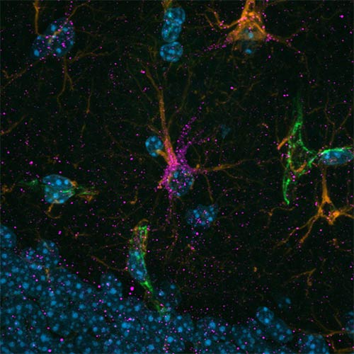
Scientists identified a distinct subtype of brain support cells—astrocytes enriched in the protein cadherin-4 (CDH4), shown in magenta, that seem to protect nerve cells against cell death. In Alzheimer’s disease, these cells become less abundant, but exercise seems to strengthen them. (Image credit: Luis Moreira)
The study focused on a part of the hippocampus – a critical region for memory and learning that is damaged early in Alzheimer’s disease. The research team leveraged single-nuclei RNA sequencing, a relatively new technologies that allow researchers to look at activity at the molecular level in single cells for an in-depth understanding of diseases like Alzheimer’s.
The researchers exercised a common mouse model for Alzheimer’s disease using running wheels, which improved their memory compared to the sedentary counterparts. They then analysed gene activity across thousands of individual brain cells, finding that exercise changed activity both in microglia, a disease-associated population of brain cells, and in a specific type of neurovascular-associated astrocyte (NVA), newly discovered by the team, which are cells associated with blood vessels in the brain. Furthermore, the scientist identified the metabolic gene Atpif1 as an important regulator to create new neurons in the brain. “That we were able to modulate newborn neurons using our new target genes set underscores the promise our study,” said lead author Joana Da Rocha, PhD, a postdoctoral fellow working in Dr Wrann’s lab.
To ensure the findings were relevant to humans, the team validated their discoveries in a large dataset of human Alzheimer’s brain tissue, finding striking similarities.
“This work not only sheds light on how exercise benefits the brain but also uncovers potential cell-specific targets for future Alzheimer’s therapies,” said Nathan Tucker, a biostatistician at SUNY Upstate Medical University and co-senior of the study. “Our study offers a valuable resource for the scientific community investigating Alzheimer’s prevention and treatment.”
A new way of thinking about Alzheimer’s disease has yielded a discovery that could be the key to stopping the cognitive decline seen in Alzheimer’s and other neurodegenerative diseases.
University of Virginia School of Medicine scientists have been investigating the possibility that Alzheimer’s is caused, at least in part, by the immune system’s wayward attempts to fix DNA damage in the brain. Their research reveals that an immune molecule called STING drives the formation of the harmful plaques and protein tangles thought responsible for Alzheimer’s. Blocking the molecule protected lab mice from mental decline, the researchers say.
An important player in the brain’s immune system, STING also may be a key contributor to Parkinson’s disease, amyotrophic lateral sclerosis (ALS or Lou Gehrig’s disease), dementia and other memory-robbing conditions. That means that developing treatments to control its activity could have far-reaching benefits for many patients facing now-devastating diagnoses.
“Our findings demonstrate that the DNA damage that naturally accumulates during aging triggers STING-mediated brain inflammation and neuronal damage in Alzheimer’s disease,” said researcher John Lukens, PhD, director of UVA’s Harrison Family Translational Research Center in Alzheimer’s and Neurodegenerative Diseases. “These results help to explain why aging is associated with increased Alzheimer’s risk and uncover a novel pathway to target in the treatment of neurodegenerative diseases.”
Alarming Trends in Alzheimer’s
Alzheimer’s is a growing problem, with researchers working frantically to find ways to better understand and treat the condition.
The causes of Alzheimer’s remain murky, but scientists are increasingly coming to appreciate the role of the immune system in the disease’s development. STING is part of that immune response; the molecule helps direct the clearance of viruses and stressed cells harboring DNA damage.
While STING is an important defender of the brain, it can also become hyperactive and cause harmful inflammation and tissue damage. That had Lukens and his team eager to determine what part it could be playing in Alzheimer’s. Blocking the molecule’s activity in lab mice, they found, helped prevent Alzheimer’s plaque formation, altered the activity of immune cells called microglia and redirected the workings of important genes, among other effects.
“We found that removing STING dampened microglial activation around amyloid plaques, protected nearby neurons from damage and improved memory function in Alzheimer’s model mice,” said researcher Jessica Thanos, part of UVA’s Department of Neuroscience and Center for Brain Immunology and Glia (BIG Center). “Together, these findings suggest that STING drives detrimental immune responses in the brain that exacerbate neuronal damage and contribute to cognitive decline in Alzheimer’s disease.”
Promising Treatment Target
While scientists have been investigating other molecules thought to be important in Alzheimer’s, STING makes for a particularly attractive target for developing new treatments, the UVA Health researchers say. That’s because blocking STING appears to slow both the buildup of amyloid plaques and the development of tau tangles, the two leading candidates for the cause of Alzheimer’s. Other molecules lack that robust involvement, and, further, could be targeted only at very specific – and very limited – stages in the disease’s progression.
“We are only beginning to understand the complex role of innate immune activation in the brain, and this is especially true in both normal and pathological aging,” Thanos said. “If we can pinpoint which cells and signals sustain that activation, we will be in a much better position to intervene effectively in disease.”
While Lukens’ pioneering research has opened new doors in the fight against Alzheimer’s, much more work will need to be done to translate the findings into treatments. For example, scientists will need to better understand STING’s roles in the body – such as in the immune system’s response to cancer – to ensure any new treatment doesn’t cause unwanted side effects.
But those are the types of big questions that Lukens and his collaborators at the Harrison Family Translational Research Center are eager to tackle as part of their efforts to fast-track new treatments and, eventually, they hope, cures.
“Our hope is that this work moves us close to finding safer and more effective ways to protect the aging brain, as there is an urgent need for treatments that can slow or prevent neuronal damage in Alzheimer’s,” Lukens said. “Shedding light on how STING contributes to that damage may help us target similar molecules and ultimately develop effective disease-modifying treatments.”
A new study has uncovered a surprising link between Alzheimer’s disease and Herpes Simplex Virus-1 (HSV-1).
Neurons in the brain of an Alzheimer’s patient, with plaques caused by tau proteins. Credit: NIH
A new study led by Dr Or Shemesh at the Hebrew University of Jerusalem has uncovered a surprising connection between Alzheimer’s disease and the Herpes Simplex Virus-1 (HSV-1). The research team used advanced techniques to identify 19 HSV-1-related proteins in the brains of people with Alzheimer’s, across all stages of the disease. This discovery, published in Cell Reports, strengthens the growing evidence that infections like HSV-1 might play a role in the development and progression of Alzheimer’s.
One key finding was the increased activity of a herpesvirus protein called ICP27, which became more prominent as the disease advanced. This protein was found to occupy the same space as tau, a brain protein that becomes harmful when it undergoes changes in Alzheimer’s disease, but it did not appear near amyloid plaques, another hallmark of the illness. This suggests that HSV-1 may directly affect tau and contribute to the changes seen in Alzheimer’s.
The team’s experiments with human brain organoids derived from stem cells revealed that HSV-1 infection can increase tau modifications at specific sites linked to Alzheimer’s disease.
Remarkably, these modifications seem to help protect brain cells early on by reducing the amount of virus and preventing cell death. However, as the disease progresses, these same processes may contribute to the brain damage associated with Alzheimer’s. The study also highlighted the role of Alzheimer’s pathologies as part of the brain’s natural immune system in this process, focusing on a pathway called cGAS-STING, which influences tau changes.
Dr Shemesh explained, “Our research shows how HSV-1 interacts with the brain and influences the pathologies of Alzheimer’s disease. Early on, the changes in tau may protect brain cells by limiting the virus, but as the disease advances, these same changes could lead to more harm and accelerate neurodegeneration.”
This study provides new insights into how infections and the brain’s immune response may be involved in Alzheimer’s disease. It suggests that targeting viral activity or modifying the immune system’s response could offer new treatment possibilities. While more research is needed to fully understand these processes, these findings open the door to innovative ways to slow or stop the progression of this devastating disease.