Total joint replacement is a proven treatment to relieve chronic knee pain, but there have been concerns raised that particles from the device may migrate to the brain and cause memory problems. New research from Rush University shows that may not be a worry.
In a study published by Acta Biomaterialia, researchers found that particles in some people with total joint replacements travelled to the brain but did not cause cognitive decline.
Researchers did find an association of cobalt, a key element of most implants, with more Alzheimer’s disease pathology in the brain.
The particles, called wear debris, are caused by friction and movement in the joint, or, in some cases, corrosion. The newer materials used in joint replacements today make this less likely to occur.
“Implants have a very specific combination of metals such as cobalt, and when they show up together in one particle in the brain in the exact same composition that the implant is, we know it can’t come from any other source,” said Robin Pourzal, PhD, associate professor and director of implant materials analysis.
“For years, we’ve known that particles can deposit in tissues surrounding the joint, but this is the first study to look at the particles deposited in the brain.”
In working with Puja Agarwal, PhD, in the Rush Alzheimer’s Disease Center, the team took a closer look at what was happening cognitively in this group of people
The study included data collected among 701 deceased participants in the ongoing Memory and Aging Project conducted by David Bennett, MD, which include older adults residing in the greater Chicago area. The participants had no dementia at the beginning of the study and were followed annually with cognitive assessments using a 19-test battery, a standard collection of memory and thinking skill tests.
A total of 229 people in the study had a total hip, knee or shoulder replacement. The control group was made up of 472 people who had no total joint replacement. Due to a higher likelihood of the metal cobalt being found in total hip replacement, the participants were studied in two groups: 146 with hip replacements and 83 with knee or shoulder replacements.
Pourzal notes that the collaboration was led by faculty members from the Department of Orthopedic Surgery, including leaders from Rush Orthopedic Research, in collaboration with colleagues from the Rush Alzheimer’s Disease Center and international partners.
“Total joint arthroplasty remains one of the most successful and life-changing interventions in modern medicine. But it does suggest that wear particles from the implant, particularly in certain hip replacements involving accelerated wear, may travel beyond the joint and warrant further study.”
Researchers explore brain patterns through different ages to pinpoint where memories get mixed up
Photo by Fakurian Design on Unsplash
Memory loss is usually thought of as something to address as a person ages. Slip-ups like forgetting someone’s name or recognising a familiar face but not remembering where you know them from are often viewed as issues that arise later in life.
New research by a team at Binghamton University reveals why memory shifts as we age – and that shift happens earlier in life than most people think, potentially at middle age.
“This is one of a growing list of studies highlighting that the period of middle age is really important for memory functioning and shouldn’t be ignored. For a long time, most studies have been focused on young adults versus older adults,” said Binghamton Associate Professor of Psychology Ian McDonough, co-author of a new study published in Cerebral Cortex.
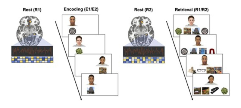
McDonough and postdoctoral associate Destaw Mekbib tested a group of approximately 60 adults ages 18 to 74 by showing them faces paired with various objects and scenes. After a five-minute rest period, participants completed a memory test where they were prompted to pick the correct object or scene for each face – all while an MRI machine took readings of their hippocampal activity.
McDonough said the test mirrors how memories are formed. First, the brain records the new memory. Then, over a short period, the hippocampus replays the memory to stabilise it. Later, the brain retrieves the memory when needed.
The question the researchers had, however, was how aging affects the continuity of the information passing between those stages.
“We’re seeing a big decline in memory accuracy from the 20- to 30-year-old age group, to people in their 50s. Some of these hippocampal processes already start to decline by midlife,” McDonough said. “That suggests middle age is really a transition point.”
Image Credit: Figure 1. Overview of the experiment. Reprinted from Mekbib and McDonough (2026), Cerebral Cortex, 36(7), bhag114, https://doi.org/10.1093/cercor/bhag114, used under CC BY-NC 4.0..
In the context of this research, middle-aged participants often paired a face with the wrong object or scene. They had recalled that they had seen it before, but not which pairing was correct, almost as an “I’ve seen something like this before” phenomenon.
“It becomes hard because now all of these images on the screen during the test seem familiar,” McDonough said. “They know they’ve seen all of these before, but now what they have to remember is that specific link. And that’s where, as people age, they start to really show these errors.”
In comparing how younger and older people access memories, the researchers assumed that older adults would show weaker hippocampal activity, but this wasn’t exactly the case. What they did find was more interesting. When young adults made mistakes in the memory tests, it was because their brains were not activating the original memory patterns, which was expected.
However, when older adults made mistakes, their brains showed strong reactivation of those memory patterns.
“The more they reactivate the hippocampus that’s consistent with encoding, the more likely they are to make these memory errors,” McDonough said. “So instead of that reactivation pattern being associated with better memory, it’s associated with those errors.”
McDonough said it’s not yet known why older adults can show strong hippocampal reactivation while making memory errors. Future follow-up studies could look at memory encoding, consolidation, and retrieval at the individual level, and explore how brain stimulation after learning impacts memory.
He noted that researchers should focus more on people in middle age, following them over many years, given this new information that memory can shift earlier in life.
“We really don’t have a good scientific understanding of what is happening in middle age, because the brain is not declining uniformly across this time, with some regions declining faster than others,” McDonough said. “Finding when those tipping points are is going to be important.”
Quitting smoking in middle age or later is linked to slower age-related cognitive decline over the long term, according to a new study by UCL researchers.
The study, published in The Lancet Healthy Longevity, looked at data from 9436 people aged 40 or over (average age: 58) in 12 countries, comparing cognitive test results among people who quit smoking with those of a matched control group who kept smoking.
The research team found that the cognitive scores of those who had quit smoking declined significantly less than their smoking counterparts in the six years after they quit. For verbal fluency, the rate of decline roughly halved, while for memory it slowed by 20%.
Since slower cognitive decline is related to reduced dementia risk, their findings add to a growing body of evidence suggesting quitting smoking might be a preventative strategy for the disease. Still, more research is needed to confirm this.
Lead author Dr Mikaela Bloomberg (UCL Institute of Epidemiology & Health Care) said: “Our study suggests that quitting smoking may help people to maintain better cognitive health over the long term even when we are in our 50s or older when we quit.
“We already know that quitting smoking, even later in life, is often followed by improvements in physical health and well-being. It seems that, for our cognitive health too, it is never too late to quit.
“This finding is especially important because middle-aged and older smokers are less likely to try to quit than younger groups, yet they disproportionately experience the harms of smoking. Evidence that quitting may support cognitive health could offer new compelling motivation for this group to try and quit smoking.
“Also, as policymakers wrestle with the challenges of an ageing population, these findings provide another reason to invest in tobacco control.”
Smoking is thought to harm brain health in part because it affects cardiovascular health – smoking causes damage to blood vessels that supply oxygen to the brain. Smoking is also thought to affect cognitive health by causing chronic inflammation and directly damaging brain cells through oxidative stress (due to the creation of unstable molecules called free radicals).
Co-author Professor Andrew Steptoe (UCL Institute of Epidemiology & Health Care) said: “Slower cognitive decline is linked to lower dementia risk. These findings add to evidence suggesting that quitting smoking might be a preventative strategy for the disease. However, further research will be needed that specifically examines dementia to confirm this.”
Previous studies, the researchers noted, had found a short-term improvement in cognitive function after people stopped smoking. But whether this improvement was sustained over the longer term – particularly when people quit smoking later in life – was not known.
To answer this question the research team looked at data from three ongoing studies* where a nationally representative group of participants answered survey questions every two years. The studies covered England, the US, and 10 other European countries.
More than 4,700 participants who quit smoking were compared with an equal number of people who carried on smoking. The two groups were matched in terms of their initial cognitive scores and other factors such as age, sex, education level, and country of birth.
The research team found that the two groups’ scores in memory and verbal fluency tests declined at a similar rate in the six years prior to participants of one group quitting smoking. These trajectories then diverged in the six years following smoking cessation.
For the smokers who quit, the rate of decline was about 20% slower for memory and 50% slower for verbal fluency. In practical terms, this meant that with each year of ageing, people who quit experienced three to four months less memory decline and six months less fluency decline than those who continued smoking.
This was an observational analysis, so unmeasured differences between smokers who quit and continuing smokers could remain; while the trends before quitting were similar, the study cannot prove cause and effect.
However, the research team noted their findings were consistent with earlier studies showing that adults aged over 65 who quit smoking during early- or mid-life have comparable cognitive scores to never smokers, and that former and never smokers have a similar risk of dementia a decade or longer after quitting.
*The longitudinal studies were the English Longitudinal Study of Ageing (ELSA), the Survey of Health, Ageing and Retirement in Europe (SHARE), and the Health and Retirement Study (HRS).
Anyone who has taken a yoga class knows how relaxing it can be to set aside the day’s worries and focus on breathing, gentle movements, healing stretches and guided meditation, even if just for an hour.
A growing body of research suggests the soothing powers of yoga may go further than temporarily easing the day’s stress. Yoga is emerging as a potential prescription to boost brain power, offset cognitive decline and help prevent dementia.
“The evidence behind yoga has really picked up,” said Dr Neha Gothe, an associate professor and director of the PhD in Human Movement and Rehabilitation Sciences programme at Bouvé College of Health Sciences at Northeastern University in Boston. “So far, it points toward the potential for it to protect brain health as we are aging.”
Exercise for an aging brain
Research into the health benefits of yoga – the origins of which trace back to 2500 to 5000 years ago – didn’t begin in earnest until the 2000s, when the practice began to surge in popularity in the US, Gothe said. Since then, yoga practice has been shown to have a positive influence on physical as well as mental health, with studies finding it may benefit cardiovascular function, musculoskeletal conditions and overall mental well-being.
More recently, researchers have turned their attention to yoga’s potential benefits on brain health, an area of growing interest as the population ages and the number of adults developing dementia and cognitive decline rises. In the U.S., about 1 in 5 people 65 and older are living with mild cognitive impairment, and 1 in 7 have some type of dementia. Researchers predict a doubling of new dementia cases in the U.S. over the next several decades.
While there is strong evidence that physical activity can benefit brain health and help slow cognitive decline, aging adults are not always able to reach the recommended 150 minutes of moderate-intensity exercise or 75 minutes of vigorously intense physical activity needed to reap these benefits. Federal guidelines also recommend muscle-strengthening activities at least two days a week.
What the research shows about yoga
Yoga – which combines physical movement with breath work and meditation – may offer a more accessible alternative or supplement to other types of exercise, Gothe said.
Studies have shown yoga may have a positive effect on both brain structure and function. In a 2019 analysis of the evidence, Gothe found yoga could hold promise as a means of offsetting age-related and neurodegenerative declines in several regions of the brain. And in another small study comparing yoga practitioners to age- and sex-matched controls, she found women who practiced yoga regularly had more grey matter – the part of the brain that controls memory, thought and movement – and better working memory than those who didn’t.
In some cases, the ancient practice may even be better for the brain than other types of physical activity. In another small study, Gothe found cancer survivors who practiced yoga for 12 weeks reported greater cognitive improvement than those who engaged in aerobic and stretching-toning exercises.
For people who can’t engage in more vigorous activities, it’s certainly more accessible, Gothe said.
“Yoga is just as good as any other form of physical activity, such as walking or stretching,” she said. “For individuals who may not be able to engage in those activities, especially older adults who have other conditions, such as knee pain or arthritis, yoga is a neat alternative to traditional forms of exercise and is very modifiable to accommodate an individual’s abilities.”
How does yoga help?
An explanation for yoga’s brain health benefits may be the close connection yoga forms between the mind and body.
Gothe and her colleagues found the cognitive benefits of yoga may stem from limiting prolonged exposure to stress and inflammation, improving stress regulation and helping the brain communicate better with the body to work more efficiently.
“We have a lot of evidence at this point telling a cohesive story about a mind-body connection with brain health,” said Dr Helen Lavretsky, a professor of psychiatry in-residence and director of integrative psychiatry at the David Geffen School of Medicine at the University of California, Los Angeles.
Lavretsky has led numerous studies on the cognitive benefits of yoga, looking specifically at Kundalini yoga. This type of yoga blends physical postures with meditation and breathing techniques that focus on relaxation, healing and self-awareness.
In several studies, Lavretsky’s team compared Kundalini yoga to memory enhancement training in postmenopausal women: those who practised yoga experienced greater improvements in memory and cognitive function, including executive function, and were able to better prevent grey matter atrophy.
In a separate analysis of published research, Lavretsky looked more broadly at mind-body practices, including yoga and meditation. The review suggested that these practices improved brain function because they were targeting the area of the brain involved in regulating attention, emotional control, mood and cognition.
“Yoga and other mind-body therapies have an effect on stress reduction and other things that underlie brain health,” Lavretsky said. “Our research shows they are well equipped to reduce inflammation, stress, improve sleep and mental health.”
Making yoga a regular practice
How much and what type of yoga is needed to accrue these benefits remains unclear.
While Lavretsky’s studies involved Kundalini yoga, Gothe said her studies mostly involved Hatha yoga, the most widely practiced form. Both blend physical postures with breathing exercises, while Kundalini incorporates more spiritual and meditation elements.
Most studies involve at least eight weeks of yoga, with hourlong classes at least two or three times a week, Gothe said. But “there are no rigorous dose-response studies. So we don’t know exactly what dose is necessary to get an improvement in cognitive performance.”
Even so, yoga shouldn’t be considered a quick fix, Gothe said. To maintain benefits, it’s important to keep up the practice.
“It is a ‘use it or lose it’ phenomena,” she said. “If you continue practicing, you will continue to see improvement. But if you stop, you go back to square one.”
The good news is it’s never too late to begin accruing those benefits, Lavretsky said. She encourages people to begin at a young age, so they have a tool for stress management whenever it’s needed.
“The benefit of starting earlier is that it becomes a lifelong skill,” she said. “But yoga has benefits no matter what your age is.”
New USC research indicates how iron-related oxidative damage and cell death may hasten the development of Alzheimer’s disease in people with Down syndrome
Scientists at the University of Southern Carolina have discovered a key connection between high levels of iron in the brain and increased cell damage in people who have both Down syndrome and Alzheimer’s disease.
In the study, researchers found that the brains of people diagnosed with Down syndrome and Alzheimer’s disease (DSAD) had twice as much iron and more signs of oxidative damage in cell membranes compared to the brains of individuals with Alzheimer’s disease alone or those with neither diagnosis. The results, published in Alzheimer’s & Dementia: The Journal of the Alzheimer’s Association, point to a specific cellular death process that is mediated by iron, and the findings may help explain why Alzheimer’s symptoms often appear earlier and more severely in individuals with Down syndrome.
“This is a major clue that helps explain the unique and early changes we see in the brains of people with Down syndrome who develop Alzheimer’s,” said Max Thorwald, lead author of the study and a postdoctoral fellow in the laboratory of University Professor Emeritus Caleb Finch at the USC Leonard Davis School. “We’ve known for a long time that people with Down syndrome are more likely to develop Alzheimer’s disease, but now we’re beginning to understand how increased iron in the brain might be making things worse.”
Down syndrome and Alzheimer’s
Down syndrome is caused by having an extra third copy, or trisomy, of chromosome 21. This chromosome includes the gene for amyloid precursor protein, or APP, which is involved in the production of amyloid-beta (Aβ), the sticky protein that forms telltale plaques in the brains of people with Alzheimer’s disease.
Because people with Down syndrome have three copies of the APP gene instead of two, they tend to produce more of this protein. By the age of 60, about half of all people with Down syndrome show signs of Alzheimer’s disease, which is approximately 20 years earlier than in the general population.
“This makes understanding the biology of Down syndrome incredibly important for Alzheimer’s research,” said Finch, the study’s senior author.
Key findings point to ferroptosis
The research team studied donated brain tissue from individuals with Alzheimer’s, DSAD, and those without either diagnosis. They focused on the prefrontal cortex — an area of the brain involved in thinking, planning, and memory — and made several important discoveries:
Iron levels much higher in DSAD brains: Compared to the other groups, DSAD brains had twice the amount of iron in the prefrontal cortex. Scientists believe this buildup comes from tiny brain blood vessel leaks called microbleeds, which occur more frequently in DSAD than in Alzheimer’s and are correlated with higher amounts of APP.
More damage to lipid-rich cell membranes: Cell membranes are made of fatty compounds called lipids and can be easily damaged by chemical stress. In DSAD brains, the team found more byproducts of this type of damage, known as lipid peroxidation, compared to amounts in Alzheimer’s-only or control brains.
Weakened antioxidant defense systems: The team found that the activity of several key enzymes that protect the brain from oxidative damage and repair cell membranes was lower in DSAD brains, especially in areas of the cell membrane called lipid rafts.
Together, these findings indicate increased ferroptosis, a type of cell death characterised by iron-dependent lipid peroxidation, Thorwald explained: “Essentially, iron builds up, drives the oxidation that damages cell membranes, and overwhelms the cell’s ability to protect itself.”
Lipid rafts: a hotspot for brain changes
The researchers paid close attention to lipid rafts — tiny parts of the brain cell membrane that play crucial roles in cell signalling and regulate how proteins like APP are processed. They found that in DSAD brains, lipid rafts had much more oxidative damage and fewer protective enzymes compared to Alzheimer’s or healthy brains.
Notably, these lipid rafts also showed increased activity of the enzyme β-secretase, which interacts with APP to produce Aβ proteins. The combination of more damage and more Aβ production may promote the growth of amyloid plaques, thus speeding up Alzheimer’s progression in people with Down syndrome, Finch explained.
Rare Down syndrome variants offer insight
The researchers also studied rare cases of individuals with “mosaic” or “partial” Down syndrome, in which the third copy of chromosome 21 is only present in a smaller subset of the body’s cells. These individuals had lower levels of APP and iron in their brains and tended to live longer. In contrast, people with full trisomy 21 and DSAD had shorter lifespans and higher levels of brain damage.
“These cases really support the idea that the amount of APP — and the iron that comes with it — matters a lot in how the disease progresses,” Finch said.
Looking ahead
The team says their findings could help guide future treatments, especially for people with Down syndrome who are at high risk of Alzheimer’s. Early research in mice suggests that iron-chelating treatments, in which medicine binds to the metal ions and allows them to leave the body, may reduce indicators of Alzheimer’s pathology, Thorwald noted.
“Medications that remove iron from the brain or help strengthen antioxidant systems might offer new hope,” Thorwald said. “We’re now seeing how important it is to treat not just the amyloid plaques themselves but also the factors that may be hastening the development of those plaques.”
Healthy hearts are adaptable, and heartbeats exhibit complex variation as they adjust to tiny changes in the body and environment. Mass General Brigham researchers have applied a new way to measure the complexity of pulse rates, using data collected through wearable pulse oximetry devices. The new method, published in the Journal of the American Heart Association, provides a more detailed peek into heart health than traditional measures, uncovering a link between reduced complexity and future cognitive decline.
“Heart rate complexity is a hallmark of healthy physiology,” said senior author Peng Li, PhD, of the Department of Anesthesia, Critical Care and Pain Medicine at Massachusetts General Hospital (MGH) and the Division of Sleep and Circadian Disorders at Brigham and Women’s Hospital (BWH). “Our hearts must balance between spontaneity and adaptability, incorporating internal needs and external stressors.”
The study used data from 503 participants (average age 82, 76% women) in the Rush Memory and Aging Project. The researchers analysed overnight pulse rate measurements – collected by a fingertip pulse oximetry device known as the Itamar WatchPAT 300 device – and comprehensive measures of cognitive functions, collected around the same time as the pulse rate measurement and at least one annual follow-up visit up to 4.5 years later.
The team found that people with greater complexity in their heartbeats at baseline tend to experience slower cognitive decline over time. They determined that the conventional measures of heart rate variability did not predict this effect, indicating their measure was more sensitive in capturing heart functions predictive of cognitive decline.
The researchers plan to investigate whether pulse rate complexity can predict development of dementia, which would make it useful for identifying people at an early stage who might benefit from therapeutic interventions.
“The findings underscore the usefulness of our approach as a noninvasive measure for how flexible the heart is in responding to nervous system cues,” said lead author Chenlu Gao, PhD, also in the Department of Anesthesia, Critical Care and Pain Medicine at MGH. “It is suitable for future studies aimed at understanding the interplay between heart health and cognitive aging.”
Past research has suggested that inflammation may contribute to the development and progression of dementia and that non-steroidal anti-inflammatory (NSAID) medications may help protect against dementia due to their anti-inflammatory effects. A new large prospective study published in the Journal of the American Geriatrics Society provides additional evidence, showing that long-term NSAID use is linked to a decreased risk of developing dementia.
In the population-based study of 11 745 adults with an average follow-up of 14.5 years, 9520 participants had used NSAIDs at any given time, and 2091 participants developed dementia. Long-term NSAID use was associated with a 12% reduced risk of developing dementia. Short- and intermediate-term use did not provide benefits. Also, the cumulative dose of NSAIDs was not associated with decreased dementia risk.
The findings suggest that prolonged, rather than intensive, use of anti-inflammatory medications may help protect against dementia.
“Our study provides evidence on possible preventive effects of anti-inflammatory medication against the dementia process. There is a need for more studies to further consolidate this evidence and possibly develop preventive strategies,” said corresponding author M. Arfan Ikram, MSc, MD, PhD, of Erasmus MC University Medical Center Rotterdam, in the Netherlands.
Meeting the minimum requirement for vitamin B12, needed to make DNA, red blood cells and nerve tissue, may not actually be enough – particularly if for older adults. It may even put them at risk for cognitive impairment, according to a study published in Annals of Neurology.
The research found that older, healthy volunteers, with lower concentrations of B12, but still in the normal range, showed signs of neurological and cognitive deficiency. These levels were associated with more damage to the brain’s white matter – the nerve fibres that enable communication between areas of the brain – and test scores associated with slower cognitive and visual processing speeds, compared to those with higher B12.
The UC San Francisco researchers, led by senior author Ari J. Green, MD, of the Departments of Neurology and Ophthalmology and the Weill Institute for Neurosciences, said that the results raise questions about current B12 requirements and suggest the recommendations need updating.
“Previous studies that defined healthy amounts of B12 may have missed subtle functional manifestations of high or low levels that can affect people without causing overt symptoms,” said Green, noting that clear deficiencies of the vitamin are commonly associated with a type of anaemia. “Revisiting the definition of B12 deficiency to incorporate functional biomarkers could lead to earlier intervention and prevention of cognitive decline.”
Lower B12 correlates with slower processing speeds, brain lesions
In the study, researchers enrolled 231 healthy participants without dementia or mild cognitive impairment, whose average age was 71. They were recruited through the Brain Aging Network for Cognitive Health (BrANCH) study at UCSF.
Their blood B12 amounts averaged 414.8pmol/L, well above the U.S. minimum of 148pmol/L. Adjusted for factors like age, sex, education and cardiovascular risks, researchers looked at the biologically active component of B12, which provides a more accurate measure of the amount of the vitamin that the body can utilize. In cognitive testing, participants with lower active B12 were found to have slower processing speed, relating to subtle cognitive decline. Its impact was amplified by older age. They also showed significant delays responding to visual stimuli, indicating slower visual processing speeds and general slower brain conductivity.
MRIs revealed a higher volume of lesions in the participants’ white matter, which may be associated with cognitive decline, dementia or stroke.
While the study volunteers were older adults, who may have a specific vulnerability to lower levels of B12, co-first author Alexandra Beaudry-Richard, MSc, said that these lower levels could “impact cognition to a greater extent than what we previously thought, and may affect a much larger proportion of the population than we realize.” Beaudry-Richard is currently completing her doctorate in research and medicine at the UCSF Department of Neurology and the Department of Microbiology and Immunology at the University of Ottawa.
“In addition to redefining B12 deficiency, clinicians should consider supplementation in older patients with neurological symptoms even if their levels are within normal limits,” she said. “Ultimately, we need to invest in more research about the underlying biology of B12 insufficiency, since it may be a preventable cause of cognitive decline.”
Subtle changes in the brain, detectable through advanced imaging, blood and spinal fluid analysis, happen approximately twenty years before a clinical motor diagnosis in people with Huntington’s disease, finds a new study led by UCL researchers which appears in Nature Medicine.
The team found that although functions such as movement, thinking or behaviour remained normal for a long time before the onset of symptoms in Huntington’s disease, subtle changes to the brain were taking place up to two decades earlier. These findings pave the way for future preventative clinical trials, offer hope for earlier interventions that could preserve brain function and improve outcomes for individuals at risk of Huntington’s disease.
Huntington’s disease is a devastating neurodegenerative condition affecting movement, thinking and behaviour. It is a genetic disease and people with an affected parent have a 50% chance of inheriting the Huntington’s disease mutation, meaning they will develop disease symptoms – typically in mid-adulthood.
The disease is caused by repetitive expansions of three DNA blocks (C, A and G) in the huntingtin gene. This sequence tends to continually expand in certain cells over a person’s life, in a process known as somatic CAG expansion. This ongoing expansion accelerates neurodegeneration, making brain cells more vulnerable over time.
For the new study, the researchers studied 57 people with the Huntington’s disease gene expansion, who were calculated as being on average 23.2 years from a predicted clinical motor diagnosis.
They were examined at two time points over approximately five years to see how their bodies and brains changed over time. Their results were compared to 46 control participants, matched closely for age, sex and educational level.
As part of the study, all participants volunteered to undergo comprehensive assessments of their thinking, movement and behaviour, alongside brain scans and blood and spinal fluid sampling.
Importantly, the group with Huntington’s disease gene expansion showed no decline in any clinical function (thinking, movement or behaviour) during the study period, compared to the closely matched control group.
However, compared to the control group, subtle changes were detected in brain scans and spinal fluid biomarkers of those with Huntington’s disease gene expansion. This indicates that the neurodegenerative process begins long before symptoms are evident and before a clinical motor diagnosis.
Specifically, the researchers identified elevated levels of neurofilament light chain (NfL), a protein released into the spinal fluid when neurons are injured, and reduced levels of proenkephalin (PENK), a neuropeptide marker of healthy neuron state that could reflect changes in the brain’s response to neurodegeneration.
Lead author, Professor Sarah Tabrizi (UCL Huntington’s Disease Research Centre, UCL Queen Square Institute of Neurology, and UK Dementia Research Institute at UCL), said: “Our study underpins the importance of somatic CAG repeat expansion driving the earliest neuropathological changes of the disease in living humans with the Huntington’s disease gene expansion. I want to thank the participants in our young adult study as their dedication and commitment over the last five years mean we hope that clinical trials aimed at preventing Huntington’s disease will become a reality in the next few years.”
The findings suggest that there is a treatment window, potentially decades before symptoms are present, where those at risk of developing Huntington’s disease are functioning normally despite having detectable measures of subtle, early neurodegeneration. Identifying these early markers of disease is essential for future clinical trials in order to determine whether a treatment is having any effect.
Co-first author of the study, Dr Rachael Scahill (UCL Huntington’s Disease Research Centre and UCL Queen Square Institute of Neurology) said: “This unique cohort of individuals with the Huntington’s disease gene expansion and control participants provides us with unprecedented insights into the very earliest disease processes prior to the appearance of clinical symptoms, which has implications not only for Huntington’s disease but for other neurodegenerative conditions such as Alzheimer’s disease.”
This study is the first to establish a direct link between somatic CAG repeat expansion, measured in blood, and early brain changes in humans, decades before clinical motor diagnosis in Huntington’s disease.
While somatic CAG expansion was already known to accelerate neurodegeneration, this research demonstrates how it actively drives the earliest detectable changes in the brain: specifically in the caudate and putamen, regions critical to movement and thinking.
By showing that somatic CAG repeat expansion changes measured in blood predicts brain volume changes and other markers of neurodegeneration, the findings provide crucial evidence to support the hypothesis that somatic CAG expansion is a key driver of neurodegeneration.
With treatments aimed at suppressing somatic CAG repeat expansion currently in development, this work validates this mechanistic process as a promising therapeutic target and represents a critical advance towards future prevention trials in Huntington’s disease.
Co-first author of the study, Dr Mena Farag (UCL Huntington’s Disease Research Centre and UCL Queen Square Institute of Neurology) added: “These findings are particularly timely as the Huntington’s disease therapeutic landscape expands and progresses toward preventive clinical trials.”
The research was done in collaboration with experts at the Universities of Glasgow, Gothenburg, Iowa, and Cambridge.
People who eat more red meat, especially processed red meat like bacon, sausage and bologna, are more likely to have a higher risk of cognitive decline and dementia when compared to those who eat very little red meat, according to a study published in the January 15, 2025, online issue of Neurology®, the medical journal of the American Academy of Neurology.
“Red meat is high in saturated fat and has been shown in previous studies to increase the risk of type 2 diabetes and heart disease, which are both linked to reduced brain health,” said study author Dong Wang, MD, ScD, of Brigham and Women’s Hospital in Boston. “Our study found processed red meat may increase the risk of cognitive decline and dementia, but the good news is that it also found that replacing it with healthier alternatives, like nuts, fish and poultry, may reduce a person’s risk.”
To examine the risk of dementia, researchers included a group of 133 771 people (65.4% female) with an average age of 49 who did not have dementia at the start of the study. They were followed up to 43 years. Of this group, 11 173 people developed dementia.
Participants completed a food diary every two to four years, listing what they ate and how often.
Researchers defined processed red meat as bacon, hot dogs, sausages, salami, bologna and other processed meat products. They defined unprocessed red meat as beef, pork, lamb and hamburger. A serving of red meat is three ounces (85gm), about the size of a deck of cards.
For processed red meat, they divided participants into three groups. The low group ate an average of fewer than 0.10 servings per day; the medium group ate between 0.10 and 0.24 servings per day; and the high group, 0.25 or more servings per day.
After adjusting for factors such as age, sex and other risk factors for cognitive decline, researchers found that participants in the high group had a 13% higher risk of developing dementia compared to those in the low group.
For unprocessed red meat, researchers compared people who ate an average of less than one half serving per day to people who ate one or more servings per day and did not find a difference in dementia risk.
To measure subjective cognitive decline, researchers looked at a different group of 43,966 participants with an average age of 78. Subjective cognitive decline is when a person reports memory and thinking problems before any decline is large enough to show up on standard tests.
The subjective cognitive decline group took surveys rating their own memory and thinking skills twice during the study.
After adjusting for factors such as age, sex and other risk factors for cognitive decline, researchers found that participants who ate an average of 0.25 servings or more per day of processed red meat had a 14% higher risk of subjective cognitive decline compared to those who ate an average of fewer than 0.10 servings per day.
They also found people who ate one or more servings of unprocessed red meat per day had a 16% higher risk of subjective cognitive decline compared to people who ate less than a half serving per day.
To measure objective cognitive function, researchers looked at a different group of 17 458 female participants with an average age of 74. Objective cognitive function is how well your brain works to remember, think and solve problems.
This group took memory and thinking tests four times during the study.
After adjusting for factors such as age, sex and other risk factors for cognitive decline, researchers found that eating higher processed red meat was associated with faster brain aging in global cognition with 1.61 years with each additional serving per day and in verbal memory with 1.69 years with each additional serving per day.
Finally, researchers found that replacing one serving per day of processed red meat with one serving per day of nuts and legumes was associated with a 19% lower risk of dementia and 1.37 fewer years of cognitive aging. Making the same substitution for fish was associated with a 28% lower risk of dementia and replacing with chicken was associated with a 16% lower risk of dementia.
“Reducing how much red meat a person eats and replacing it with other protein sources and plant-based options could be included in dietary guidelines to promote cognitive health,” said Wang. “More research is needed to assess our findings in more diverse groups.”
A limitation of the study was that it primarily looked at white health care professionals, so the results might not be the same for other race, ethnic and non-binary sex and gender populations.