Study Reveals How a Stubborn Lung Infection Evolves Inside Patients over Years
Researchers wanted to know what allows the infection to hang on or come back, and whether it develops new tricks or resistances while living inside the lungs.

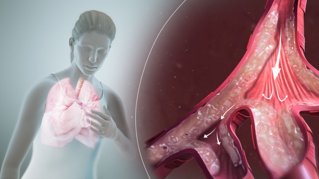
Researchers at Trinity Translational Medicine Institute (TTMI) and the Irish Mycobacterial Reference Laboratory at St James’s Hospital have uncovered how the bacterium Mycobacterium avium – a leading cause of difficult-to-treat chronic lung infections – changes and adapts inside patients over many years of illness. Their findings, published in Genome Medicine, could help doctors understand why M. avium infections come back and why antibiotics sometimes fail.
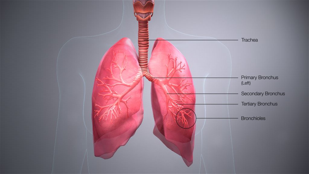
The team undertook this research to understand how M. avium manages to survive for years in people’s lungs, even during long courses of antibiotics. This bacterium causes a type of chronic lung infection that’s becoming more common around the world. By looking closely at its genetic code, the team hoped to see how it changes inside the body and why it can be so difficult to clear.
Treating M. avium lung disease is difficult – patients often need 12 months or more of several antibiotics, and treatment still fails in up to half of cases. Many patients get sick again even after therapy.
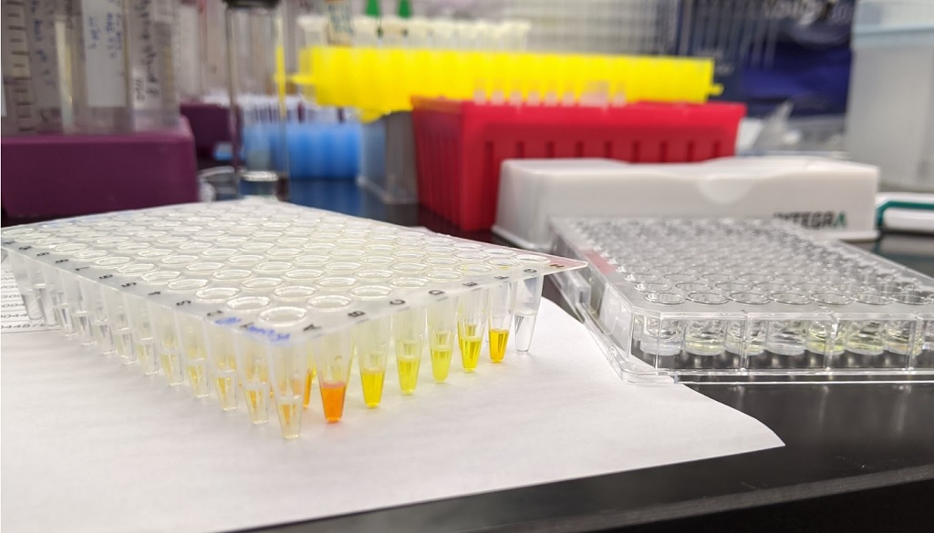
The team used whole-genome sequencing to analyse nearly 300 bacterial samples from patients in Ireland, the UK and Germany, including 20 Irish patients treated at St James’s Hospital. By reading the DNA of these bacteria over time, the scientists tracked how M. avium evolves, swaps strains and develops resistance while living in the human lung.
They found that infection is often not caused by one single long-term strain, but by repeated reinfection with new ones, sometimes closely related to strains seen in other European countries—hinting at shared environmental sources. The bacterium acquires roughly one new genetic change per year, and most importantly, the team found that thirteen specific genes showed signs of adaptation to antibiotics, immune attack and low-oxygen stress.
Lead author Dr Aaron Walsh, researcher in the Trinity Translational Medicine Institute said:
“Our study shows that M. avium can evolve in real time inside the lung. Understanding which genes help it survive may point us towards new treatment targets for this increasingly common and stubborn infection.”
This is the first study to use whole-genome sequencing to follow M. avium infections inside patients over many years, revealing how the germ evolves within the lungs.
Key findings from study
- Reinfection is common: Many patients picked up new strains over time, suggesting they were reinfected from the environment rather than suffering relapse of the same infection.
- International connections: Some Irish strains were genetically almost the same as ones from UK and Germany.
- Thirteen key genes changed under pressure: These genes help the bacterium cope with antibiotics, low oxygen, or attack by the immune system.
- Resistance can appear during treatment: We saw changes in a gene linked to rifampicin resistance in two patients receiving that drug.
Uniquely, in this study researchers found that thirteen genes under “positive selection” was new for M. avium.
Dr Emma Roycroft, Specialist Medical Scientist in the Irish Mycobacterial Reference Laboratory. “Some of those genes weren’t previously linked to survival of M. avium inside the body. For example, one involved in handling oxidative stress and another in forming biofilms. This has highlighted important pathways that could be targeted with new treatments. It was also striking that Irish, British and German samples were so closely related, even though the patients had never met.”
The team’s next steps are:
- Test in the lab how those thirteen genes help the bacterium survive.
- Use long-read sequencing to see genetic changes that short-read methods miss.
- Study environmental samples to find where reinfections come from.
- Expand their research to other patient groups to see if the same patterns occur.
Source: Trinity College Dublin