Category: Respiratory Diseases

Monthly Injection Helps Severe Asthma Patients Safely Stop or Reduce Daily Steroids

Photo by Elen Sher on Unsplash

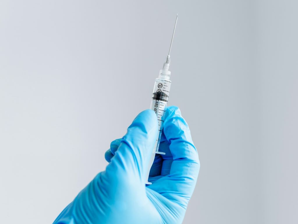
A monthly injection has helped 90% of severe asthma patients reduce daily steroid tablets, which are associated with long-term side effects. More than half of the participants who had received the injection were able to stop their daily steroid tablets entirely, without any impact on their symptoms.

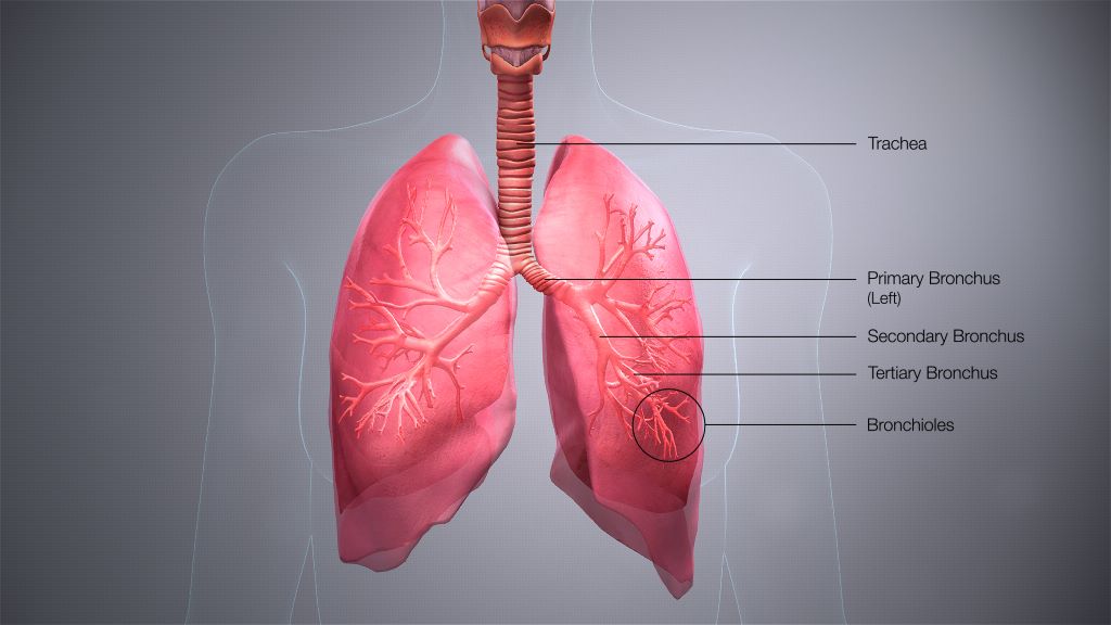
The clinical trial led by a King’s College London academic followed patients who had been injected with tezepelumab every four weeks for a year. Tezepelumab is a type of antibody which targets parts of the immune system, reducing lung inflammation.

Treatment with tezepelumab was also shown to significantly improve asthma symptoms, lung function, and overall quality of life. During the study, two-thirds of patients stopped having any asthma attacks. These improvements were seen as early as two weeks into treatment and lasted for the duration of the study.

Scientists are trying to identify alternative treatments for managing severe asthma, as long-term daily steroid use can lead to serious health problems, including osteoporosis, diabetes, and increased vulnerability to infections.

The WAYFINDER study, published in The Lancet Respiratory Medicine, is among long-standing research into severe asthma at King’s College London. Last year, another team at King’s discovered that another antibody, benralizamab, could be injected during some asthma and COPD attacks to reduce the need for further treatment. The latest discovery could help people manage their asthma long term.

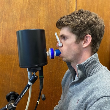
Participants in the trial had a diagnosis of severe asthma and were recruited from 68 clinical centres across 11 countries. They received tezepelumab every four weeks and completed questionnaires on their asthma symptoms and medication at 28 and 52 weeks.

Professor David Jackson, Respiratory Medicine expert at King’s College London, and Clinical Lead of the asthma services across Guy’s and Royal Brompton Hospitals, Guy’s and St Thomas’ NHS Foundation Trust, said: “The WAYFINDER study is an important step forward for patients with the most severe form of asthma who require daily oral steroids in order to achieve reasonable disease control.

“In this International, multicentre clinical trial of more than 300 patients, the NICE-approved asthma treatment tezepelumab, a biologic therapy that targets asthma-related inflammation but without all the side effects of steroids, was capable of allowing the vast majority of patients to wean their steroids down to a low dose with over half able to stop their steroids altogether.

“As tezepelumab also suppresses allergy related symptoms and improves chronic rhinosinusitis as well, the results are particularly exciting for patients with severe asthma who suffer with both upper and lower airway symptoms.”

Dr Samantha Walker, Director of Research & Innovation at Asthma + Lung UK, said: “This study is a promising sign that tezepelumab injections support certain people with severe asthma to reduce or stop taking steroid tablets, which can have serious unwanted health consequences. Tezepelumab, an injectable biologic, significantly improves asthma symptoms, lung function and overall quality of life for participants.

“This is an incredibly encouraging development for the future of asthma care that could transform the lives of people with severe asthma. It’s vital that research into new types of treatment continues but we know current funding for lung health research is on life-support, despite lung conditions remaining the third biggest cause of death in the UK. Studies like this show the positive impact that research can make on providing potentially life-changing treatment for people with asthma and other lung conditions.”

The findings of the WAYFINDER study will be presented at the British Thoracic Society Winter Meeting 2025 on Thursday 27th November 2025.

Read the study here: https://www.thelancet.com/journals/lanres/article/PIIS2213-2600(25)00359-5/fulltext

Metabolic Hormone Found to Boost Resilience Against Flu Symptoms

Photo by Andrea Piacquadio on Pexels

A hormone known for regulating energy balance also helps the body cope with influenza by triggering protective responses in the brain, a study led by UT Southwestern Medical Center researchers shows. The findings, published in the Proceedings of the National Academy of Sciences (PNAS)suggest that targeting this pathway could offer a new pharmacological approach for treating the flu.

“Our work demonstrates that FGF21, a stress-induced hormone that regulates whole-body metabolism, acts on the brain to protect against the hypothermia and weight loss caused by influenza infection,” said senior author Steven Kliewer, PhD, Professor of Molecular Biology and Pharmacology at UT Southwestern.

The study found that levels of fibroblast growth factor 21 (FGF21) rose in both humans and mice during flu infection. In mice, the hormone activated a brain region that regulates the noradrenergic nervous system, prompting heat production from tissues that help regulate body temperature in mice.

This thermogenic response helped stabilise body temperature and improved the response to flu infection. Mice lacking FGF21 or its receptor in these neurons recovered more slowly, while treatment with pharmacologic FGF21 improved recovery. The hormone did not change viral levels, indicating that it protects the body by mitigating the physiological stress of infection rather than directly targeting the virus. Collectively, these results suggest FGF21 could help the body respond more effectively to a range of infections, not just influenza. 

“For serious cases of influenza infection, the care is mostly supportive,” Dr Kliewer said. “Our findings suggest a new pharmacological approach for treating the flu. Further studies are required to determine if these findings are applicable to other infections.”

The research builds on decades of work from the Mangelsdorf/Kliewer Lab at UTSW, which previously identified FGF21 as a hormone produced by the liver in response to metabolic stresses such as fasting and alcohol exposure. The new study extends that work to infection, showing that FGF21 uses the same liver-to-brain signalling pathway to help the body maintain metabolic balance during illness. 

“These findings demonstrate that the immune system is not the only critical part of the response to infection,” said corresponding author Kartik Rajagopalan, M.D., Ph.D., Assistant Professor of Internal Medicine in the Division of Pulmonary and Critical Care Medicine and in Children’s Medical Center Research Institute at UT Southwestern. “There are signals that are sent to the brain that reprogram metabolism for an optimal response.”

Source: UT Southwestern Medical Center

Lung Study Reveals Why Deep Sighs Are Actually Good for Us  

Photo by Julian Jagtenberg on Pexels

The surface of the lungs is covered with a fluid that increases their deformability. This fluid has the greatest effect when you take deep breaths from time to time, as researchers at ETH Zurich have discovered using sophisticated measurement techniques in the laboratory.  In their study, published in Science Advances, they found that it reorders the thin films created by lung fluids.

More than half of all premature babies born before the 28th week of pregnancy develop respiratory distress syndrome shortly after birth. As their lungs are not yet fully developed, they produce too little of the seemingly magical fluid that reduces surface tension in the lungs. As a result, some alveoli collapse – and the lungs are unable to get enough oxygen.  

Lungs become more deformable

Until 40 years ago, this usually spelled death. But then, in the late 1980s, pediatricians developed a life-saving procedure: they extracted the fluid from animal lungs and injected it into the lungs of premature babies. “This works very well in newborns,” says Jan Vermant, Professor of Soft Materials at ETH Zurich. “The fluid coats the entire surface, making the lungs more deformable or – with a more technical word – compliant.”  

But even in adults, lungs can fail. During the coronavirus pandemic, around 3000 people in Switzerland developed acute respiratory distress syndrome. Injecting surface-active fluid from animal lungs into the lungs of adults, however, does not help. “This shows that it’s not just about reducing surface tension,” as Vermant states. “We believe that mechanical stresses within the fluid also play an important role.”  

In collaboration with scientists from Spain, Belgium and the USA, his research group harnessed sophisticated measurement techniques to investigate precisely how lung fluid behaves when it is stretched and recompressed in the laboratory. The fluid in our bodies is also subjected to similar movements when the lungs expand during inhalation and contract again during exhalation.

Explanation for the feeling of relief in the chest  

In their experiments, the researchers simulated the movements of normal and particularly deep breaths – measuring the surface stress of the fluid in each case. “This surface stress influences how compliant the lungs are,” explains Vermant. The more compliant the lungs are, the less resistance there is to expansion and contraction – and the easier it is to breathe.  

With the help of their measuring instruments, the researchers found that surface stress decreases significantly after deep breaths. Apparently, there is a physical explanation for the feeling of relief experienced in the chest that often occurs after a deep sigh. The explanation starts from realising that the thin film formed by the lung fluid on the surface of the lungs actually consists of several layers.  

“Directly at the boundary with the air, there is a slightly stiffer surface layer. Underneath, there are several layers that should be softer than the surface layer,” explains Maria Novaes-Silva, a doctoral student in Vermant’s research group and first author of the study. As she has proven in experiments, this layering returns to the equilibrium configuration over time when the fluid does not move at all or moves only slightly during shallow breathing.  

Reconstructing multilayered structures  

A deep breath is needed from time to time to restore this ideal layering. Based on their analyses, the researchers have discovered that the pronounced stretching and compression of the pulmonary fluid causes the composition of the outer layer to change. ” There is an enrichment of saturated lipids, this results in a more densely packed interface,” says Novaes-Silva. Vermant adds: “This is a state outside of the boundaries of the thermodynamic equilibrium that can only be maintained through mechanical work.”  

It is also known from clinical practice that lung compliance gradually changes over time – and that breathing becomes increasingly difficult in connection with constant shallow breathing. The measurements in the laboratory therefore seems to reflect observations from the clinic. Novaes-Silva concludes: “These similarities are indications that we have captured real properties with our experimental setup.”  

Can the new insights gained by materials scientists also be used to derive expedient conclusions and insights for lung failure in adults? “A promising approach is to identify components that can artificially reconstruct multilayered structures,” the researchers note in their technical article. In conversation, Vermant points to therapies involving foam that are currently being developed and researched in greater depth by other groups. 

Source: ETH Zurich

Mortality Risk is Six Times Higher in Hospital Patients with Dyspnoea

Photo by engin akyurt on Unsplash

The risk of dying is six times higher among patients who become short of breath after being admitted to hospital, according to research published on Monday in ERJ Open Research. Patients who were in pain were not more likely to die.

The study of nearly 10 000 people suggests that asking patients if they are feeling short of breath could help doctors and nurses to focus care on those who need it most.

The study is the first of its kind and was led by Associate Professor Robert Banzett from Beth Israel Deaconess Medical Center, Harvard Medical School, Boston, USA. He said: “In hospital, nurses routinely ask patients to rate any pain they are experiencing, but this is not the case for dyspnoea. In the past, our research has shown that most people are good at judging and reporting this symptom, yet there is very little evidence on whether it’s linked to how ill hospital patients are.”

Working with nurses at Beth Israel Deaconess Medical Center, who documented patient-reported dyspnoea twice per day, the researchers found that it was feasible to ask hospital patients to rate their dyspnoea from 0 to 10, in the same way they are asked to rate their pain. Asking the question and recording the answer only took 45 seconds per patient.

Researchers analysed patient-rated shortness of breath and pain for 9 785 adults admitted to the hospital between March 2014 and September 2016. They compared this with data on outcomes, including deaths, in the following two years.

This showed that patients who developed shortness of breath in hospital were six times more likely to die in hospital than patients who were not feeling short of breath. The higher patients rated their shortness of breath the higher their risk of dying. Patients with dyspnoea were also more likely to need care from a rapid response team and to be transferred to intensive care.

Twenty-five per cent of patients who were feeling short of breath at rest when they were discharged from hospital died within six months, compared to seven per cent mortality among those who felt no dyspnoea during their time in hospital.

Conversely, researchers found no clear link between pain and risk of dying.

Professor Banzett said: “It is important to note that dyspnoea is not a death sentence – even in the highest risk groups, 94% of patients survive hospitalisation, and 70% survive at least two years following hospitalisation. But knowing which patients are at risk with a simple, fast, and inexpensive assessment should allow better individualised care. We believe that routinely asking patients to rate their shortness of breath will lead to better management of this often-frightening symptom.

“The sensation of dyspnoea is an alert that the body is not getting enough oxygen in and carbon dioxide out. Failure of this system is an existential threat. Sensors throughout the body, in the lungs, heart and other tissues, have evolved to report on the status of the system at all times, and provide early warning of impending failure accompanied by a strong emotional response.

“Pain is also a useful warning system, but it does not usually warn of an existential threat. If you hit your thumb with a hammer, you will probably rate your pain 11 on a scale of 0-10, but there is no threat to your life. It is possible that specific kinds of pain, for instance pain in internal organs, may predict mortality, but this distinction is not made in the clinical record of pain ratings.”

The researchers say their findings should be confirmed in other types of hospital elsewhere in the world, and that research is needed to show whether asking patients to rate their shortness of breath leads to better treatments and outcomes.

Professor Hilary Pinnock is Chair of the European Respiratory Society’s Education Council, based at the University of Edinburgh and was not involved in the research. She said: “Historically, the monitoring of vital signs in hospitalised patients includes respiratory rate along with temperature and pulse rate. In a digital age, some have questioned the value of this workforce-intensive routine, so it is interesting to read about the association of subjective breathlessness with mortality and other adverse outcomes.

“Breathlessness was assessed on a 0-10 scale which took less than a minute to administer. These noteworthy findings should trigger more research to understand the mechanisms underpinning this association and how this ‘powerful alarm’ can be harnessed to improve patient care.”

Source: EurekAlert!

Study Reveals How a Stubborn Lung Infection Evolves Inside Patients over Years

Researchers wanted to know what allows the infection to hang on or come back, and whether it develops new tricks or resistances while living inside the lungs.

Photo by Anna Shvets

Researchers at Trinity Translational Medicine Institute (TTMI) and the Irish Mycobacterial Reference Laboratory at St James’s Hospital have uncovered how the bacterium Mycobacterium avium – a leading cause of difficult-to-treat chronic lung infections – changes and adapts inside patients over many years of illness. Their findings, published in Genome Medicine, could help doctors understand why M. avium infections come back and why antibiotics sometimes fail.

The team undertook this research to understand how M. avium manages to survive for years in people’s lungs, even during long courses of antibiotics. This bacterium causes a type of chronic lung infection that’s becoming more common around the world. By looking closely at its genetic code, the team hoped to see how it changes inside the body and why it can be so difficult to clear.

Treating M. avium lung disease is difficult – patients often need 12 months or more of several antibiotics, and treatment still fails in up to half of cases. Many patients get sick again even after therapy.

The team used whole-genome sequencing to analyse nearly 300 bacterial samples from patients in Ireland, the UK and Germany, including 20 Irish patients treated at St James’s Hospital. By reading the DNA of these bacteria over time, the scientists tracked how M. avium evolves, swaps strains and develops resistance while living in the human lung.

They found that infection is often not caused by one single long-term strain, but by repeated reinfection with new ones, sometimes closely related to strains seen in other European countries—hinting at shared environmental sources. The bacterium acquires roughly one new genetic change per year, and most importantly, the team found that thirteen specific genes showed signs of adaptation to antibiotics, immune attack and low-oxygen stress.

Lead author Dr Aaron Walsh, researcher in the Trinity Translational Medicine Institute said:

 “Our study shows that M. avium can evolve in real time inside the lung. Understanding which genes help it survive may point us towards new treatment targets for this increasingly common and stubborn infection.”

This is the first study to use whole-genome sequencing to follow M. avium infections inside patients over many years, revealing how the germ evolves within the lungs.

Key findings from study

  1. Reinfection is common: Many patients picked up new strains over time, suggesting they were reinfected from the environment rather than suffering relapse of the same infection.
  2. International connections: Some Irish strains were genetically almost the same as ones from UK and Germany.
  3. Thirteen key genes changed under pressure: These genes help the bacterium cope with antibiotics, low oxygen, or attack by the immune system.
  4. Resistance can appear during treatment: We saw changes in a gene linked to rifampicin resistance in two patients receiving that drug.

Uniquely, in this study researchers found that thirteen genes under “positive selection” was new for M. avium.

Dr Emma Roycroft, Specialist Medical Scientist in the Irish Mycobacterial Reference Laboratory“Some of those genes weren’t previously linked to survival of M. avium inside the body. For example, one involved in handling oxidative stress and another in forming biofilms. This has highlighted important pathways that could be targeted with new treatments. It was also striking that Irish, British and German samples were so closely related, even though the patients had never met.”

The team’s next steps are:

  • Test in the lab how those thirteen genes help the bacterium survive.
  • Use long-read sequencing to see genetic changes that short-read methods miss.
  • Study environmental samples to find where reinfections come from.
  • Expand their research to other patient groups to see if the same patterns occur.

Source: Trinity College Dublin

Combination Inhaler Cuts Asthma Attacks in Children by Nearly Half

Photo by cottonbro studio from Pexels

Findings from a trial comparing the real-world effectiveness of asthma inhalers could reshape how children with asthma are treated.

In the first randomised controlled trial to investigate the use of a 2-in-1 inhaler as the sole reliever therapy for children aged 5 to 15, an international team found the combined treatment to be more effective than salbutamol, the current standard for asthma symptom relief in children, with no additional safety concerns.

The results show that using a single 2-in-1 anti-inflammatory reliever inhaler – which combines the inhaled corticosteroid (ICS) budesonide and the fast-acting bronchodilator formoterol – reduced children’s asthma attacks by an average of 45%, compared to the widely-used salbutamol inhaler.

Asthma attacks in children may be life-threatening and reducing their frequency and severity is a public health priority.

The 2-in-1 budesonide-formoterol inhaler is widely recommended as the preferred reliever treatment for adults, but children are still usually prescribed salbutamol.

Researchers say the findings, published in The Lancet, provide the evidence needed to bring children’s global asthma guidelines into line with adults’, which could benefit millions of children around the world with mild-to-moderate asthma.

The CARE study (Children’s Anti-inflammatory REliever) was designed and led by the Medical Research Institute of New Zealand (MRINZ), in collaboration with Imperial College London, University of Otago Wellington, Starship Children’s Hospital, and the University of Auckland. It recruited 360 children across New Zealand who were then randomly assigned to receive either budesonide-formoterol or salbutamol for on-demand symptom relief.

The trial lasted a year and the budesonide-formoterol reliever resulted in a lower rate of asthma attacks than salbutamol reliever, with rates of 0.23 versus 0.41 per participant per year. This means that for every 100 children with mild asthma who are switched from salbutamol to a 2-in-1 budesonide-formoterol inhaler, there would be 18 fewer asthma attacks per year. Importantly, the study also confirmed the safety of the combined-inhaler approach, with no significant differences in children’s growth, lung function, or asthma control between the two groups.

Dr Lee Hatter, lead author of the study and Senior Clinical Research Fellow at the MRINZ, said: “This is a key step in addressing the evidence gap that exists between asthma management in adults and children. For the first time, we have demonstrated that the budesonide-formoterol 2-in-1 inhaler, used as needed for symptom relief, can significantly reduce asthma attacks in children with mild asthma. This evidence-based treatment could lead to improved asthma outcomes for children worldwide.”

Professor Richard Beasley, Director of MRINZ and senior author of the study, said: “Implementing these findings could be transformative for asthma management on a global scale. The evidence that budesonide-formoterol is more effective than salbutamol in preventing asthma attacks in children with mild asthma has the potential to redefine the global standard of asthma management.”

The burden of asthma in the estimated 113 million children and adolescents with asthma worldwide is substantial. The latest study builds on previous studies in adults led by MRINZ researchers which shaped international asthma treatment guidelines. These findings contributed to the recommended use of the 2-in-1 ICS–formoterol reliever inhaler as the preferred reliever treatment for adults with asthma around the world.

The incorporation of findings from the CARE study into global asthma treatment strategies could help reduce disparities in care and ensure that more children access effective, evidence-based treatments.

The researchers say that global health organisations have long advocated for child-targeted asthma interventions, and their findings provide crucial evidence to support those efforts.

However, the authors acknowledge some limitations of the clinical trial. It was undertaken during the COVID-19 pandemic, during which stringent public health measures and fewer circulating respiratory viruses contributed to the lower than predicted rate of severe asthma attacks. The authors also acknowledge the challenges with the identification of asthma attacks in children, and the potential bias with the lack of blinding of the randomised treatments. They say though that the study’s findings are generalisable to clinical practice due to its pragmatic, real-world design.

Professor Andrew Bush, from Imperial College London, senior respiratory paediatrician and co-author of the CARE study, said: “Having an asthma attack can be very scary for children and their parents. I’m so pleased that we’ve been able to prove that an inhaler that significantly reduces attacks – already a game-changer for adults – is safe for children with mild asthma too. We believe this will transform asthma care worldwide and are excited to be building on this work with the CARE UK study.”

Professor Helen Reddel, Chair of the Science Committee of the Global Initiative for Asthma (GINA), commented on the global significance of the study, saying that it fills a critically important gap for asthma management globally. Professor Reddel said: “Asthma attacks have a profound impact on children’s physical, social and emotional development and their prevention is a high priority for asthma care. It is in childhood, too, that lifelong habits are established, particularly reliance on traditional medications like salbutamol that only relieve symptoms and don’t prevent asthma attacks.”

Professor Bob Hancox, Medical Director of the New Zealand Asthma and Respiratory Foundation, said: “This is a very important study for children with mild asthma. We have known for some time that 2-in-1 budesonide/formoterol inhalers are better than the traditional reliever treatment in adults, but this had not been tested in children. This research shows that this 2-in-1 inhaler is effective and safe for children as young as 5. This information will help to reduce the burden of asthma for many children, and both they and their families will breathe easier because of it.”

Source: Imperial College London

First Drug Treatment for Sleep Apnoea Succeeds in Clinical Trial

Photo by Mert Kahveci on Unsplash

The drug sulthiame reduces the number of breathing pauses and improves sleep quality in patients with obstructive sleep apnoea, according to a European clinical study in which the University of Gothenburg played a significant role. The findings offer hope for a drug-based treatment for people who cannot tolerate CPAP breathing masks.

The study, published in The Lancet, included 298 patients with moderate to severe sleep apnoea. One quarter received a placebo, while the remaining participants were given sulthiame in different doses. The double-blind clinical trial was conducted in four European countries.

A possible breakthrough

The study found that patients receiving the higher doses of the drug had up to 47% fewer breathing interruptions and better oxygenation compared with those given placebo. Sulthiame works by stabilizing the body’s breathing control and increasing respiratory drive, thereby reducing the risk of the upper airway collapsing during sleep. Most side effects were mild and transient.

Jan Hedner, senior professor of pulmonary medicine at the Sahlgrenska Academy, University of Gothenburg, has played a leading role in the study. 

“We have been working on this treatment strategy for a long time, and the results show that sleep apnoea can indeed be influenced pharmacologically. It feels like a breakthrough, and we now look forward to larger and longer studies to determine whether the effect is sustained over time and whether the treatment is safe for broader patient groups,” says Jan Hedner.

Many struggle with CPAP

Sleep apnoea occurs when the upper airways collapse during sleep, causing repeated breathing pauses, oxygen deprivation, and disrupted sleep. Over time, untreated sleep apnoea increases the risk of high blood pressure, cardiovascular disease, stroke, and type 2 diabetes.

Although the condition is common, there is still no drug that treats the underlying cause. The standard treatment – continuous positive airway pressure (CPAP) – is highly effective but difficult for many to tolerate. Up to half of all patients discontinue CPAP within a year, as the mask can feel uncomfortable or disturb sleep.

Sulthiame is an existing medication previously approved for treating a form of childhood epilepsy, and is now being evaluated for sleep apnoea.

Source: University of Gothenburg

Targeted Steroid Use Might Offer a Universal Complementary Treatment for TB

Tuberculosis bacteria. Credit: CDC

While steroids like dexamethasone are used in certain tuberculosis cases (eg, TB meningitis), their impact on immune cells is not well understood. Given the renewed interest in the steroid dexamethasone, as a host-directed treatment during the COVID-19 pandemic, a Trinity College Dublin team provides evidence that treating patients with steroids may enhance the function of their macrophages to kill the mycobacteria, while diminishing pathways of inflammatory damage. The study is published now in the journal Scientific Reports.

The team’s goal was to determine whether dexamethasone impacts the macrophage’s ability to fight TB. Although glucocorticoids can reactivate TB, they are paradoxically the only adjunctive host-directed therapies that are recommended by the World Health Organization for TB. Steroids are given to patients alongside antimicrobials in certain circumstances, however, scientists don’t fully understand the effect of these drugs on the immune system, especially innate immune cells such as macrophages.

The researchers studied immune cells called macrophages derived from the blood of healthy volunteers or isolated from lung fluid donated by patients undergoing routine bronchoscopies. By treating and infecting these macrophages in the lab with Mycobacterium tuberculosis (Mtb), the scientists could examine and understand how dexamethasone affects the immune response that protects the lungs during infection.

Key findings from the study

  • Dexamethasone a potent glucocorticoid reduces glycolysis in human lung and blood derived macrophages. This reduces the amount of energy available in the cell.
  • Dexamethasone reduced the production of both pro and anti-inflammatory cytokines measured in the study, IL-1β, TNF, IL-6, IL-8 and IL-10. Although helpful for immunity, limiting the production can also limit damage from excessive inflammation.
  • Mtb-infected macrophages have increased survival when they were treated with dexamethasone. This suggests that dexamethasone may protect macrophages from dying due to the harmful effects of infection or detrimental immune responses to infection.
  • Dexamethasone reduces bacterial burden in infected macrophages, and we have identified that this is at least partly mediated by autophagy and phagosomal acidification. Dexamethasone can enhance the macrophages’ ability to degrade and clear bacteria helping to overcome infection with Mtb.

This study identifies that macrophages from different sources have differential responses to glucocorticoids. This highlights that tissue origin can influence how macrophages react to drugs, which may be important for targeting treatment strategies. This is one of the first studies to show that dexamethasone can reduce inflammation while preserving or enhancing antimicrobial function in primary human lung macrophages infected with Mtb.

How could this research change a patient’s life?

The findings support the use of steroids as an extra therapy in conjunction with existing antimicrobial therapies in TB treatment, especially in cases with excessive inflammation. Steroids might also be useful with antimicrobials in TB preventative therapy, to reduce progression from latent TB infection to active TB disease. This study opens avenues for macrophage-targeted steroid therapies that balance inflammation control with antimicrobial defence.

For now, researchers hope this study will hasten the recovery of TB patients who experience debilitating symptoms, often for months into existing therapy.

Dr Donal Cox, Senior Research Fellow, Clinical Medicine, Trinity College Dublin said:

“Our study shows that dexamethasone, which is known to dampen inflammation, can also help macrophages fight tuberculosis more effectively. This challenges the assumption that steroids always suppress immunity and opens the door to smarter, targeted adjunctive therapies that balance inflammation control with antimicrobial defence.”

Prof Joseph Keane, Professor of Medicine, Trinity College Dublin and Consultant Respiratory Physician, St James Hospital said:

“In clinical practice, steroids are the most under-used adjunctive therapy for TB. We often rely on steroids to manage inflammation in tuberculosis, particularly in severe forms like TB meningitis. What’s reassuring from this study, is that dexamethasone not only tempers inflammation but also appears to support the macrophage’s ability to control infection. This study provides new evidence to help us redefine steroid use in TB care—targeting inflammation without compromising antimicrobial defence.”

Next steps for this research

Developing steroid therapies that can be specifically targeted to lung macrophages via mechanisms such as inhaled nanoparticles might be an option to translating this into better therapy. The team also wants to identify how steroids altered different metabolic pathways in human lung macrophages and not in blood derived macrophages so they can exploit this to make steroid therapies better in the future.

Source: Trinity College Dublin

Researchers Pioneer New Breathing Test to Detect Lung Disease Earlier

Credit: Scientific Animations CC4.0

A new project led by Oxford University aims to develop a novel breathing test that could detect asthma and COPD earlier, more accurately, and closer to home – reducing pressure on the NHS and improving outcomes for patients.

This work is included in a portfolio of research funded by the Engineering and Physical Sciences Research Council (EPSRC), part of UK Research and Innovation, to make diagnostic testing more accessible by designing simple, affordable tools that can be used in everyday settings. The project, called ACCESS (A Community-based diagnostiC for early airwayS disease), focuses on airway diseases, and will receive £1.3 million over three years.

Chronic respiratory diseases such as asthma and chronic obstructive pulmonary disease (COPD) affect more than half a billion people worldwide and cause over four million deaths every year. In the UK, these conditions hit disadvantaged communities hardest, with people in areas of socioeconomic deprivation facing particular challenges in accessing hospital-based diagnostic care.

A volunteer undertaking a computed cardiopulmonography based breath test. (The volunteer is Dr Nick Smith, a member of the research team developing the CCP technology). Credit: Department of Chemistry, University of Oxford.

Currently, asthma and COPD diagnosis relies on a test called spirometry. This measures how much air a person can forcefully breathe out, but it is difficult for many patients to perform and often fails to detect disease in its earliest stages. By the time spirometry picks up abnormalities, damage to the lungs is usually irreversible, resulting in late diagnoses, missed chances for early treatment, and poorer outcomes.

The project is enabled by the strong ethos for collaborative and innovative research in this University and will be delivered by a multi-disciplinary team, working across university departments and the NHS.

Project lead Professor Grant Ritchie

To address these issues, Oxford researchers have developed a new test, known as computed cardiopulmonography (CCP), that could transform the way lung diseases are diagnosed. CCP is performed while a patient breathes normally for 12 minutes through a mouthpiece linked to a highly accurate gas analyser. Using cutting-edge laser technology and advanced mathematical modelling, the test captures a detailed picture of how evenly air flows through the lungs. This “fingerprint” of lung function changes early in the course of disease, making CCP a powerful tool for spotting subtle problems that spirometry can miss.

Early studies in people with asthma, COPD, and even in otherwise healthy smokers suggest CCP is a highly sensitive marker of small airways damage. But while the results are promising, the test currently takes too long and requires specialist gas supplies, restricting its use to hospitals and research labs.

The goal of the ACCESS project is to adapt CCP for community use, for instance in GP surgeries, pharmacies, and community diagnostic hubs. Specifically, the research team will work to reduce the time duration and volume of gas needed for each test, and speed up the data analysis so that results can be shared with the patient during the same appointment. Towards the end of the project, CCP will be trialled in a community diagnostic centre to gather feedback from patients and healthcare professionals.

The long-term goal is to support earlier diagnosis and treatment, helping reduce hospital visits and tackle health inequalities. By enabling earlier intervention and delivering care closer to home, CCP has the potential to improve outcomes for millions of people living with lung disease.

Source: Oxford University

Inhaling Cannabis May Greatly Increase Risk of Lung Disease

Photo by Rodnae Productions on Pexels

For those looking to reduce their chances of developing lung disease, say experts at UC San Francisco, then it may be smart to avoid inhaling cannabis.

A new study in the Journal of General Internal Medicine found that inhaling marijuana every day is associated with a 44% increased chance of developing asthma. It also increases the odds of developing a common set of lung diseases known as chronic obstructive pulmonary disease (COPD) by 27%.

The risk of COPD, which includes emphysema and chronic bronchitis, may be understated. The disease takes decades to develop, and the researchers did not have detailed information on how long people in the study had been using cannabis.

Researchers defined “inhaling” as smoking, vaping, and so-called “dabbing,” which involves breathing in the vapors of concentrated marijuana. The study found an association between elevated risks to a person’s lungs and doing any of those things with cannabis even for those who had never smoked cigarettes.

For those who never smoked cigarettes, inhaling marijuana every day was linked to a 51% increased likelihood of developing asthma. The association with COPD was also elevated, but it was not statistically significant.

The study is the largest yet to examine the association between inhaling cannabis and risks to respiratory health among people who have not smoked cigarettes. Of the 380 000 adult participants, nearly 222 000 had never smoked tobacco. The data comes from the Behavioral Risk Factor Surveillance System, a national survey by the Centers for Disease Control and Prevention (CDC).

Experts say the broad legalization of marijuana across much of the country and the perception that it is healthier than tobacco has led people to minimize the risks.

“The message about smoking tobacco being bad for you has gotten out there, but for cannabis, it’s much less clear,” said Alison Rustagi, MD, PhD, assistant professor at UCSF and first author of the paper.

“If people are looking to reduce their likelihood of developing a chronic lung disease, they should not start using cannabis,” she said. “And if they already smoke cannabis, they should do it less often.”

Source: University of California – San Francisco