Severe pneumococcal disease requiring hospitalisation, most commonly pneumonia, can be a sign of previously undiagnosed blood cancer and immunodeficiency in adult patients, according to a study from the University of Gothenburg.
The study followed 156 individuals who developed invasive pneumococcal disease and required hospital care in the Region Västra Götaland between 2018 and 2023. The median age of the patients was 70 years.
The researchers examined the patients’ immune system by measuring antibody levels and studied the presence of M protein, known as a risk marker for blood cancer. The control group consisted of 64 individuals matched for age and sex who did not have invasive pneumococcal disease.
In the pneumococcal disease group, one in four individuals had M protein in their blood, a marker that may be present in blood cancer or in precursor conditions. The examinations led to seven patients being diagnosed with blood cancer, while another twelve were diagnosed with a condition that can, in some cases, progress to blood cancer.
Routine screening should be considered
Eight patients were found to have immunodeficiency, seven of whom were able to start preventive treatment against new, severe infections. These findings were considerably less common in the control group.
The results, published in the journal Scientific Reports, suggest that screening for M proteins and antibody levels in adults with invasive pneumococcal disease should be considered, according to the researchers, as the disease can reveal previously undiagnosed blood cancer and immunodeficiency.
“Currently, these tests are not routinely performed after a severe pneumococcal infection. As a result, we may miss patients with undiagnosed blood cancer or immunodeficiency and therefore missing the opportunity to initiate treatment,” says Tor Härnqvist, a doctoral student at the University of Gothenburg, an infectious disease physician at NU Hospital Group, and one of the lead authors.
Importance of tailoring vaccinations
Karin Bergman is a doctoral student in the same research group and an infectious disease physician at Södra Älvsborg Hospital. The study is part of her doctoral thesis, which she will soon defend at the University of Gothenburg, in which she also demonstrates how the pneumococcal bacteria causing severe disease have changed since the pneumococcal vaccine was introduced into the Swedish childhood vaccination programme.
The bacterial serotypes covered by the childhood vaccine have decreased sharply but have largely been replaced by other serotypes against which the vaccine offers no protection. Older adults and individuals with underlying conditions, particularly cancer and compromised immune systems, are frequently affected by these serotypes.
“The results show that recommendations on pneumococcal vaccines for adults need to take into account which vaccines are used in children and which bacterial serotypes subsequently circulate in the community,” says Karin Bergman.
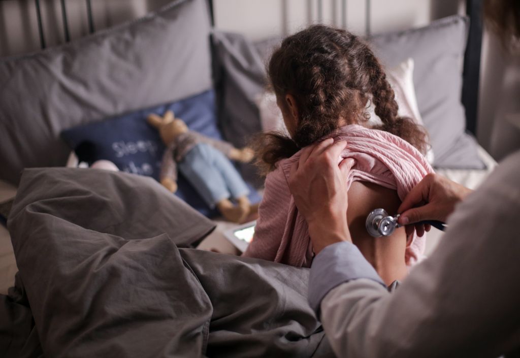
For generations, a stethoscope has been one of the most essential tools for diagnosing pneumonia. But a new study suggests that what doctors hear through it may depend on who is listening.
In this cohort study, US investigators who are part of the Pediatric Emergency Care Applied Research Network analysed data from children and teens diagnosed as having community-acquired pneumonia (CAP). The findings showed that clinicians often disagreed about their findings on physical exam, including decreased breath sounds, crackles (wet, bubble-likes sounds when air flow is blocked), and rhonchi (abnormal breathing that sounds like snoring or rattling) – symptoms frequently associated with pneumonia.
Current US guidelines recommend diagnosing CAP based on clinical findings rather than chest X-rays in children who are treated as outpatients.
Same patient, different findings
For the study, published in JAMA Network Open, researchers analysed data from 252 participants, ages 3 months to 17 years, diagnosed with CAP at one of seven academic paediatric emergency departments across the United States. Two clinicians independently examined each patient within 60 minutes of one another and recorded their findings.
None of the physical exam findings met the researchers’ predetermined threshold for acceptable interrater reliability, a measure of how much different clinicians agree when analysing the same data or event. Wheezing (kappa value [κ], 0.50) and chest retractions (κ, 0.49) were the exam findings clinicians agreed on the most. Even so, agreement was modest.
Agreement was significantly lower for decreased breath sounds and crackles, which both had κ values under 0.25.
The findings raise questions about how heavily clinicians should rely on listening to the lungs when diagnosing pneumonia.
“This variability is not a trivial concern,” write Susan Lipsett, MD, of Boston Children’s Hospital, and Mark Neuman, MD, MPH, of Harvard Medical School, in an accompanying commentary. When examination findings vary depending on the observer, “their utility as diagnostic anchors is diminished” and their ability to appropriately guide treatment is compromised.
“If physicians cannot consistently agree on the presence of auscultatory findings, treatment decisions may hinge more on examiner interpretation than underlying pathology,” Lipsett and Neuman write. “This variability may contribute to well-documented differences in antibiotic prescribing and chest radiograph use across institutions.”
Why lung sounds can be hard to interpret
Several factors could explain the disagreement. Accurately interpreting lung sounds can depend on background noise, patient cooperation, and clinician experience—factors that “become even more challenging in a busy emergency department,” write Lipsett and Neuman. Children also have more flexible chest walls, which may make it harder to determine exactly where the sounds originate. What’s more, clinicians may also use terms like “crackles” to refer to slightly different things.
The commentators argue that the results strengthen the case for objective risk-assessment tools that combine factors such as fever, oxygen saturation, demographics, and selected clinical findings rather than relying on individual lung sounds.
“By quantifying risk and reducing dependence on subjective auscultatory interpretation, clinical prediction tools may mitigate interobserver variability and promote more consistent decision-making regarding imaging and antibiotic therapy,
Children hospitalised with severe pneumonia can safely switch from injectable to oral antibiotics once they begin to recover, allowing many to return home sooner and complete treatment outside hospital, according to a major clinical trial involving 13 hospitals in Southern Africa.
The new results were published in The Lancet. The trial involved partners across Europe and Africa and was led with researchers at City St George’s, University of London.
Pneumonia remains one of the leading infectious killers of children worldwide, particularly in low- and middle-income countries. Current World Health Organization (WHO) guidelines recommend five days of injectable antibiotics for children hospitalised with severe community-acquired pneumonia, often requiring them to stay in hospital even after they have already substantially improved.
Longer hospital stays are more expensive, placing a higher burden on already pressurised healthcare systems and facilities, whilst increasing the risk of hospital-acquired antibiotic-resistant infections and impacting the wellbeing of the children and their families.
The PediCAP trial is one of the largest studies to assess antibiotic treatment for severe childhood pneumonia in Africa. The study enrolled 1101 children aged two months to six years with community-acquired pneumonia that developed outside hospital but was severe enough to require hospital treatment. Thirteen hospitals across South Africa, Uganda, Zambia, Zimbabwe and Mozambique contributed to the study.
All children in the trial began treatment with a WHO-recommended injectable antibiotic. Some were assigned to switch to either oral amoxicillin or oral amoxicillin-clavulanate when their condition had improved, as confirmed by a healthcare worker. Researchers compared these children to those who received the WHO-recommended injectable treatment for the full five days.
Children who switched to oral antibiotics recovered just as well as those who remained on injectable treatment for five days. Rates of hospital readmission or death within 28 days were similar across all groups – 6% for oral amoxicillin, 7% for oral amoxicillin-clavulanate and 6% for injectable antibiotics – showing that an early switch to oral treatment is a safe and effective strategy.
The standard amoxicillin performed just as well as the broader-spectrum antibiotic amoxicillin-clavulanate, supporting the use of a treatment that is cheaper and widely available.
Researchers also compared how well children recovered with different durations of antibiotic treatment, ranging from four to eight days in total. A total antibiotic course of four to five days was as effective as longer courses of seven or eight days, suggesting many children can be treated successfully with substantially less antibiotic exposure than is often used in practice.
Children who switched to oral antibiotics left hospital around one day earlier compared to those who remained on injectable treatment for the full five days.
Co-lead author Dr Michelle Clements, based at UCL Innovative Clinical Trials Unit, said: “PediCAP is the first large-scale study to use an innovative multi-arm trial design, which we developed here at UCL, to evaluate different antibiotics and treatment durations at the same time. Rather than simply comparing one short course with one longer course, this approach allowed us to establish that the shortest studied treatment strategy was effective and safe, while also helping us to understanding the relationship between treatment length and effect.
“By generating robust evidence more efficiently, this trial design has helped answer questions that we hope will support changes to global treatment guidelines and improve care for millions of children with pneumonia worldwide.”
Co-lead author Professor Julia Bielicki, from City St George’s, University of London, said: “Every year millions of children around the world are admitted to hospital with severe pneumonia. Our study shows that once a child is clinically improving, it is safe to switch from injectable to oral antibiotics, and complete treatment at home.
“This simple change could help children get back to their families sooner, reduce pressure on busy hospitals, lower healthcare costs and avoid sometimes catastrophic financial impacts on families from lost caregiver earnings. Because amoxicillin is affordable and widely available, these findings have the potential to change clinical practice and improve care for children around the world.”
The trial was funded by the European Union’s EDCTP2 programme and sponsored by the Penta Foundation.
Cambridge researchers have shown that severe pneumonia has three different subtypes, helping explain why some patients in intensive care units (ICUs) recover from their illness faster than others, while for other patients the disease can be life-threatening.
Their findings could in future help inform tailored treatments, allowing individual patients to receive the most appropriate therapies.
The current approach of classifying patients by their clinical syndromes without looking at the underlying biology risks missing what’s key
Andrew Conway Morris
Pneumonia is the commonest infectious cause of death worldwide, responsible for an estimated 2.5 million deaths per year. In severe cases, patients may need to be admitted to an ICU and given mechanical ventilation. Severe pneumonia accounts for six in 10 infections managed in intensive care, and spread of the infection within ICUs is a significant concern.
Doctors have long struggled to understand why patients whose condition looks similar clinically can have very different recoveries. Some respond quickly to treatment, while others remain critically ill for weeks or even die.
Dr Andrew Conway Morris from the Department of Medicine at the University of Cambridge and an ICU consultant at Addenbrooke’s Hospital, Cambridge, said: “Even though we’re able to treat the initial infection, many patients with severe pneumonia still struggle to come off the ventilator and can develop lung failure. Therapies to tackle inflammation in the lungs have had mixed results in clinical trials – some suggest they are beneficial, others that they’re harmful.
“The current approach of classifying patients by their clinical syndromes – sepsis, acute respiratory distress syndrome and so on – without looking at the underlying biology risks missing what’s key. Instead of asking ‘Does this patient have pneumonia?’, we should be asking ‘What’s the inflammatory pattern in this patient’s lungs?’”
In findings published today in Nature Communications, Professor Conway Morris and team recruited patients admitted with suspected severe pneumonia to the ICU at Addenbrooke’s Hospital, part of Cambridge University Hospitals NHS Foundation Trust.
Severe pneumonia is usually diagnosed through a combination of symptoms, imaging and blood tests. Symptoms typically include fever or hypothermia, low oxygen levels, breathing difficulties and confusion.
Instead of relying only on blood tests or scans, however, the Cambridge team analysed immune cells, inflammatory signals, and gene activity in fluid taken from the lungs of the patients. They discovered that there are three distinct biological types – or ‘pneumotypes’ – of severe pneumonia, none of which could be reliably detected using standard blood tests, even though they were strongly linked to how patients recovered.
The most common pneumotype – accounting for almost half (49%) of cases – was characterised by immune suppression, significant damage to the lining of the lungs, and bleeding in the alveoli (tiny air sacs within the lungs). There were fewer signs of inflammation, which may explain why treatments targeting inflammation can fail or even harm some patients.
The second pneumotype – accounting for just under a quarter (23%) of cases – was characterised by a balanced immune response and active repair of damage to the lungs. Patients were most likely to recover faster from this pneumotype and require the shortest time on the ventilator, even though they initially looked just as ill as the others.
Patients with the most dangerous pneumotype – the one that most resembles ‘classic’ pneumonia – spent longest on mechanical ventilation and had prolonged critical illness. They had severe and persistent inflammation, with a flood of immature immune cells in the lung. This group may be most likely to respond to anti-inflammatory therapies, say the team.
Dr Mark Jeffrey from the Department of Medicine at the University of Cambridge, the study’s first author, said: “Even though on the surface, all of the patients seemed to have similar types of pneumonia, with comparable illness severity, oxygen levels and clinical diagnoses, their outcomes were very different.
“It was only when we drilled down and looked at patterns of inflammation that the differences became apparent. Severe pneumonia is not a single disease, but several biologically distinct conditions that happen to look alike. This helps explain why ‘one-size-fits-all’ treatments – including some immune-modulating drugs – have often failed in clinical trials.”
The tests used to determine the pneumotypes are too complex to enable rapid classification, but the researchers hope to develop a simplified tool that could help them stratify the patients and ultimately offer tailored treatments.
Dr Vilas Navapurkar from the John Farman Intensive Care Unit at Addenbrooke’s Hospital said: “If we know which subtype of pneumonia an individual has, we can potentially tailor their treatment more precisely, boosting the immune response in some, while calming harmful inflammation in others. This has the potential to help critically ill patients, reduce deaths from pneumonia, shorten ICU stays and cut unnecessary antibiotic use.”
The study was funded by Addenbrooke’s Charitable Trust, the National Institute for Health and Care Research Cambridge Biomedical Research Centre, and The Forster Foundation. Professor Conway Morris is a Fellow at Emmanuel College, Cambridge.
This illustration depicts a 3D computer-generated image of a group of Gram-positive, Streptococcus pneumoniae bacteria. The artistic recreation was based upon scanning electron microscopic (SEM) imagery. Credit: CDC on Unsplash
In a recent multicentre prospective study conducted at three hospitals in Tennessee and Georgia, including Vanderbilt University Medical Center, researchers at VUMC found a substantial burden of hospitalisations for community-acquired pneumonia (CAP) among adults.
Community-acquired pneumonia refers to a case of the disease contracted without prior exposure to a health care setting, otherwise known as hospital-acquired pneumonia (HAP).
The study, published in JAMA Network Open, included data from 2018 to 2022 and used a novel serotype-specific urinary test that can identify infections caused by 30 different Streptococcus pneumoniae serotypes. A serotype refers to a distinct strain of microorganism, such as bacteria.
An important aspect of the study was the identification of noninvasive pneumococcal infections, said Carlos Grijalva, MD, MPH, professor of Health Policy and Biomedical Informatics and the study’s lead author.
“Standard clinical diagnostic methods such as bacterial cultures of blood are helpful for identifying invasive cases of pneumococcal disease, but the majority of pneumococcal pneumonias are thought to be noninvasive,” Grijalva added. “Using a novel and more sensitive urinary antigen detection method allowed us to identify a number of pneumococcal infections that may have otherwise passed unrecognised.”
Based on current population estimates, some 114 800 U.S. adults may be hospitalised for pneumococcal pneumonia each year, a figure made up in large part by older adults. And according to the study’s findings, each year sees approximately 340 hospitalisations for community-acquired pneumonia per 100 000 adults, approximately 14% of which had evidence of Streptococcus pneumoniae infection.
“Our study results show that Streptococcus pneumoniae remains an important cause of severe community-acquired pneumonia,” said Wesley Self, MD, MPH, professor of Emergency Medicine, Senior Vice President for Clinical Research and the paper’s senior author.
Many of the serotypes identified by pneumococcal detections corresponded with those covered by a recently licensed adult-specific pneumococcal conjugate vaccine, V116, which includes 21 serotypes but was not commercially available during the study period.
“Vaccines with coverage of additional pneumococcal serotypes could be quite beneficial in lessening the burden of severe pneumonia on the U.S. population, especially among older adults,” added Self, who holds the Directorship in Emergency Care Research.
Pneumonia diagnoses are marked by pronounced uncertainty, according to an AI-based analysis of over 2 million hospital visits. The study, published in Annals of Internal Medicine, found that more than half the time, a pneumonia diagnosis made in the hospital will change from a patient’s entrance to their discharge – either because someone who was initially diagnosed with pneumonia ended up with a different final diagnosis, or because a final diagnosis of pneumonia was missed when a patient entered the hospital (not including cases of hospital-acquired pneumonia).
Understanding that uncertainty could help improve care by prompting doctors to continue to monitor symptoms and adapt treatment accordingly, even after an initial diagnosis.
Barbara Jones, MD, pulmonary and critical care physician at University of Utah Health and the first author on the study, found the results by searching medical records from more than 100 VA medical centres across the country, using AI-based tools to identify mismatches between initial diagnoses and diagnoses upon discharge from the hospital. More than 10% of all such visits involved a pneumonia diagnosis, either when a patient entered the hospital, when they left, or both.
“Pneumonia can seem like a clear-cut diagnosis,” Jones says, “but there is actually quite a bit of overlap with other diagnoses that can mimic pneumonia.” A third of patients who were ultimately diagnosed with pneumonia did not receive a pneumonia diagnosis when they entered the hospital. And almost 40% of initial pneumonia diagnoses were later revised.
The study also found that this uncertainty was often evident in doctors’ notes on patient visits; clinical notes on pneumonia diagnoses in the emergency department expressed uncertainty more than half the time (58%), and notes on diagnosis at discharge expressed uncertainty almost half the time (48%). Simultaneous treatments for multiple potential diagnoses were also common.
When the initial diagnosis was pneumonia, but the discharge diagnosis was different, patients tended to receive a greater number of treatments in the hospital, but didn’t do worse than other patients as a general rule. However, patients who initially lacked a pneumonia diagnosis, but ultimately ended up diagnosed with pneumonia, had worse health outcomes than other patients.
A path forward
The new results call into question much of the existing research on pneumonia treatment, which tends to assume that initial and discharge diagnoses will be the same. Jones adds that doctors and patients should keep this high level of uncertainty in mind after an initial pneumonia diagnosis and be willing to adapt to new information throughout the treatment process. “Both patients and clinicians need to pay attention to their recovery and question the diagnosis if they don’t get better with treatment,” she says.
Professor Shabir Madhi of Wits University. Photo: supplied.
The Sabin Vaccine Institute presented the Albert B. Sabin Gold Medal to physician-researchers Keith Klugman and Shabir Madhi.
Nicole Basta, an associate professor at Canada’s McGill University and Canada Research Chair in Infectious Disease Prevention, received Sabin’s 2024 Rising Star Award.
The awards were made on 18 April 2024 at a ceremony in the National Academy of Sciences building in Washington D.C.
Formidable Wits alumni are world leaders in vaccinology
Klugman and Madhi received the Sabin Gold Medal, one of the highest recognitions for vaccinologists globally, for their seminal combined contributions to the development of vaccines against pneumonia and diarrhoeal disease – major causes of death in children in low- and middle-income countries (LMICs).
Klugman is a Wits University alumnus who received an honorary doctorate from his alma mater in 2023.
Madhi, also a Wits alumnus, is currently Professor of Vaccinology and Dean of the Faculty of Health Sciences at Wits University.
The Gold Medal is Sabin’s highest scientific honour. It has been given annually for more than three decades to a distinguished member of the global health community who has made exceptional contributions to vaccinology or a complementary field.
Klugman first met his then-graduate student Madhi at Wits University, where Klugman established, and Madhi expanded, a now globally renowned infectious diseases research institute. Apart from pneumonia, their work focused on maternal and children’s vaccines including influenza, respiratory syncytial virus (RSV), typhoid, and Group B streptococcus (GBS).
The evidence produced by these two awardees has and continues to inform the World Health Organization’s recommendations for vaccines. Klugman and Madhi’s research has helped pave the way for the introduction of lifesaving vaccines in public immunization programs – including the pneumococcal conjugate vaccine where their findings were pivotal in influencing vaccination policy in many low- and middle-income countries (LMICs).
Klugman’s efforts help prevent babies from dying of pneumonia
Fuelled by an early interest in science as a child in South Africa – in part due to a physician father – Klugman holds both a medical as well as a science doctorate degree from Wits University and was the first student in the school’s history to obtain them simultaneously.
He began his research career nearly five decades ago investigating the typhoid vaccine and has since distinguished himself as a formidable infectious diseases’ scientist.
Klugman is widely known for his work on pneumonia, which still kills a child under five every 43 seconds, many in the world’s poorest countries.
As the director of the pneumonia programme at the Seattle-based Bill & Melinda Gates Foundation, Klugman orchestrates strategic initiatives aimed at reducing deaths from pneumonia, RSV, neonatal sepsis, and meningitis.
He has authored hundreds of publications that have been cited over 50 000 times to date and has been elected to the National Academy of Medicine in the United States. He is also a professor emeritus of global health at Atlanta’s Emory University.
His scientific achievements aside, Klugman has long championed the need for the world’s poorest children to have equitable access to vaccines. While in South Africa he joined in Wits University’s struggle to allow access to the institution for all students.
“It is absolutely wonderful to be receiving this award, especially together with Shabir,” he says. “When I look down the list of previous awardees, I recognize the great majority of them, and it is extraordinary to now be numbered among them.”
Past award recipients include leaders of vaccinology and vaccine advocacy such as Drs. Barney Graham, Carol Baker, Bill Foege, Anne Gershon, Stanley Plotkin, and Kathrin Jansen.
Madhi’s research informed WHO recommendations on universal rotavirus vaccination
With a career spanning more than 25 years, Madhi, also from South Africa, is a trained paediatrician whose research continues to be instrumental in prioritising the rollout of vital vaccines and guiding global public health policies. At Wits University, he led clinical trials focused on respiratory and meningeal pathogens, including vaccines targeted at pregnant women and their unborn babies.
Madhi led the first study showing that a rotavirus vaccine could significantly prevent severe diarrhoea during the first year of life in African infants. That research served as a key piece of evidence for the WHO’s recommendation of universal rotavirus vaccination. In addition, he also led the first two COVID-19 vaccine trials in Africa, and a number of COVID-19 epidemiology studies which led to the first evidence suggesting that infection-induced immunity and vaccinations played a role in reducing severity of disease.
In addition to serving as Professor of Vaccinology and Dean of Health Sciences at Wits University, Madhi heads South Africa’s widely respected South African Medical Research Council (SAMRC) Vaccines and Infectious Diseases Analytics Research Unit (Wits VIDA). He is also the co-founder and co-Director of the African Leadership Initiative for Vaccinology Expertise (ALIVE).
He has co-authored hundreds of publications which have been cited over 59 000 times. Madhi is a recipient of numerous lifetime achievement awards in South Africa, as well being bestowed an Honorary Commander of the Order of the British Empire (CBE) from the British Government for his services to science and public health in a global pandemic.
“It is really humbling for me to be acknowledged for my contributions in the field of vaccinology along with those who have received the Gold Medal award,” says Madhi. “It makes me realise that the work my team and I have done is acknowledged by my peers as being of substance. Most significantly, we contributed to protecting lives in those settings where a majority of death and suffering occurs, and that is in LMICs.”
Amy Finan, Sabin’s chief executive officer, says, “I am honoured to award the Sabin Gold Medal to Dr Klugman and D. Madhi for their extraordinary work on vaccines that have saved lives in communities most in need of these interventions. Their pneumonia research has been particularly transformative in shaping our understanding of the disease and strengthening global health strategies to protect children from this vaccine-preventable disease.”
The World Health Organization (WHO) noted an upsurge of unidentified pneumonia-like respiratory illnesses among children in Northern China, and asked China for more information. This is significant as previous outbreaks of severe respiratory illnesses have started out in this fashion, but such WHO requests for more information on disease clusters are routine as part of its monitoring. No “unusual or novel pathogens” have been found, according to China, which attributed it to an increase in multiple pathogens and the lifting of COVID restrictions.
Earlier this month, China’s National Health Commission reported a nationwide increase in respiratory disease incidence, mostly among children. This increase was attributed to lifting of COVID restrictions and the arrival of the cold season, and due to circulating known pathogens including Mycoplasma pneumonia and RSV, which are known to affect children more than adults.
On 22 November 2023, the WHO identified media and ProMED reports about clusters of undiagnosed pneumonia in children’s hospitals in Beijing, Liaoning and other places in China. The WHO requested from China additional epidemiologic and clinical information, as well as lab results from these cases and data about recent trends in circulating respiratory pathogens.
The WHO held a teleconference with Chinese health authorities and received data indicating an increase in outpatient consultations and hospital admissions of children due to Mycoplasmapneumoniae pneumonia since May, and RSV, adenovirus and influenza virus since October. Some of these increases are earlier in the season than usual, but not unexpected given the lifting of COVID restrictions, as similarly experienced in other countries. No changes in the disease presentation were reported by the Chinese health authorities, who said no unusual or novel pathogens or unusual clinical presentations had been detected, but only the general increase in respiratory illnesses by known pathogens. Local hospitals had not been overloaded by new cases.
Risk assessment
In the current outbreak of respiratory illness, the reported symptoms are common to several respiratory diseases and, as of now, at the present time, Chinese surveillance and hospital systems report that the clinical manifestations are caused by known pathogens in circulation. M. pneumoniae is a common respiratory pathogen and a common cause of paediatric pneumonia, and is readily treated with antibiotics.
China has stepped up its influenza-like illness (ILI) and severe acute respiratory infections (SARI) sentinel surveillance system since mid-October, including for M. pneumoniae.
There is limited detailed information available to fully characterize the overall risk of these reported cases of respiratory illness in children. However, due to the arrival of the winter season, the increasing trend in respiratory illnesses is expected; co-circulation of respiratory viruses may increase burden on health care facilities.
According to surveillance data reported to WHO’s FluNet and published by the National Influenza Centre in China, ILI was above usual levels for this time of year and increasing in the northern provinces. Influenza detections were predominantly A(H3N2) and B/Victoria lineage viruses.
WHO advice
The WHO advice was for people in China to take measures against respiratory illnesses, including vaccines, masking and social distancing. It also does not recommend any specific measures for travellers to China.
A new study published in The Lancet Global Health, highlights the impact indoor air pollution can have on the development of child pneumonia, showing that increases in airborne particulate matter results in greater carriage of Streptococcus pneumoniae.
Streptococcus pneumoniae is a major human pathogen causing more than two million deaths per year; more than HIV/AIDS, measles and malaria combined, but it is also part of the normal microbial community of the nasopharynx. It is the leading cause of death due to infectious disease in children under five years of age; in sub-Saharan Africa, the burden of pneumococcal carriage and pneumonia is especially high.
Household air pollution from solid fuels increases the risk of childhood pneumonia. Nasopharyngeal carriage of S. pneumoniae is a necessary step in the development of pneumococcal pneumonia. More than 2.6 billion people are exposed to household air pollution worldwide. Inefficient indoor biomass burning is estimated to cause 3.8 million premature deaths annually and approximately 45% of all pneumonia deaths in children aged younger than five years. However, a causal pathway between household air pollution and pneumonia had not yet been identified.
In order to understand the connection between exposure to household air pollution and the risk of childhood pneumonia researchers from the UK, Malawi and the United States conducted the MSCAPE (Malawi Streptococcus pneumoniae Carriage and Air Pollution Exposure) study embedded in the ongoing CAPS (Child And Pneumonia Study) trial. The MSCAPE study assessed the impact of PM2.5, the single most important health-damaging pollutant in household air pollution, on the prevalence of pneumococcal carriage in a large sample of 485 Malawian children.
Through exposure-response analysis, a statistically significant 10% increase in risk of S. pneumoniae carriage in children was observed for a unit increase (deciles) of exposure to PM2.5 (ranging from 3.9 μg/m³ to 617.0 μg/m³).
Dr. Mukesh Dherani, the study principal investigator, indicated: “This study provides us with greater insight into the impact household air pollution can have on the development of child pneumonia. These findings provide important new evidence of intermediary steps in the causal pathway of household air pollution exposure to pneumonia and provide a platform for future mechanistic studies.”
Study author Professor Dan Pope said: “Moving forward further studies, particularly new randomized controlled trials comparing clean fuels (e.g. liquefied petroleum gas) with biomass fuels, with detailed measurements of PM2.5 exposure, and studies of mechanisms underlying increased pneumococcal carriage, are required to strengthen causal evidence for this component of the pathway from household air pollution exposure to ALRI in children.”
Professor Nigel Bruce, co-principal investigator, stated: “This study provides further important evidence that emphasises the need to accelerate to cleaner fuels, such as LPG, which are now being promoted by many governments across the continent in order to meet SDG7 by 2030.”
A newly installed solar-powered medical oxygen system at a hospital in central Somalia is proving effective in saving lives, Somali and World Health Organization doctors told Voice of America.
The innovative solar oxygen system, the first of its kind in the country, was installed at Hanaano hospital, in the central town of Dhusamareb a year ago. Doctors say the system is having an impact and helping save the lives of very young patients.
“This innovation is giving us promise and hopes,” says Dr Mamunur Rahman Malik, WHO Somalia Representative.
According to Dr Malik, 171 patients received oxygen at the hospital from the solar-powered system from February to October 2021. Of these, only three patients died, and five others were referred to other hospitals.
Every year some 15 000 to 20 000 deaths occur in Somalia among children under five years of age due to pneumonia, said Dr Malik, making it the deadliest disease among under-fives.
The director of Hanaano hospital, Dr Mohamed Abdi, said the innovation is making a difference.
“It has helped a lot, it has saved more than a hundred people who received the service,” he said to VOA Somali.
“It was a problem for the children under one year and the children who are born six months to get enough oxygen. Now we are not worried about oxygen availability if the electricity goes out because there are the oxygen concentrators.”
One patient was Abdiaziz Omar Abdi, admitted to the hospital on January 16 with severe pneumonia and was struggling to breathe normally. The oxygen rate in his body had dropped to 60%, Dr Abdi said. Doctors immediately put him on oxygen along with ampicillin and dexamethasone medications. When discharged three days later, he was breathing normally. His oxygen was up to 90%.
Dr Malik said the oxygen is being used to treat a wide range of medical conditions – asphyxia, pneumonia, injuries, trauma, and road traffic accidents.
“We have seen in other countries that use of solar-powered medical oxygen (if applied in a timely manner) can save up to 35% of deaths from childhood pneumonia,” he said, adding that it could save the lives of at least 7000 children who die “needlessly” due to pneumonia.
The initiative to install solar-powered bio-medical equipment at Hanaano hospital emerged during the height of COVID in 2020, at a time when people were dying due to respiratory problems. Hospitals were unable to keep up with case loads and the cost of a cylinder of oxygen rose to between $400 to $600, and only 20% of health facilities had any kind of access to oxygen, said Dr Malik.
“If you look at the current situation, as of today Somalia needs close to 3000 or 4000 cubic metres of oxygen per day. So, oxygen was the biggest need in all the hospitals.”
Solar power can also be used for medical refrigerators, and their use is becoming widespread in Africa.