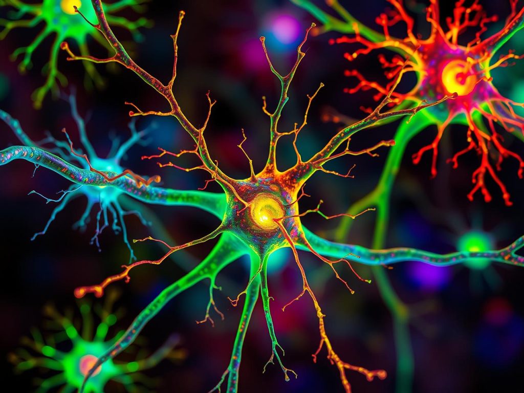
Electrotherapy using injectable nanoparticles delivered directly into the tumour could pave the way for new treatment options for glioblastoma, according to a new study from Lund University in Sweden.
Glioblastoma is the most common and most aggressive form of brain tumour among adults. Even with intensive treatment, the average survival period is 15 months. The tumour has a high genetic variation with multiple mutations, which often makes it resistant to radiation therapy, chemotherapy and many targeted drugs. The prognosis for glioblastoma has not improved over the past few decades despite extensive research.
Electrotherapy – a new treatment method
Electrotherapy offers another strategy to combat solid tumours. Using short, strong electric pulses (irreversible electroporation), non-reversible pores are created in the cancer cells leading to their death. The body’s immune system is simultaneously stimulated. The problem is that surgery is required to place the stiff metal electrodes that are necessary for the treatment. In sensitive tissue, in the brain for example, this often entails a very difficult procedure, which has led to strict criteria regarding which patients can be treated. Johan Bengzon is a researcher in glioblastoma and adjunct professor at Lund University, and consultant in neurosurgery at the Skåne University Hospital. He regularly treats patients with glioblastoma and is frustrated by the limited treatment options.
“The short distance between the hospital and the University in Lund facilitates cooperation and that’s why I contacted research colleagues to find out if injectable electrodes could be an alternative solution in electrotherapy,” says Johan Bengzon.
Said and done. The research team, with Amit Singh Yadav, Martin Hjort, and Roger Olsson at the helm, had previously used nanoparticles to form injectable and electrically conductive hydrogels to control brain signalling and heart contractions. It is aminimally invasive method in which the particles are injected using a thin syringe directly into the body. The particles break down after the treatment and thus do not need to be surgically removed. Perhaps the same technology could be used to destroy tumour cells in glioblastoma.
“After surgical treatment, unfortunately the glioblastoma tumour often returns on the outer edge of the area operated on. By drop casting the nanoparticles into the tumour cavity after an operation, we could electrify the edges while the immune system is also activated. In animal models the procedure, due to this irreversible electroporation, led to tumours being wiped out within three days,” says Roger Olsson, professor of chemical biology and drug development at Lund University, who led the study.
Promising results – but a long way to the patient
The prospects are good and the researchers are very hopeful for the future, even though there is a long way to go before it becomes a clinical reality. The challenge is now to test the method on larger tumours.
“We have seen that the electrode is well received in the brain. We have not noted any problems relating to side effects and after 12 weeks the electrode disappeared by itself as it’s biodegradable. The technology combines direct tumour destruction with activation of the immune system and can be an important step towards more effective treatment of glioblastoma,” concludes Amit Singh Yadav, researcher at Lund University and first author of the study.
For more than a century, maps of the brain have been based on how brain tissue looks under the microscope. These anatomical maps divide the brain into regions according to structural variations in the tissue. But do these divisions really reflect how the brain works? A new study on mice from Karolinska Institutet, published in Nature Neuroscience, suggests that this is often not the case.
By describing the brain in terms of electrical activity of its neurons, the researchers have found a new way to understand the functional organisation of the prefrontal cortex, the brain region responsible for planning, decision-making, and other advanced cognitive functions.
“Considering that deviations in prefrontal cortex function have been linked to virtually all psychiatric disorders, it is surprising how little is known about how this region actually works,” says Marie Carlén, Professor at the Department of Neuroscience at Karolinska Institutet.
Did not align with previous maps
Her research group recorded and analysed the activity of more than 24 000 neurons in awake mice and created the first activity-based maps of the prefrontal cortex. The maps of spontaneous and cognition-related neuron activity did not match the traditional, tissue-based maps.
“Our findings challenge the traditional way of defining brain regions and have major implications for understanding brain organisation overall,” says Marie Carlén.
The researchers found that the activity patterns of neurons reflected the hierarchy of information flow in the brain rather than the structure of the tissue. Neurons with slow, regular activity turned out to be characteristic of the prefrontal cortex, which sits at the top of this hierarchy. The same activity pattern also marked regions at the top of the prefrontal cortex’s own internal hierarchy. Slow, regular activity is thought to characterise the integration of information flows, a process that is central to cognitive functions such as planning and reasoning.
Different neuronal activity patterns work together
Carlén and her colleagues discovered that neurons involved in decision-making were concentrated in regions high up in the prefrontal hierarchy. Surprisingly, these neurons were characterised by very fast activity patterns.
“This suggests that cognitive processes rely on local collaboration between neurons whose activity patterns complement one another. Some neurons appear to specialise in integrating information streams, while others have high spontaneous activity that supports quick and flexible encoding of information, for instance, information needed to make a specific decision,” says Marie Carlén.”
Eating cheese and cream with a high fat content may be linked to a lower risk of developing dementia. This is shown by a new large-scale study from Lund University. The researchers analysed the dietary habits of more than 27 000 people and linked these to the occurrence of dementia over a follow-up period of up to 25 years.
The debate about low-fat diets has long shaped our health advice and influenced how we view food and health. For several decades, fear of saturated fat and its link to cardiovascular disease has dominated. The MIND diet1 is a diet developed with the aim of reducing the risk of dementia. The diet includes protective foods such as vegetables, nuts, fruits, berries, whole grains, and fish, while cheese is one of the foods that should be limited.
Emily Sonestedt, researcher in nutritional epidemiology at Lund University in Sweden, and her colleagues, therefore wanted to investigate whether there was any link between dairy products and dementia. They collected dietary data from 27,670 people using the Malmö Diet Cancer population study, in which the participants respond about their dietary and cooking habits. The average age at the start of the study was 58, and the participants were followed for an average of 25 years, during which time 3,208 people developed dementia. The dementia diagnoses were obtained from the Swedish patient registry. For cases diagnosed up to 2014, additional validation studies were conducted in which dementia specialists reviewed medical records, brain scans, and cognitive test results.
After adjusting for lifestyle factors such as physical activity, diet, smoking, and alcohol consumption, the researchers found that people who ate 50 grams of cheese (with more than 20 percent fat) daily had a 13 percent lower risk of developing dementia than those who ate less than 15 grams daily. 50 grams is equivalent to about five regular slices of cheese. In total, about a quarter of the participants ate more than 50 grams or more daily.
”When we went on to look at specific types of dementia, we found that there was a 29 percent lower risk of vascular dementia in people who ate more full-fat cheese. We also saw a lower risk of Alzheimer’s disease, but only among those who did not carry the APOE e4 gene variant—a genetic risk factor for Alzheimer’s disease.”
The researchers also investigated the link between high-fat cream (30-40 percent fat) and dementia. People who consumed 20 grams or more daily had a 16 percent lower risk of dementia than those who did not consume any at all.
The results of the cheese studies support the link between vascular health and brain health.
”The updated dietary guidelines in Sweden from this year say that we can eat dairy products every day, preferably fermented varieties such as yogurt or kefir. Both we and other researchers have found in observational studies that fermented dairy products in particular may be linked to a slightly reduced risk of cardiovascular disease 2,” says Emily Sonestedt.
In previous studies3, the research team has seen links to vascular health, with cheese and fermented dairy products in particular protecting against cardiovascular disease.
”Although higher-fat cheese and cream were associated with a reduced risk of dementia, other dairy products and low-fat alternatives did not show the same effect. Therefore, not all dairy products are equal when it comes to brain health. The few studies that have investigated this have found a correlation with cheese, so more research is needed to confirm our results and investigate whether certain high-fat dairy products really do provide some protection for the brain.”
The MIND diet stands for Mediterranean–DASH Intervention for Neurodegenerative Delay – a combination of the Mediterranean diet and the DASH diet. DASH (Dietary Approaches to Stop Hypertension) is a diet developed primarily to lower high blood pressure and improve cardiovascular health.
Our experiences leave traces in the brain, stored in small groups of cells called “engrams”. Engrams are thought to hold the information of a memory and are reactivated when we remember, which makes them very interesting to research on memory and age- or trauma-related memory loss.
At the same time, scientists know that the biology of learning is accompanied by epigenetic changes, which refers to the ways the cell regulates genes by adding chemical “post-it notes” on DNA.
But the question of whether the epigenetic state of a single gene in turn can cause a memory to change has thus far remained unanswered.
A team led by Professor Johannes Gräff at EPFL’s Laboratory of Neuroepigenetics combined CRISPR-based gene control with a technique that tags engram cells in mice. They focused on Arc, a gene that helps neurons adjust their connections to other neurons. By targeting the control region of Arc, the team asked whether flipping its epigenetic “switch” could directly change memory. They published their findings in Nature Genetics.
An “epigenetic switch”
The researchers developed specialised, CRISPR-based tools that could either dial down or boost Arc activity in memory neurons. Some, like the KRAB-MeCP2 tool, were designed to switch off gene activity by adding repressive marks that make the DNA less accessible, while others opened the DNA and turned the gene on. These tools were essentially an “epigenetic switch” for the Arc gene.
They then used harmless viruses to deliver these tools directly into the hippocampus of mice, a brain region central for storing and retrieving memory. The mice were then trained to link a specific place with a mild foot shock. By changing the epigenetic state of Arc in the neurons, the scientists could see whether the animals remembered the shock or not. They also added a “safety switch” that could undo the editing and reset the memory state.
The study showed that epigenetically silencing Arc in engram cells made the mice not learn, while boosting it made their memory stronger. These changes could be reversed in the same animal, showing that this epigenetic “switch” can dial memory expression up or down. Even memories that were already several days old, which are usually hard to change, could be modified. On the molecular level, the editing caused changes in gene activity and DNA packaging that matched the behavioural effects.
Controlling memory expression
The study is the first direct demonstration that changing the epigenetic state in memory cells is necessary and sufficient to control memory expression. It points to new ways of exploring how memories are stored and altered, which could eventually also be relevant in humans.
In the future, similar approaches could help researchers better understand conditions where memory processing goes awry, such as traumatic memories in PTSD, drug-related memories in addiction, or the memory problems that appear in neurodegenerative diseases.
First-of-its-kind study offers evidence that microbes from different primate species influence physiology in ways linked to brain size and function
Source: Pixabay
Humans have the largest relative brain size of any primate, but little is known about how mammals with larger brains evolved to meet the intense energy demands required to support brain growth and maintenance.
A new study from Northwestern University provides the first empirical data showing the direct role the gut microbiome plays in shaping differences in the way the brain functions across different primate species.
“Our study shows that microbes are acting on traits that are relevant to our understanding of evolution, and particularly the evolution of human brains,” said Katie Amato, associate professor of biological anthropology and principal investigator of the study, which was published in PNAS.
The study builds upon previous findings from Amato’s lab that showed the microbes of larger-brained primates, when introduced in host mice, produced more metabolic energy in the microbiome of the host – a prerequisite for larger brains, which are energetically costly to develop and function. This time, the researchers wanted to look at the brain itself to see if the microbes from different primates with different relative brain sizes would change how the brains of host mice functioned.
What they found
In a controlled lab experiment, the researchers implanted gut microbes from two large-brain primate species (human and squirrel monkey) and one small-brain primate species (macaque) into microbe-free mice.
Within eight weeks of making changes to the hosts’ microbiomes, they observed that the brains of mice with microbes from small-brain primates were indeed working differently than the brains of mice with microbes from large-brain primates.
In the mice with large-brain primate microbes, the researchers found increased expression of genes associated with energy production and synaptic plasticity, the physical process of learning in the brain. In the mice with smaller-brain primate microbes, there was less expression of these processes.
“What was super interesting is we were able to compare data we had from the brains of the host mice with data from actual macaque and human brains, and to our surprise, many of the patterns we saw in brain gene expression of the mice were the same patterns seen in the actual primates themselves,” Amato said. “In other words, we were able to make the brains of mice look like the brains of the actual primates the microbes came from.”
Another surprising discovery the researchers made was a pattern of gene expression associated with ADHD, schizophrenia, bipolar and autism in the genes of the mice with the microbes from smaller-brained primates.
While there is existing evidence showing correlations between conditions like autism and the composition of the gut microbiome, there is a lack of data showing the gut microbes contribute to these conditions.
“This study provides more evidence that microbes may causally contribute to these disorders —specifically, the gut microbiome is shaping brain function during development,” Amato said. “Based on our findings, we can speculate that if the human brain is exposed to the actions of the ‘wrong’ microbes, its development will change, and we will see symptoms of these disorders, i.e., if you don’t get exposed to the ‘right’ human microbes in early life, your brain will work differently, and this may lead to symptoms of these conditions.”
Implications and next steps
Amato sees clinical implications for further exploration of the origins of some psychological disorders and for taking an evolutionary perspective on the way microbes affect brain physiology.
“It’s interesting to think about brain development in species and individuals and investigating whether we can look at cross-sectional, cross-species differences in patterns and discover rules for the way microbes are interacting with the brain, and whether the rules can be translated into development as well.
Ischaemic and haemorrhagic stroke. Credit: Scientific Animations CC4.0
When a person suffers a stroke, physicians must restore blood flow to the brain as quickly as possible to save their life. But, ironically, that life-saving rush of blood can also trigger a second wave of damage — killing brain cells, fuelling inflammation and increasing the odds of long-term disability.
Now, Northwestern University scientists have developed an injectable regenerative nanomaterial that helps protect the brain during this vulnerable window.
In a new preclinical study, the team delivered a single intravenous dose, immediately after restoring blood flow, in a mouse model of ischemic stroke, the most common type of stroke. The therapy successfully crossed the blood-brain barrier — a major challenge for most drugs — to reach and repair brain tissue. The material significantly reduced brain damage and showed no signs of side effects or organ toxicity.
Published in the journal Neurotherapeutics, the findings suggest the new therapy could eventually complement existing stroke treatments by limiting secondary brain injury and supporting recovery.
“Current clinical approaches are entirely focused on blood flow restoration,” said co-corresponding author Dr Ayush Batra, associate professor at Northwestern and a neurocritical care physician with Northwestern Medicine. “Any treatment that facilitates neuronal recovery and minimises injury would be very powerful, but that holy grail doesn’t yet exist. This study is promising because it’s leading us down a pathway to develop these technologies and therapeutics for this unmet need.”
The injectable therapy is based on supramolecular therapeutic peptides (STPs), a platform developed by Northwestern’s Samuel I. Stupp. A study published in 2021 in the journal Science demonstrated the use of an STP technology — nicknamed “dancing molecules” — because of the highly dynamic nature of its therapeutic agents that could reverse paralysis and repair tissue in mice after a single injection at the site of severe spinal cord injury. The new study found scientists can administer similar dynamic assemblies of molecules intravenously, without requiring surgery or an invasive injection directly into the brain.
“One of the most promising aspects of this study is that we were able to show this therapeutic technology, which has shown incredible promise in spinal cord injury, can now begin to be applied in a stroke model and that it can be delivered systemically,” said Stupp, co-corresponding author. “This systemic delivery mechanism and the ability to cross the blood-brain barrier is a significant advance that could also be useful in treating traumatic brain injuries and neurodegenerative diseases such as ALS.”
Study mimicked real-world stroke treatment
Acute ischaemic stroke is a devastating condition and is one of the leading causes of morbidity and mortality worldwide, Batra said, severely impacting a patient’s quality of life and engagement in society.
“It has not only a significant personal and emotional burden on patients, but also a financial burden on families and communities,” he said. “Reducing this level of disability with a therapy that could potentially help in restoring function and minimising injury would really have a powerful long-term impact.”
The findings are highly relevant for future clinical applications because the scientists tested the approach in a mouse model that closely mimics real-world ischemic stroke treatment, Batra said. They first blocked blood flow to simulate a major ischaemic stroke and then restored it (a process called reperfusion), just as whem doctors restore blood flow acutely for ischaemic stroke patients.
The scientists monitored the mice for seven days and didn’t observe any significant side effects or biocompatibility issues such as toxicity or immune system rejection. They used advanced imaging techniques, such as real-time intravital intracranial microscopy seen in this video, to confirm the therapy localised to the stroke injury site. Compared to untreated mice, those treated with the “dancing molecules” had significantly less brain tissue damage, reduced signs of inflammation and reduced signs of excessive, damaging immune response.
Stupp said the therapy has pro-regenerative and anti-inflammatory properties, both of which contributed to the positive results.
“You get an accumulation of harmful molecules once the blockage occurs and then suddenly you remove the clot and all those ‘bad actors’ get released into the bloodstream, where they cause additional damage,” Stupp said. “But the dancing molecules carry with them some anti-inflammatory activity to counteract these effects and at the same time help repair neural networks.”
Dynamic ‘dancing molecules’ can be dialed down in concentration
The secret behind Stupp’s “dancing molecules” breakthrough therapeutic is tuning the collective motion of molecules, so they can find and properly engage constantly moving cellular receptors. The treatment sends signals that encourage nerve cells to repair themselves. For example, it can help nerve fibres (called axons) grow again and reconnect with other nerve cells, restoring lost communication through neural plasticity.
In previous studies, scientists injected the dancing molecules as a liquid, and when used to treat spinal cord injury, the therapy immediately gels into a complex network of nanofibres that mimic the dense, extracellular matrix of the spinal cord. By matching the matrix’s structure, mimicking the motion of biological molecules and incorporating signals for receptors, the synthetic materials are able to communicate with cells.
In the new study, the scientists dialled down the concentration of supramolecular peptide assemblies to prevent possible clotting as the therapy enters the bloodstream. Smaller aggregates of peptides easily crossed the blood-brain barrier. Once enough molecules cross, larger nanofibre assemblies can form in brain tissue to produce a more potent therapeutic effect, Stupp said.
“We chose for this stroke study one of the most dynamic therapies we had in terms of its molecular structure so that supramolecular assemblies would have a better probability of crossing the blood-brain barrier,” Stupp said.
Optimiaing therapeutic targeting
The fact that seemingly effective therapies cannot cross the blood-brain barrier has plagued the neuroscience field for decades, Batra said. This new therapy could change that.
When a physician acutely restores blood flow to a region of the brain in a stroke patient, the blood-brain barrier permeability is locally increased, naturally creating a transient opening and opportunity for therapeutic intervention, Batra said.
“Add to that a dynamic peptide that is able to cross more readily, and you’re really optimising the chances that your therapy is going where you want it to go,” Batra said.
Next steps
Further studies will need to assess whether this treatment can support longer-term, functional recovery, Batra said. For instance, many stroke patients suffer from significant cognitive decline throughout the subsequent year after a stroke. The new therapy is primed to address that secondary injury, Batra said, but the studies will require a longer follow-up period and more sophisticated behavioral testing.
In addition, the team is interested in testing whether additional regenerative signals could be incorporated into the therapeutic peptides to produce even better results.
Dopamine neurons, the cells that drive reward and motivation while we’re awake, become surprisingly active during nonrapid eye movement sleep right after we learn something new.
According to a new University of Michigan study, this night surge that is synchronised with memory-boosting sleep spindles, helps strengthen motor memories and improves motor skills.
The findings challenge long-held assumptions about dopamine’s role in the brain, showing that these neurons don’t just support learning during the day – they actively help lock in new skills while we sleep, said study co-author Ada Eban-Rothschild, U-M associate professor of psychology.
“As alterations in dopamine signalling are associated with neurodegenerative diseases that also involve motor deficits and sleep disturbances, understanding these links could pave the way for improved therapeutics and advancements in human health,” she said.
The study focused on specific midbrain dopamine neurons that become active after learning, but only during nonrapid eye movement, or NREM, sleep. This burst of activity helps the brain fine-tune and reinforce newly learned movements, contributing to more precise motor performance once awake.
Understanding how dopamine supports motor learning at night also sheds light on the broader importance of sleep in shaping behavior, said Eban-Rothschild and colleagues.
“The findings highlight that sleep is an active biological period during which key neural circuits strengthen the skills and patterns we rely on every day,” she said.
By revealing how dopamine helps consolidate motor memories during sleep, the researchers say the findings open a new window into brain health: It may eventually guide the development of therapies that target both sleep and dopamine pathways, offering new hope for improving motor function and quality of life in affected individuals.
The study was published in the Journal Science Advances. In addition to Eban-Rothschild, the study’s authors are Bibi Alika Sulaman, Eric Chen, Aaron Crane, Sangjin Lee and Gideon Rothschild
View of the spinal cord. Credit: Scientific Animations CC4.0
After a spinal cord injury, cells in the brain and spinal cord change to cope with stress and repair tissue. A new study from Karolinska Institutet, published in Nature Neuroscience, shows that this response is controlled by specific DNA sequences. This knowledge could help develop more targeted treatments.
When the central nervous system is damaged – for example, in a spinal cord injury – many cells become reactive. This means they change their function and activate genes that protect and repair tissue. However, how this process is regulated has long been unclear.
Researchers at Karolinska Institutet have now mapped thousands of so-called enhancers; small DNA sequences that act like “switches” for genes, turning them on or boosting their activity. By analysing individual cell nuclei from mice with spinal cord injuries using AI models, the researchers discovered that these genetic switches are activated after injury and instruct specific cell types to respond. The main cells affected were glial cells such as astrocytes and ependymal cells – support cells that help protect and repair the nervous system.
New opportunities for precision treatments
“We have shown how cells read these instructions through a code that tells them how to react to injury. This code combines signals from general stress factors with the cell’s own identity,” explains Enric Llorens-Bobadilla, researcher at the Department of Cell and Molecular Biology at Karolinska Institutet.
“This opens up the possibility of using the code to target treatments specifically to the cells affected by the injury,” says Margherita Zamboni, researcher at the same department and first author of the study.
The study is a collaboration between researchers at Karolinska Institutet and SciLifeLab, supported by the European Research Council (ERC), the Swedish Research Council, and the Swedish Foundation for Strategic Research. Some researchers have reported consultancy roles and patent applications related to the technology.
Microplastics could be fuelling neurodegenerative diseases like Alzheimer’s and Parkinson’s, with a new study highlighting five ways microplastics can trigger inflammation and damage in the brain.
More than 57 million people live with dementia, and cases of Alzheimer’s and Parkinson’s are projected to rise sharply. The possibility that microplastics could aggravate or accelerate these brain diseases is a major public health concern.
Pharmaceutical scientist Associate Professor Kamal Dua, from the University of Technology Sydney, said it is estimated that adults are consuming 250 grams of microplastics every year – enough to cover a dinner plate.
“We ingest microplastics from a wide range of sources including contaminated seafood, salt, processed foods, tea bags, plastic chopping boards, drinks in plastic bottles and food grown in contaminated soil, as well as plastic fibres from carpets, dust and synthetic clothing.”
“Common plastics include polyethylene, polypropylene, polystyrene and polyethylene terephthalate or PET. The majority of these microplastics are cleared from our bodies, however studies show they do accumulate in our organs, including our brains.”
The systematic review, recently published in Molecular and Cellular Biochemistry, was an international collaboration led by researchers from the University of Technology Sydney and Auburn University in the US.
The researchers highlighted five main pathways through which microplastics can cause harm to the brain, including triggering immune cell activity, generating oxidative stress, disrupting the blood–brain barrier, impairing mitochondria and damaging neurons.
“Microplastics actually weaken the blood–brain barrier, making it leaky. Once that happens, immune cells and inflammatory molecules are activated, which then causes even more damage to the barrier’s cells,” said Associate Professor Dua.
“The body treats microplastics as foreign intruders, which prompts the brain’s immune cells to attack them. When the brain is stressed by factors like toxins or environmental pollutants this also causes oxidative stress,” he said.
Microplastics cause oxidative stress in two main ways: they increase the amount of “reactive oxygen species” or unstable molecules that can damage cells, and they weaken the body’s antioxidant systems, which normally help keep those molecules in check.
“Microplastics also interfere with the way mitochondria produce energy, reducing the supply of ATP, or adenosine triphosphate, which is the fuel cells need to function. This energy shortfall weakens neuron activity and can ultimately damage brain cells,” said Associate Professor Dua.
“All these pathways interact with each other to increase damage in the brain.”
The paper also explores specific ways in which microplastics could contribute to Alzheimer’s, including triggering increased buildup of beta-amyloid and tau; and in Parkinson’s through aggregation of α-Synuclein and damage to dopaminergic neurons.
First author UTS Master of Pharmacy student Alexander Chi Wang Siu is a currently working in the lab of Professor Murali Dhanasekaran at Auburn University, in collaboration with Associate Professor Dua, Dr Keshav Raj Paudel and Distinguished Professor Brian Oliver from UTS, to better understand how microplastics affect brain cell function.
Previous UTS research has examined how microplastics are inhaled and where they are deposited in the lungs. Dr Paudel, a visiting scholar in the UTS Faculty of Engineering, is also currently investigating the impact of microplastic inhalation on lung health.
While evidence suggests microplastics could worsen diseases like Alzheimer’s and Parkinson’s, the authors emphasise that more research is needed to prove a direct link. However, they recommend taking steps to reduce microplastic exposure.
“We need to change our habits and use less plastic. Steer clear of plastic containers and plastic cutting boards, don’t use the dryer, choose natural fibres instead of synthetic ones and eat less processed and packaged foods,” said Dr Paudel.
The researchers hope the current findings will help shape environmental policies to cut plastic production, improve waste management and reduce long-term public health risks posed by this ubiquitous environmental pollutant.
Headache disorders affected almost 3 billion people worldwide in 2023 – nearly one in every three people, a figure unchanged since 1990 – and ranked sixth among causes of health loss, according to new research published in The Lancet Neurology. The analysis is part of the Global Burden of Disease (GBD) 2023 study and estimated health loss from migraine, tension-type headache, and medication-overuse headache from 1990 through 2023.
The study, led by researchers at IHME and the Norwegian University of Science and Technology (NTNU), examined the health loss resulting from headache disorders, and how long people have headache across different ages and sexes. Health loss was measured in years lived with disability (YLDs), which captures the total time people spend living with health conditions that limit daily activities and overall well-being. Drawing on population-based studies worldwide, the analysis provides the most comprehensive picture to date of how headache disorders affect daily life and overall health.
Headache disorders rank among the world’s most disabling conditions, disproportionately affecting women
In 2023, headache disorders accounted for an age-standardised rate of 541.9 YLDs per 100 000 people, ranking sixth among all causes of disability globally. The burden of headache disorders was more than twice as high among women as men, with rates of 739.9 and 346.1 YLDs per 100 000, respectively. Across every age group, women consistently spent more time experiencing headache symptoms than men.
“Our analysis shows that headache disorders have remained unchanged in three decades,” said Yvonne Xu, co-author and research scientist at IHME. “And women experience significantly higher levels of headache-related disability because they have headaches more frequently and for longer durations than men. Recognizing this is essential for improving how we prevent and manage headache disorders worldwide.”
Migraine and medication overuse drive most of the global burden from headache disorders
Although tension-type headache is nearly twice as prevalent as migraine, migraine accounts for about 90% of headache-attributed YLDs. In 2023, migraine alone caused an estimated 40.9 million YLDs globally, with an age-standardised rate of 487.5 YLDs per 100 000. Tension-type headache accounted for 54.4 YLDs per 100 000, showing that migraine, though less common, is far more disabling and drives most of the overall burden of headache disorders. While the highest rates of disability from migraine were seen in North Africa and the Middle East, closely followed by high-income regions such as Europe and North America, the burden remains high worldwide.
Medication-overuse headache, defined as the worsening of an existing headache due to excessive use of medication (e.g., pain medication) mainly used to treat migraine or tension-type headache, further amplifies this burden. While this condition affects relatively few, its impact on population-level disability is substantial because of the high individual burden. For migraine, medication overuse accounted for 22.6% of YLDs in men and 14.1% in women, while for tension-type headache, it contributed 58.9% and 56.1%, respectively. Overall, medication overuse was responsible for more than one-fifth of all headache-related disability globally.
“Our findings show that a large part of the global headache burden is preventable,” said Andreas Kattem Husøy, lead author and post-doctoral fellow in the Department of Neuromedicine and Movement Science at NTNU and Norwegian Centre for Headache Research (NorHead). “Integrating headache services into primary care, especially in low- and middle-income countries where effective treatments remain scarce, could reduce lost productivity and improve quality of life for hundreds of millions.”
Improved care and education are key to reducing the global burden of headache disorders.
Headache disorders remain one of the most common and disabling health conditions worldwide. The burden is unevenly distributed by sex and further intensified by overuse of pain medication, a preventable cause of long-term pain and disability. Although effective and affordable treatments are available, access to appropriate care and education on safe medication use remain limited in many settings.
The findings highlight an urgent need to strengthen prevention, management, and access to care for headache disorders worldwide. With greater awareness and coordinated action, much of the global burden of headache disorders can be prevented.