Tag: fibroblasts

Fibroblasts Have Hidden Powers That Could Heal Brain Injuries

A mouse brain cortex seven days after a stroke that caused injury. Fibroblasts (green) have created collagen (pink) to form a protective scar layer around the injury. All images by Molofsky Lab, UCSF

Healing from any injury involves a delicate balance between scarring and inflammation – two processes that can wreak havoc as well as make repairs.

When the injury is to the brain, the balance is that much more important, yet scientists know almost nothing of how this process works.

Now, a study from UC San Francisco spotlights how a cell type called a fibroblast, that plays a healing role in other parts of the body also performs a similar function in the brain. The discovery is a step toward finding new ways to treat brain injuries, which are the nation’s leading cause of death and disability and for which there aren’t any drugs that can intervene.

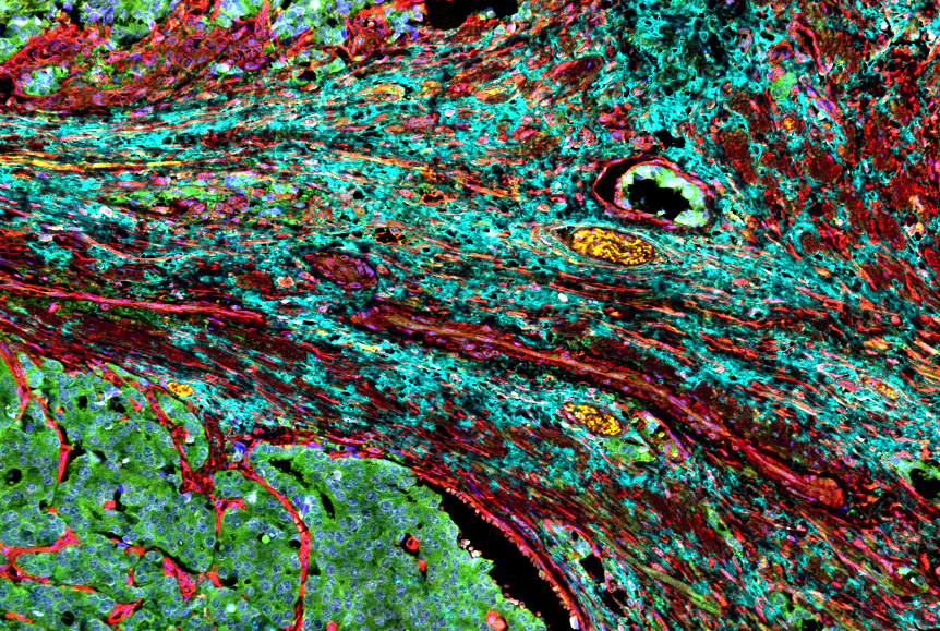
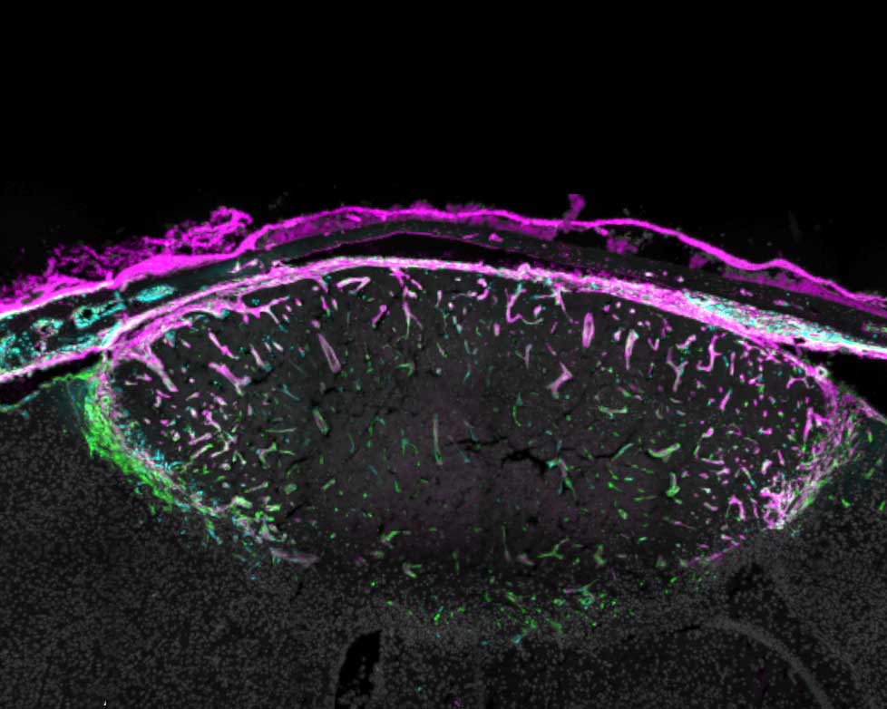
Fibroblasts were only identified in the brain in the last decade. They reside mostly in the meninges, a set of protective membranes that surround the brain and spinal cord. Until now, scientists thought they mostly served to maintain the structure of the meninges and its network of blood vessels.

Ari Molofsky, MD, PhD, a professor of laboratory medicine, suspected the fibroblasts might be doing much more than that. He and Tom Arnold, MD, a professor of paediatrics, discovered that when the brain is injured – whether from a blow or a stroke – fibroblasts navigate from the meninges and surround the injured tissue where they create a protective barrier, or scar.

The same injury 14 days after the stroke. The scar now surrounds the whole injury, which is less swollen. Some fibroblasts have returned to their usual location in the meninges. Those that remain have switched roles and are now recruiting immune cells to moderate inflammation.

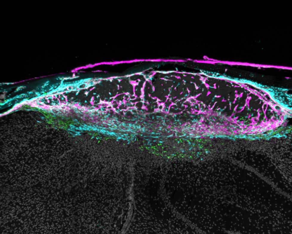
Then, about a week later, after the scar has formed, the fibroblasts adopt new roles. Some recruit immune cells that are required for healing; others ensure that the immune response doesn’t cause too much inflammation; and still others return to the meninges. Understanding these distinct stages could spur new interventions to help people with serious injuries.

Various views of a mouse brain cortex seven days after a stroke that caused injury. Green dots show fibroblast cells; pink areas show collagen produced by the fibroblasts to create a protective scar layer; and blue shows blood vessels with fibroblasts.

“Our study reveals opportunities to enhance the natural repair process,” said Molofsky, the senior author of the study, which appeared in Nature. “The goal is to give someone who’s experienced a traumatic brain injury or stroke the best outcome possible, based on the stage of healing they’re in.”

Therapies currently in clinical trials for lung and liver fibrosis target a molecule that prompts fibroblasts to create scarring. This suggests that other similar drugs could enhance healing in the early stages of a brain injury.

Molofsky’s study also offers an ideal venue for scientists to learn how fibroblasts are doing their work elsewhere in the body. Being largely devoid of immune cells, the brain offers a much clearer view than other organs like the lungs or liver, where immune cells may be too crowded around fibroblasts to see what they are doing.

“There’s a lot of potential here,” Molofsky said. “These overlooked cells seem adept at solving the common challenge of balancing healing and inflammation.”

Source: University of California – San Francisco

Immunotherapy Blocks Scarring, Improves Cardiac Function in Heart Failure

Right side heart failure. Credit: Scientific Animations CC4.0

A new study from Washington University School of Medicine in St. Louis suggests that a type of immunotherapy also may be an effective treatment strategy for heart failure by using an FDA-approved drug to block the signalling protein IL-1 beta. The study is published in Nature.

After a heart attack, viral infection or other injury to the heart, scar tissue often forms in the heart muscle, where it interferes with the heart’s normal contractions and plays a leading role in heart failure, a chronic condition which can only be slowed, not cured.

Studying human tissue samples as part of the new study, the researchers identified a type of fibroblast cell in the heart as the main culprit responsible for the formation of scar tissue in heart failure. To see if they could prevent scar formation, the scientists turned to mouse models of heart failure that have the very same type of fibroblasts. They used a therapeutic monoclonal antibody that blocks the formation of this harmful type of fibroblast, and succeeded in reducing the formation of scar tissue and improving heart function in the mice.

“After scar tissue forms in the heart, its ability to recover is dramatically impaired or impossible,” said cardiologist and senior author Kory Lavine, MD, PhD, a professor of medicine in the Cardiovascular Division at WashU Medicine. “Heart failure is a growing problem in the US and globally, affecting millions of people. Current treatments can help relieve symptoms and slow the progression, but there is a tremendous need for better therapies that actually stop the disease process and prevent the formation of new scar tissue that causes a loss of heart function. We are hopeful our study will lead to clinical trials investigating this immunotherapy strategy in heart failure patients.”

Fibroblasts have many roles in the heart, and parsing out the differences between various populations of these cells has been challenging. Some types of fibroblasts support the heart’s structural integrity and maintain good blood flow through the heart’s blood vessels, while others are responsible for driving inflammation and the development of scar tissue. Only recently, with the wide availability of the most advanced single cell sequencing technologies, could scientists peg which groups of cells are which.

“These various types of fibroblasts highlight newly recognised opportunities to craft treatment strategies that specifically block the type of fibroblasts that promote scarring and protect fibroblasts that maintain the structure of the heart, so the heart doesn’t rupture,” Lavine said. “Our research suggests that the fibroblasts that promote scarring in the injured heart are very similar to fibroblasts associated with cancer and other inflammatory processes. This opens the door to immunotherapies that potentially can stop the inflammation and resulting scar tissue.”

The research team, co-led by Junedh Amrute, a graduate student in Lavine’s lab, used genetic methods to demonstrate that a signaling molecule called IL-1 beta was important in a chain of events driving fibroblasts to create scar tissue in heart failure. With that in mind, they tested a mouse monoclonal antibody that blocks IL-1 beta and found beneficial effects in the mouse hearts. The mouse monoclonal antibody was provided by Amgen, whose scientists were also co-authors of the study. Monoclonal antibodies are proteins manufactured in the lab that modulate the immune system. The treatment reduced the formation of scar tissue and improved the pumping capacity of the mouse hearts, as measured on an echocardiogram.

At least two FDA-approved monoclonal antibodies, canakinumab and rilonacept, can block IL-1 signalling. These immunotherapies are approved to treat inflammatory disorders such as juvenile idiopathic arthritis and recurrent pericarditis, which is inflammation of the sac surrounding the heart.

One of these antibodies also has been evaluated in a clinical trial for atherosclerosis, a buildup of plaque that hardens the arteries. The trial, called CANTOS (Canakinumab Anti-inflammatory Thrombosis Outcome Study), showed a benefit for study participants with atherosclerosis.

“Even though this trial was not designed to test this treatment in heart failure, there are hints in the data that the monoclonal antibody might be beneficial for patients with heart failure,” Lavine said. “Secondary analyses of the data from this trial showed that the treatment was associated with a sizable reduction in heart failure admissions compared with standard care. Our new study may help explain why.”

Even so, the IL-1 antibody used in the CANTOS study had some side effects, such as increased risk of infection, that could perhaps be reduced with a more targeted antibody that specifically blocks IL-1 signaling in cardiac fibroblasts, according to the researchers.

“We are hopeful that the combination of all of this evidence, including our work on the IL-1 beta pathway, will lead to the design of a clinical trial to specifically test the role of targeted immunotherapy in heart failure patients,” Lavine said.

Source: Washington University School of Medicine

Cancer-associated Fibroblasts Sometimes Aid Certain Drugs

Cancer-associated fibroblasts surrounding a prostate tumour. Credit: Moscat and Diaz Meco labs

Cancer-associated fibroblasts in the tumour environment have typically been linked to tumour progression and therapy resistance, but some studies suggest that these fibroblasts may also sensitise cancer cells to therapy. In a new article published in Science Signaling, researchers shed light on these conflicting studies and demonstrate that cancer associated fibroblasts can promote or inhibit drug sensitivity based on the type of tumour cell and the drug used for treatment.

Through a series of laboratory experiments, the research team from Moffitt Cancer Center determined the impact of cancer associated fibroblasts on drug responses among different non-small cell lung cancer (NSCLC) cell lines. They discovered that the presence of cancer-associated fibroblasts had varying effects on tumour cells based on both the type of NSCLC and the drug used for treatment. For example, the presence of cancer associated fibroblasts induced resistance to two different MEK inhibitors in non-small cell lung cancer cell lines with a mutant KRAS protein. However, cancer associated fibroblasts sensitised NSCLC cell lines with a mutant EGFR protein to EGFR inhibitors. Interestingly, normal lung associated fibroblasts never sensitised cells to drug treatment, suggesting that cancer associated fibroblasts secrete a factor that causes differential responses to drug treatment in a cell-context manner.

The researchers compared cancer associated fibroblasts to normal fibroblasts to identify factors that would produce these disparate effects. They found that cancer associated fibroblasts had alterations in the levels of secreted proteins that are part of the insulin-like growth factor (IGF) signalling pathway, which is involved in cell growth, death and migration. Specifically, cancer-associated fibroblasts secreted higher levels of proteins called IGF binding proteins (IGFBPs), which inhibit IGF signalling, and lower levels of IGFs, which activate IGF signalling. In combination, these alterations result in inhibitory effects on the IGF signalling pathway.

In further analyses, the researchers found that IGFBPs sensitised lung cancer cell lines to EGFR inhibitor treatment, while IGF proteins induced resistance to EGFR inhibitor treatment. They identified that survival signalling in response to EGFR inhibitor treatment was dependent on the proteins IGF1R and FAK, which are both part of the IGFBP signalling pathway. Importantly, they discovered that drugs that blocked the activity of IGF1R and FAK sensitised mutant EGFR lung cancer cells to EGFR inhibitors, suggesting that this combination approach may be effective in the clinic.

“These results highlight tumour suppressive effects competing with otherwise tumour promoting effects of cancer associated fibroblasts and add to the growing evidence that eliminating cancer associated fibroblasts in an undifferentiated way may be detrimental to cancer therapy,” said lead study author Lily Remsing Rix, PhD

“We show that mechanistic understanding not just of cancer associated fibroblast-mediated resistance, but also of their tumour suppressive pathways, can lead to rational design of improved therapeutic approaches that mimic these effects and may delay the onset of drug resistance,” added Uwe Rix, PhD, principal investigator of the study.

Source: H. Lee Moffitt Cancer Center & Research Institute