Category: Neurology

Making Neurosurgeons Even Better at Removing Brain Tumours

In a leap for personalised medicine, scientists have discovered a simple and valuable way to improve brain cancer surgeries.

Taylor Furst, MD, observes a brain mapping procedure in progress at the University of Rochester’s Strong Memorial Hospital. Credit: Matt Wittmeyer

When removing cancerous tissue in the brain, neurosurgeons often use “awake brain mapping” to minimise the risk of causing unintended disruptions to a patient’s quality of life while removing as much tumour as possible. This practice, which has been used for decades, involves waking a patient up mid-surgery to test their neurocognitive functions in real time by stimulating the brain surface and assessing for functional changes.

A new study published in the journal Science Advances details a promising new avenue toward improving awake brain mapping results by investigating the tiny, nearly imperceptible variabilities in patient behaviour that occur during the procedure. This work, led by Carnegie Mellon University researchers, points to a future where brain surgeries are not just safer, but more precisely tailored to protect each patient’s speech, movement and quality of life.

How awake brain mapping works

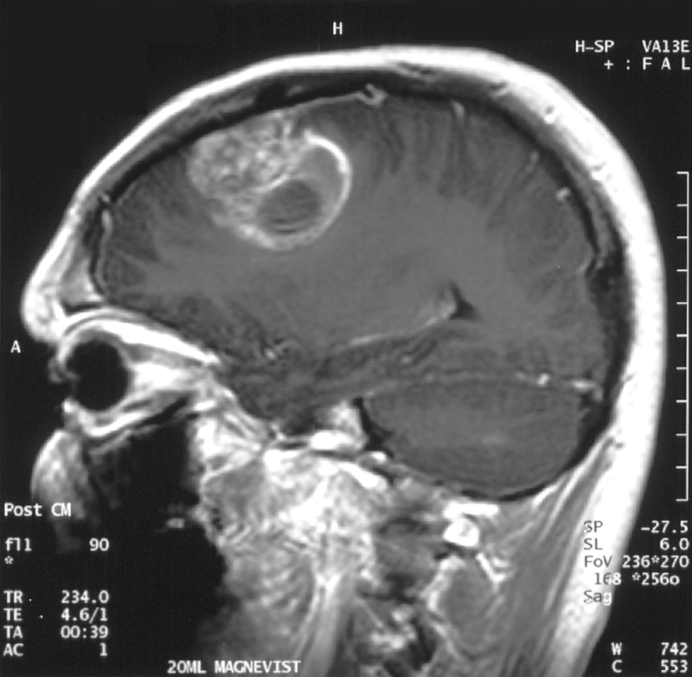
As cancer grows in the brain, it rarely keeps to itself. Cancerous cells can be found in the seemingly healthy brain tissue surrounding a tumour, presenting neurosurgeons with a dilemma. They need to remove as much tissue infiltrated by cancer as possible, but they also need to avoid the removal of too much tissue since it can cause permanent harm to a patient’s ability to hold a fork or a conversation.

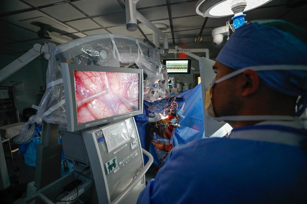
During awake brain mapping, surgeons gently stimulate the brain with small electrical impulses while the patient completes planned tasks. One of the most common applications of awake brain mapping is to identify where language is represented in a patient’s brain, which is done by having the patient name pictures or read words while their brain is being stimulated. If the patient can respond quickly and correctly, the clinicians know the part of the brain they stimulated can be safely removed. If the patient slurs or becomes unable to speak, then that part of the brain may be essential for language. Surgeons require a significant amount of experience to understand the nuances of this complex technique.  

While the method may sound extreme, the brain has no sensory nerves, so patients do not feel their brain surgery as it is happening. Recent research also shows that for some types of brain cancer, improving a patient’s quality of life after surgery extends their expected survival into the future. This means that anything that can make awake brain mapping even more effective will translate into improved outcomes for brain cancer patients.

New measures show how slight changes in procedure affect patient behaviour

Based on a decade of research, the study authors uncovered new insights from examining the answers patients get wrong – and right – while undergoing awake brain mapping.

“We found that if you measure both the types of errors that patients make, as well as how fast they respond even when they do not make errors, more granular inferences can be drawn about language organization from an awake brain mapping procedure,” said Bradford Mahon, a cognitive neuroscientist at CMU’s Neuroscience Institute and Department of Psychology and senior author of the study. “We also found that physical parameters of the direct electrical stimulation delivered to the patient’s brain – such as its duration, and when it started and stopped relative to the task the patient is performing – were tightly related to small changes in patient behaviour that we could measure.”

Mahon and his team don’t yet know exactly what combination of parameters should be used to maximise the effect of direct electrical stimulation mapping. But they have discovered an intriguing signal hidden inside of the data that, until now, has gone unnoticed.

“What we have measured and formalised in our study is how slight changes in the awake mapping procedure can cause slight changes in patient behaviour. This is exciting because it is a new and meaningful signal that can be extracted from the data already being generated during awake brain mapping procedures,” said Mahon.

A new level of personalised medicine

The new study suggests that awake brain mapping may offer more informative and more personalized guidance for surgery than has been possible in the past.

“Stimulation has traditionally been treated as a binary test – either it causes an error, or it doesn’t,” said Raouf Belkhir, lead author and a psycholinguist who is completing the University of Pittsburgh-Carnegie Mellon University Medical Scientist Training Program. “But in reality, these effects are often more continuous than binary.”

For example, stimulating a particular area of the brain might reliably cause an error, never affect behaviour at all, or – more subtly – slow a patient’s response without causing an obvious mistake. In some cases, stimulation may affect behaviour at one moment, but not when tested again just seconds later.

“In other words, brain mapping isn’t always black or white,” said Belkhir. “Sometimes the most important information lives in the grey area.”

The nuance matters because every brain is different, which means every surgery is different, too. Understanding why stimulation has variable effects across different patients, and even within the same patient from one part of the surgery to another part of the surgery, may be key to protecting outcomes for future patients.

“Surgeons are seeking to optimise the balance between removing all of the cancerous tissue while preserving critical functions that may be represented by nearby brain regions,” said Mahon. “This research shows that by measuring aspects of patient performance that were previously not considered relevant for awake brain mapping, even better predictive models of brain organisation can be developed.”  

If clinical teams have better predictive models personalised to each patient, then the consequences of different surgical approaches on postoperative neurocognitive function can be simulated. This allows for patients and their caregivers to personalise decisions to what is most important to the patient.

In other words, Mahon said, a business manager may consent to a surgery that may diminish their motor skills, but not their speech, whereas a concert violinist may prefer the opposite.

Bringing standardisation to awake brain mapping surgery

Another important development from this research is the startup company MindTrace, which has built an integrated software platform that supports neurocognitive testing before, during and after surgery. It is working to build a longitudinal dataset of patient outcomes that will be used to train forecasting models. 

Tyler Schmidt, MD, study co-author and neurosurgeon at the University of Rochester, has used MindTrace in over a dozen awake surgeries since its release this year.

“In the beginning of brain tumour surgery, it used to be, ‘Can we remove any of this tumour safely?’” said Schmidt. ”But now in some brain tumour cases it’s, ‘Can we get you back to work potentially? Can we keep your quality of life close to what it was prior to your diagnosis? Can we hone in on the things that are most important to you and then try and protect them while getting the same oncological outcome?’” said Schmidt. “I think it’s a positive paradigm shift in how we take care of this patient population.”

The options today are measurably better than they were even 20 years ago. Clinicians now understand how to maximise the likelihood that patients have the best possible outcomes from brain cancer surgery.

“Ultimately, we are contributing toward the set of tools that clinicians will have that will enable them to map the brain with even greater confidence and precision, and personalised to each patient,” said Mahon. “The big goal is to translate scientific insights into solutions that improve people’s lives. We will meet that goal by building tools that enable the best possible outcomes in neurosurgery patients, both in terms of neurocognitive function and quality of life, and ultimately, in terms of survival.”

Source: Carnegie Mellon University

Second Pregnancy Uniquely Alters Women’s Brains

Photo by Anna Hecker on Unsplash

A second pregnancy changes women’s brains in the same way as a first pregnancy, but in a different way than the first time. This is according to researchers from Amsterdam UMC, published in the scientific journal Nature Communications. The findings of Elseline Hoekzema and her colleagues show that a second pregnancy uniquely changes a woman’s brain, entailing both convergent and distinct neural transformations.

An earlier study by Elseline Hoekzema was the first to demonstrate that pregnancy changes the structure of the human brain. The research group also demonstrated that pregnancy changes the functioning of the brain. For the follow-up study, the results of which have now been published, 110 women were monitored: some were becoming mothers for the first time, others were having their second child, and a third group remained childless. Brain scans before and after pregnancy showed what changes occurred in the brain. “We have shown for the first time that the brain not only adapts during the first pregnancy, but also during the second,” says Hoekzema, head of the Pregnancy Brain Lab at Amsterdam UMC.

Different brain networks

The biggest changes during a first pregnancy occurred in the structure and activity of the so-called Default Mode Network. This part of the brain is important for many functions, including self-reflection and social processes. During a second pregnancy, this network changed again, but less dramatically. However, during a second pregnancy, there were more changes in brain networks related to paying attention and responding to stimuli. “It seems that during a second pregnancy, the brain changes more significantly in networks involved in responding to sensory stimuli and directing your attention,” explains researcher Milou Straathof, who analyzed the data. “These processes can be beneficial when caring for multiple children.”

Mental health of mothers

The researchers also found a connection between the changes in the brain and the bond between mother and child. This link was more prominent in the first pregnancy than during the second pregnancy. In addition, the researchers observed links between structural brain changes and peripartum depression, both during a first and a second pregnancy. This provides the first evidence that the changes that occur in a woman’s cerebral cortex during pregnancy are related to depression. In women who became mothers for the first time, this was particularly noticeable after giving birth. In women who had their second child, this was the case during pregnancy. “This knowledge can help us to better recognize and understand mental health issues in mothers. We must understand how the brain adapts to motherhood.”

The importance of research into the maternal brain

This study provides new insights into how the female brain adapts to motherhood and contributes to closing this important knowledge gap about female biology. Hoekzema: “The majority of women become pregnant one or more times in their lives, but only now are researchers beginning to unravel how this affects the female brain.” The results may also contribute to better care for mothers, for example, in the prevention and treatment of postpartum depression. The study also shows that the brain is flexible and can continually adapt to major changes in life.

Read the publication in Nature CommunicationsThe effects of a second pregnancy on women’s brain structure and function

Source: University of Amsterdam Medical Centers

Research Uncovers Factors Most Affecting Life Expectancy in Spina Bifida

Photo by National Cancer Institute

Studies have reported on survival probabilities of people born with open spina bifida, a condition where the spinal cord and nerves are exposed through an opening in the back. Research published in Developmental Medicine & Child Neurology now provides life expectancies, with results reported by age, sex, and different levels of impairment.

In the study of 1659 patients with open spina bifida who received support from the California Department of Developmental Services in 1986–2019, survival varied significantly by walking and feeding ability and by bowel/bladder continence.

As an example, at age 5, the life expectancy was 27 additional years for males in the most severely impaired group and 65 years in the least severely impaired, compared with 70 years in the general population. Life expectancies also decreased markedly with age and were modestly lower for males compared with females.

“This is the first long-term study of spina bifida patients to report life expectancies by age, sex, and severity of impairment,” the authors wrote. “We hope the results… will aid patients and caregivers alike in the proper planning for and treatment of those living with spina bifida.”

Source: Wiley

Could a Common Vitamin Treat Glioblastoma?

Findings indicate vitamin B3 looks promising to help rearm a compromised immune system

Unrestricted tumour growth in mouse brain, left, compared to the tumour growth in a mouse who received niacin treatment, right (both after 42 days). Courtesy Yong lab

Edward (Ed) Waldner had no idea why he didn’t feel well, but he knew he didn’t feel like himself. At 55 years of age, he felt exhausted all the time. It didn’t seem to matter how hard he had worked that day. He wondered if he had sleep apnoea. He noticed his walking was off. His heels would drag now and again. One day, when his symptoms were worse than usual, he decided to go to the Emergency department. 

“The doctor said I had a mass on my brain and needed to see an oncologist,” says Waldner. 

The mass was glioblastoma, a deadly brain cancer. Treatment often involves a three-pronged approach: surgery to remove as much of the tumour as possible, followed by radiation and chemotherapy. However, despite advances in cancer treatment, the aggressive cancer comes back. 

University of Calgary researchers are investigating whether adding high doses of vitamin B3 or niacin to the treatment plan could be beneficial. They approached Waldner about being in the trial.

“I have no problem trying to help anybody. I agreed. I want to help myself, too,” says Waldner. “I can tell you being part of this research helps me mentally because we’re trying. When I left the hospital after surgery I was told, that’s it, that’s all we can do.”

Hotchkiss, Charbonneau members partner for study

The research is led by two members of both the Hotchkiss Brain Institute and Arnie Charbonneau Cancer Institute – Dr Gloria Roldan Urgoiti, MD, PGME’16, an oncologist specialised in brain cancers, and Dr Wee Yong, PhD, a neuroscientist whose research focuses on immune effects on the brain. Together, they designed a study to investigate whether niacin could rejuvenate compromised immune cells to kill tumour cells. The research began in the Yong lab, with mice, where findings showed niacin prolonged survival. That work evolved into a Phase I and II clinical trial.

“Normally, the immune system will try to counter and prevent tumour growth; however, this brain cancer supresses the immune system,” says Yong, a professor at the Cumming School of Medicine (CSM). “Niacin treatment rejuvenates immune cells so they can do what they are supposed to do, attack and kill the cancer cells. I see it as an ongoing ‘battle for the brain.’”

Studying the benefits of adding niacin to chemotherapy, radiation

The clinical trial was designed to determine the maximum dose and potential benefit of controlled-release niacin that could be added to the recommended chemotherapy and radiotherapy treatments. Researchers decided the study would stop if the progression-free survival over six-months did not improve by at least 20 per cent compared with older studies. Early results involving 24 patients showed 82 per cent of the participants were free of progression of the cancer at six months; an increase of 28 per cent from previous studies. The researchers say this is a promising advancement for this incurable cancer. 

“Glioblastoma is the most aggressive brain cancer in adults. Survival of patients with this condition hasn’t changed significantly for 20 years,” says Roldan Urgoiti, a clinical associate professor at the CSM. “Anything that may help should be explored, but it requires strict protocols and safety monitoring.”

The findings were published in the Journal of Neuro-Oncology.

The researchers caution that high amounts of vitamins, like niacin, have toxicity and can have a negative impact on someone’s health if not monitored closely by medical professionals. 

The team hopes to be able to do the final analysis, that will include 48 participants by the end of 2026 or early 2027. 

Waldner says he’s feeling really good these days and is just happy to hear the word “stable” when he goes for his regular scans. 

Source: University of Calgary

Coffee Linked to Lower Dementia Risk, Better Cognitive Function

Photo by Mike Kenneally on Unsplash

A new prospective cohort study by investigators from Mass General Brigham and colleagues analysed 131 821 participants from the Nurses’ Health Study (NHS) and Health Professionals Follow-Up Study (HPFS), finding that moderate consumption of caffeinated coffee (2-3 cups a day) or tea (1-2 cups a day) reduced dementia risk, slowed cognitive decline, and preserved cognitive function. Their results are published in JAMA.

“When searching for possible dementia prevention tools, we thought something as prevalent as coffee may be a promising dietary intervention – and our unique access to high quality data through studies that has been going on for more than 40 years allowed us to follow through on that idea,” said senior author Daniel Wang, MD, ScD, associate scientist with the Channing Division of Network Medicine in the Mass General Brigham Department of Medicine and assistant professor at Harvard Medical School. Wang is also an assistant professor in the Department of Nutrition at Harvard Chan School and an associate member at the Broad Institute. “While our results are encouraging, it’s important to remember that the effect size is small and there are lots of important ways to protect cognitive function as we age. Our study suggests that caffeinated coffee or tea consumption can be one piece of that puzzle.”

Early prevention is especially crucial for dementia, since current treatments are limited and typically offer only modest benefit once symptoms appear. Focus on prevention has led researchers to investigate the influences of lifestyle factors like diet on dementia development. 

Coffee and tea contain bioactive ingredients like polyphenols and caffeine, which have emerged as possible neuroprotective factors that reduce inflammation and cellular damage while protecting against cognitive decline. Though promising, findings about the relationship between coffee and dementia have been inconsistent, as studies have had limited follow-up and insufficient detail to capture long-term intake patterns, differences by beverage type, or the full continuum of outcomes—from early subjective cognitive decline to clinically diagnosed dementia.

Data from the NHS and HPFS help to overcome these challenges. Participants repeated assessments of diet, dementia, subjective cognitive decline, and objective cognitive function and were followed for up to 43 years. Researchers compared how caffeinated coffee, tea, and decaffeinated coffee influenced dementia risk and cognitive health of each participant.

Of the more than 130 000 participants, 11 033 developed dementia. Both male and female participants with the highest intake of caffeinated coffee had an 18% lower risk of dementia compared with those who reported little or no caffeinated coffee consumption. Caffeinated coffee drinkers also had lower prevalence of subjective cognitive decline (7.8% versus 9.5%). By some measurements, those who drank caffeinated coffee also showed better performance on objective tests of overall cognitive function.

Higher tea intake showed similar results, while decaffeinated coffee did not – suggesting that caffeine may be the active factor producing these neuroprotective results, though further research is needed to validate the responsible factors and mechanisms.

The cognitive benefits were most pronounced in participants who consumed 2–3 cups of caffeinated coffee or 1–2 cups of tea daily. Contrary to several previous studies, higher caffeine intake did not yield negative effects – instead, it provided similar neuroprotective benefits to the optimal dosage.

“We also compared people with different genetic predispositions to developing dementia and saw the same results – meaning coffee or caffeine is likely equally beneficial for people with high and low genetic risk of developing dementia,” said lead author Yu Zhang, MBBS, MS, PhD student at Harvard Chan School and a research trainee at Mass General Brigham. 

Source: Mass General Brigham

Babies are Born with a Sense of Rhythm, Study Suggests

Newborns listening to Bach music predicted rhythm, but not melody, according to their brain waves  

Human newborns can predict rhythmic structure from music, while they are not as good at expecting melodic changes. Image credit: Diego Perez-Lopez, PLOS, CC-BY 4.0

Babies are born with the ability to predict rhythm, according to a study published February 5th in the open-access journal PLOS Biology by ​​Roberta Bianco from the Italian Institute of Technology, and colleagues.

It’s anticipating a beat drop, key change or chorus in a song you’ve never heard. Across all cultures, humans can inherently anticipate rhythm and melody. But are babies born with these behaviours, or are they learned? Research shows that by approximately 35 weeks of gestation, foetuses begin to respond to music with changes in heart rate and body movements. However, newborns’ ability to anticipate rhythm and melody is not fully understood.

To understand babies’ musical aptitudes, researchers played J.S. Bach’s piano compositions for an audience of 49 sleeping newborns. Musical stylings included 10 original melodies and four shuffled songs with scrambled melodies and pitches. While the babies listened, the researchers used electroencephalography – electrodes placed on the babies’ heads – to measure their brainwaves. When the babies’ brain waves showed signs of surprise, it meant they expected the song to go one way, but it went another.

The newborns tended to show neural signs of surprise when the rhythm unexpectedly changed; in other words, the miniature maestros had generated musical expectations based on rhythm. Previously, this result had been observed in non-human primates. The researchers found no evidence that the newborns tracked melody or were surprised by unexpected melodic changes, a skill that comes at an unknown exact point later in development.

According to the authors, understanding how humans become aware of rhythm can help biologists understand how our auditory systems develop. Future studies can investigate how exposure to music during gestation affects acquisition of rhythm and melody.

The authors add, “Are newborns ready for Bach? Newborns come into the world already tuned in to rhythm. Our latest research shows that even our tiniest 2-day old listeners can anticipate rhythmic patterns, revealing that some key elements of musical perception are wired from birth. But there’s a twist: melodic expectations – our ability to predict the flow of a tune – don’t seem to be present yet. This suggests that melody isn’t innate but gradually learned through exposure. In other words, rhythm may be part of our biological toolkit, while melody is something we grow into.”

Provided by PLOS

Physical Pressure on the Brain Triggers Neurons’ Self-destruction Programming

Gliobastoma (astrocytoma) WHO grade IV – MRI sagittal view, post contrast. 15 year old boy. Credit: Christaras A.

The brain and spinal cord is made up of billions of neurons connected by synapses and managed and modified by glial cells. When neurons die, this communication network is disrupted and since this loss is irreversible, neuron death causes sensory loss, motor impairment and cognitive decline.

An interdisciplinary team of researchers from the University of Notre Dame is investigating the mechanisms of neuron death caused by chronic compression – such as the pressure exerted by a brain tumour – to better understand how to prevent neuron loss.

Published in the Proceedings of the National Academy of Sciences, their study found that chronic compression triggers neuron death by a variety of mechanisms, both directly and indirectly. The research is helping lay the groundwork for identifying therapies to prevent indirect neuron death.

“The impetus for this project was to figure out those underlying mechanisms. In cancer research, most researchers are focused on the tumour itself, but in the meantime, while the tumour is sitting there and growing, it’s damaging the organ that it’s living in,” said Meenal Datta, the Jane Scoelch DeFlorio Collegiate Professor of Aerospace and Mechanical Engineering at Notre Dame and co-lead author of the study. “We fully believe that these growth-induced mechanical forces of the tumor as it expands is part of the reason we see damage in the brain.”

As an engineer who leads the TIME Lab, Datta studies the mechanics of tumors and the microenvironment, specifically for glioblastoma, an incurable brain cancer. She had found in prior work that tumors damage the surrounding brain. But to understand the mechanisms by which tumors kill neurons from compression alone, Datta needed a “hardcore neuroscientist.”

Neurons captured on screen for research experiment.
Imaging of neurons from an experiment with the control group neurons on the left and the neurons impact by chronic compression on the right. (Provided by the Patzke lab.)

That neuroscientist is Christopher Patzke, the John M. and Mary Jo Boler Assistant Professor in the Department of Biological Sciences at Notre Dame and co-lead author of the study. Patzke utilises induced pluripotent stem cells (iPSCs), which are either obtained from external sources or generated directly in his lab. These cells function like embryonic stem cells and can be differentiated or changed in the lab into any cell type in the body, including neurons.

For this study, iPSCs were used to create neural cells and develop a model system of neurons and glial cells that behave as a neuronal network would in the brain. Researchers grew the cells and then applied pressure to the system to mimic the chronic compression of a glioblastoma tumour.

After compressing the cells, graduate students Maksym Zarodniuk and Anna Wenninger, from Datta and Patzke’s labs respectively, compared how many neurons and glial cells died versus lived.

“For the neurons that are still alive, many of them have this programmed self-destruction signaling activated,” Patzke said. “We wanted to understand which molecular pathway was responsible for this; is there a way to save neurons from going down the drain to this cell death mechanism?”

By sequencing and analysing all messenger RNA from the living neuronal and glial cells, the researchers found an increase in HIF-1 molecules, signalling for stress adaptive genes to improve cell survival, which leads to inflammation in the brain. The compression also triggered AP-1 gene expression, a type of neuroinflammatory response.

Both neurological reactions are indicators that neuronal damage and death is underway.

An analysis of data from the Ivy Glioblastoma Atlas Project shows that glioblastoma patients also reflect these compressive stress patterns and gene expression changes as well as synaptic dysfunction in line with the experiment’s results. The researchers confirmed these results by mimicking force via a live compression system applied to preclinical models of brains.

Overall, the findings may help explain why glioblastoma patients experience cognitive impairments, motor deficits and elevated seizure risk. Additionally, the signalling pathways offer opportunities for researchers to explore as drug targets to reduce neuronal death.

“Our approach to this study was disease agnostic, so our research could potentially extend to other brain pathologies that affect mechanical forces in the brain such as traumatic brain injury,” Datta said. “I’m all in on mechanics. Whatever it is that you’re interested in when it comes to cancer, above your question of interest, mechanics is sitting there and many don’t even know they should be considering it.”

The mechanics of compression and its effect on neuron loss is key for future research.

“Understanding why neurons are so vulnerable and die upon compression is critical to prevent excessive sensory loss, motor impairment and cognitive decline,” Patzke said. “This is how we will help patients.”

Source: University of Notre Dame

Antidepressants not Linked to Serious Complications from TBI

Photo by Cottonbro on Pexels

Taking certain antidepressants at the time of a traumatic brain injury (TBI) is not associated with an increased risk of death, brain surgery or longer hospital stays, according to a study published on January 28, 2026, in Neurology®, the medical journal of the American Academy of Neurology.

For the study, researchers looked at serotonergic antidepressants, which treat anxiety and depression by increasing serotonin activity in the brain. These included selective serotonin reuptake inhibitors (SSRIs), serotonin and norepinephrine reuptake inhibitors (SNRIs) and tricyclic antidepressants (TCAs).

“Concerns have previously been raised that serotonergic antidepressants might increase the risk of bleeding in the brain or complicate early recovery after traumatic brain injury,” said study author Jussi P. Posti, MD, PhD, of the University of Turku in Finland. “However, our study found no evidence to support those concerns.” The study included 54 876 people in Finland who were 16 or older when hospitalised with a TBI. A total of 14% used serotonergic antidepressants at the time of the TBI.

Researchers reviewed national prescription records for preinjury antidepressant use and medical records to determine how many people died within a month, whether they needed emergency brain surgery, and how long they stayed in the hospital. A total of 4105 people died within a month. This included 7.6% of those taking antidepressants and 7.5% of people who did not. After adjusting for factors such as age, sex and other health conditions, researchers found people taking antidepressants before injury were no more likely to die within a month than those not taking them.

Antidepressant users were slightly less likely to require emergency brain surgery to relieve pressure or bleeding in the brain and prevent further damage. Of the total participants, 6.8% of the antidepressant users and 8.6% of those who did not use antidepressants needed emergency brain surgery. After adjustments, antidepressant users had an 11% lower risk. The amount of time in the hospital was the same for both groups.

“These findings provide reassurance for people who take antidepressants that antidepressant use does not appear to worsen early recovery after traumatic brain injury,” said Posti. “Future studies should examine whether these results hold true for long-term recovery and across different health care settings.”

A limitation of the study was that it was conducted only at hospitals and health care centres in Finland, so results may vary in other areas. The study was supported by the Finnish government, the Paulo Foundation, Paavo Nurmi Foundation, Research Council of Finland, Sigrid Jusélius Foundation and Finnish Foundation for Cardiovascular Research.

Source: American Academy of Neurology

The Brain Can Boost Vaccine Effectiveness, New Study Suggests

Photo by National Cancer Institute

New research reveals that activating the brain’s reward system through positive anticipation strengthens the immune response and increases antibody production

Can positive anticipation that activates the brain’s reward system strengthen the body’s immune defences? A new study by Tel Aviv University, the Technion, and Tel Aviv Medical Center (Ichilov), published in the prestigious journal Nature Medicine, provides the first evidence in humans that brain activity associated with the expectation of reward has a measurable effect on the body’s response to a specific vaccine.

Training the Brain’s Reward SystemThe study was conducted through a collaboration between two research groups: the laboratory of Prof Talma Hendler, from the School of Psychological Sciences and the Gray Faculty of Medical and Health Sciences; and the laboratory of Prof Asya Rolls from The George S. Wise Faculty of Life Sciences,.

Eighty-five healthy volunteers participated in the experiment. Some underwent special brain training using fMRI neurofeedback technology – a method that enables individuals to learn, in real time, to regulate activity in specific brain regions through reinforcing learning. The aim of the brain training was to increase activity in a key region of the brain’s reward system including the Ventral Tegmental Area (VTA), which is responsible for dopamine release in the context of mental activity related to the expectation of positive outcomes and motivation to obtain rewards. Participants were instructed to modulate their brain activity using various mental strategies (eg, thoughts, feelings, memories) while monitoring positive feedback about the strategy that was successful in regulating their brain.

From Brain Activation to Antibodies

Immediately after completing the brain training, all participants received a hepatitis B vaccine. The researchers then tracked the immune response through a series of blood tests, measuring levels of specific antibodies produced following the vaccination.

The results showed that participants who succeeded in significantly increasing activity in the brain’s reward region also demonstrated a greater increase in antibody levels after vaccination. The association was specific to the VTA and was not observed in other brain regions used for control purposes (such as the hippocampus), nor in other reward-system areas linked to different reward-related experiences such as pleasure and satisfaction. In other words, the effect was both anatomically and mentally specific.

The Role of Positive Anticipation

Furthermore, an in-depth analysis of the mental strategies participants used during training of the VTA (and not other regions) revealed that those who focused on positive anticipation, such as belief in a good outcome, or the expectation of something positive about to happen, were able to maintain higher VTA brain activity over time, which was also associated with a better immune response. In other words, the researchers identified a link between reward-system brain activity, a mental state of positive anticipation, and the body’s response to an immune challenge.

According to the research team, this is not “positive thinking” in the popular sense or a New Age slogan, but a measurable neurobiological mechanism – related, among other things, to the well-known placebo effect in medicine (a therapeutic response beyond a specific medical intervention). “We show that mental states have a clear brain signature, and that this signature can influence physiological systems such as the immune system,” explain the researchers.

While the study does not propose a substitute for vaccines or medical treatment, it opens the door to new, noninvasive approaches that may one day strengthen immune responses, improve the effectiveness of medical treatments, and even contribute to fields such as immunotherapy and the treatment of chronic immune pathologies. The researchers note that the study’s findings underscore a broader message: the mind–body connection is not merely a theoretical concept, but a real biological process that can be measured, trained, and potentially harnessed to promote better health.

Implications for Medicine and Health

The research team adds that the findings highlight the potential inherent in integrating neuroscience, psychology, and medicine. “Our study shows that the brain is not only a system that responds to the body’s state of health, but also an active player that influences it,” say the researchers. “The ability to consciously activate brain mechanisms associated with positive anticipation opens a new avenue for research and future treatments – as a complement to existing medicine, not as a replacement. In the future, it may be possible to develop simple, noninvasive tools to help strengthen immune responses and enhance the effectiveness of medical treatments by relying on the brain’s natural capacity to influence the body. However, it is important to emphasise that activation of the reward system and its effect on immune response vary between individuals. Therefore, this approach cannot replace existing medical treatments, but may well serve as an additional supportive component.”

Source: Tel Aviv University

New Neurosurgical Classification Reveals Pivotal Role of Glioma Volume Reduction

International team develops system for a standardised assessment of operative success in treating certain brain tumours

Photo by cottonbro studio

Low-grade brain tumours known as IDH-mutant gliomas CNS WHO grade 2 are life-threatening despite their slow growth. Neurosurgeons across the globe are faced with the question as to striking the correct balance between a “radical” tumour resection and avoiding further neurological damage. An international research team from the RANO working group involving Friedrich-Alexander-Universität Erlangen-Nürnberg (FAU) and Uniklinikum Erlangen has developed a new classification that records the extent to which any residual tumour tissue influences the progression of the disease. The results were published in The Lancet Oncology.

As a rule, the initial treatment for an IDH-mutated glioma CNS WHO grade 2 is surgery. The aim is to remove as much of the tumour as possible without jeopardising important neurological functions. As the results of the operation only become apparent many years later, there has been a lack of clear data, which has led to a number of different approaches. “On the one hand, this is due to the fact that we must be very careful to weigh up the chances of potentially boosting a patient’s chance of survival against avoiding neurological deficits. On the other hand, there has been a lack of clear criteria for assessing the risk of surgery until now, meaning that recommendations for treatment range from taking as little tissue as possible for diagnostic purposes to removing as much tumour tissue as possible,” explains Prof Dr Oliver Schnell from Uniklinikum Erlangen.

New basis for assessing success of surgery

In order to standardise therapeutic decisions, the RANO working group has conducted a large international study and assessed the data of 1391 patients from 16 neuro-oncological specialist units.

Based on the comprehensive data collected, the new RANO classification categorises the extent of the surgery based on the volume of the tumour that remains visible in a special MRI sequence (T2-FLAIR) after the operation. “Until now, there was no common language available for describing surgical outcomes,” explains PD Dr Philipp Karschnia from Uniklinikum Erlangen. “The new classification provides clarity, as it is guided exclusively by the residual tumour tissue.”

Less residual tumor means longer survival

The analysis of the RANO working group shows: A low volume of residual tumor after the initial operation is one of the most important factors for the further progression of the disease. A positive effect was also demonstrated for removing as much of the tumor as possible in the case of oligodendrogliomas, that tend to have a more favourable progression and are highly sensitive to chemo and radiation therapy. “We were surprised to discover that even follow-up treatments such as chemotherapy or radiation therapy were not able to replace the influence of the operation,” admits PD Dr Karschnia.

Internationally verified and useful in a wide range of scenarios

The results were confirmed in an independent patient group at the University of California in San Francisco. The new classification supports surgeons in making more accurate decisions and paves the way for future studies: “The new RANO classification is a milestone that will make a significant impact on neuro-oncological research and care in the long term,” according to Prof Schnell.

The Response Assessment in Neuro-Oncology (RANO) Working Group is an international, multidisciplinary collaboration between experts from various disciplines who have been working together to develop standardised criteria for assessing brain tumours for more than a decade now. Experts involved in the study from Erlangen were Prof Dr Oliver Schnell and PD Dr Philipp Karschnia, who has been leading the surgical focus group of the RANO Working Group since 2024, Dr Nico Teske and Alfred Gramelt from the Department of Neurosurgery at Uniklinikum Erlangen.

Source: Friedrich–Alexander University Erlangen–Nurnberg