Different Liquids Can Impact the Effectiveness of Certain Drugs
Some alkaline mineral and medicinal waters may weaken the enteric coating of medications within just a few minutes, potentially reducing their effectiveness, according to a new study by Semmelweis University. The study, published in Pharmaceutics, found that if the active ingredient is released too early in the stomach rather than in the intestinal tract, it may reduce – and, in extreme cases, even eliminate – the effectiveness of certain anti-reflux, gastroprotective, psychiatric, or anti-inflammatory pain-relief medications.
Researchers at Semmelweis University studied how different liquids could affect enteric-coated medications. The study analysed 22 commonly consumed beverages, seven of which – including several mineral and medicinal waters, tap water, filtered water, and apple juice – were examined in detail under laboratory conditions.
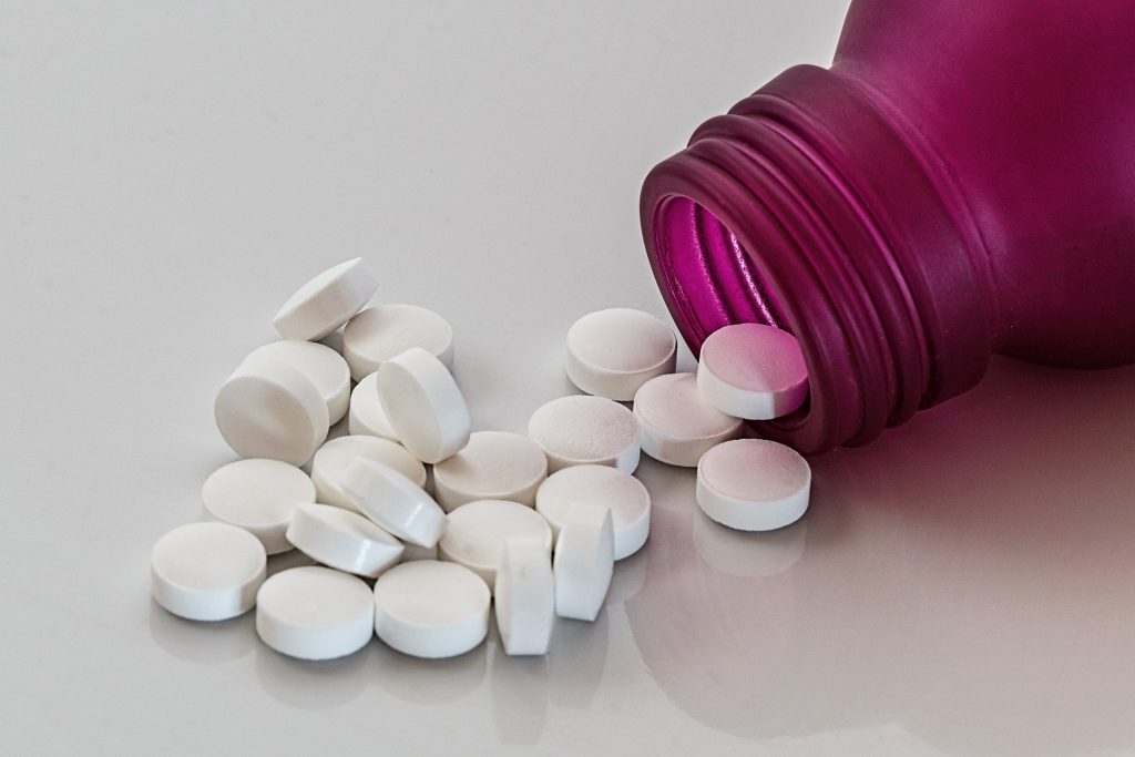
Alkaline bottled waters with high mineral content caused the greatest changes in the protective coating surrounding the active ingredient-loaded particles. According to the researchers, not only the water’s alkalinity but also its high mineral and ion content may have contributed to the faster dissolution of the protective coating, an effect that was particularly pronounced in some medicinal waters. In some cases, the enteric coating began to deteriorate after just five minutes, and after 15–30 minutes of pre-soaking, more than 90 percent of the active ingredient had been released prematurely.
Enteric-coated medications are designed so that the active ingredient is not released in the stomach but later in the intestines. This is important because some active ingredients are broken down by stomach acid, while others may irritate the stomach lining. Such coatings are commonly used on certain reflux medications, anti-inflammatory pain relievers, and digestive enzyme products.
Acidic liquids are less impactful
By contrast, more acidic liquids caused less damage to the enteric coating of the medications. In apple juice, for example, almost no premature release of the active ingredient was observed at the start of the tests, indicating that the coating remained far more stable than in alkaline waters.
“The small drug particle does not know whether it is already in the intestine or still sitting in a glass. If the pH of the surrounding environment is similar, the coating may begin to dissolve in the same way. Healthcare professionals generally assume that medications are swallowed with plain tap water, but that is not always obvious to patients today, given the wide variety of mineral and medicinal waters available on the market,” said Dr Nikolett Kállai-Szabó, Associate Professor at the Faculty of Pharmaceutical Sciences of Semmelweis University and senior author of the study.
The researchers also analysed the Summary of Product Characteristics (SmPCs) of 103 enteric-coated medications. In 42 cases, the instructions did not specify what liquid should be used to take the medication. Another 31 mentioned only “liquid,” while 21 referred simply to “water” without further clarification. Only nine SmPCs provided specific guidance on what beverage to take or mix the medication with, such as apple juice or another mildly acidic liquid.
This may be particularly important for people who open hard capsules because of swallowing difficulties and mix the capsule contents with liquids, yogurt, or applesauce. Older adults, children, and patients with a temporary sore throat or swallowing difficulties are more likely to find themselves in this situation.
“In the pharmacy, we regularly see that many patients are unaware of how much it matters what they take their medication with. This can also affect whether the treatment works as intended,” said Adrienn Demeter, PhD student at the Faculty of Pharmaceutical Sciences of Semmelweis University and first author of the study.
The researchers emphasize that the findings do not mean mineral or medicinal waters are inherently problematic. The key takeaway is that enteric-coated medications should preferably be taken with plain tap water, and patients should consult a pharmacist or physician before opening a capsule or splitting a tablet.
Source: Semmelweis University