Tag: bioprinting

Company Successfully Bioprints Kidney and Liver Tissues in Space

Weightlessness of space allows tissues to be bioprinted without collapsing
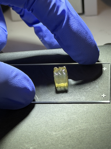
Bioprinted liver tissue
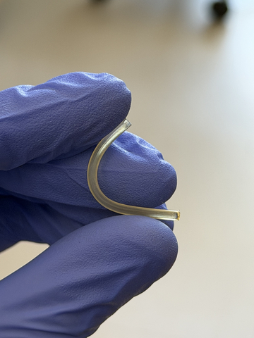
Bioprinted nerve implant

Auxilium Biotechnologies announced a major milestone in space biomanufacturing with the successful bioprinting of kidney and liver tissues aboard the International Space Station (ISS), marking the first time either tissue type has been manufactured in space. Auxilium’s bioprinted biological tissues and nerve repair implants returned to earth on Mission AXLM-3 that flew on a SpaceX capsule and returned to Earth on June 17th, 2026.

“Successfully bioprinting living liver & kidney tissue aboard the International Space Station marks an important step forward for regenerative medicine.” — Dr. Anthony Atala, MD, Professor & Director of the Wake Forest Institute for Regenerative MedicineShare

During the mission, Auxilium’s AMP-1 orbital bioprinter successfully manufactured kidney, liver, and cartilage tissues while also producing 28 nerve repair implants. The achievement represents the first demonstration of kidney tissue manufacturing in space, the first demonstration of liver tissue manufacturing in space, and the first mission to manufacture three distinct tissue types during a single spaceflight. The production of multiple tissue types and clinically relevant nerve repair implants represents the first demonstration of a scalable, multi-product biomanufacturing platform in space.

Equally important, the mission demonstrated the ability of a single autonomous manufacturing platform to produce both living tissues and implantable medical products during the same flight. The simultaneous production of multiple tissue types alongside 28 nerve repair implants highlights not only the versatility of the platform, but also its scalability and higher-throughput manufacturing in space.

The kidney and liver tissues were manufactured in support of research conducted by the Wake Forest Institute for Regenerative Medicine (WFIRM) using the institute’s cells and tissue designs. Auxilium provided the orbital manufacturing platform that enabled tissue fabrication in microgravity.

“This mission represents a significant milestone for both Auxilium and the future of space biomanufacturing,” said Jacob Koffler, PhD, MBA, CEO of Auxilium. “For the first time, we successfully bioprinted kidney and liver tissues in space, demonstrating that complex biological products can be manufactured in orbit. We also produced cartilage tissue and 28 nerve repair implants during the same mission using the same manufacturing platform. The ability to manufacture multiple tissue types alongside clinically relevant medical products highlights both the versatility and scalability of our technology. These results build on our previous demonstration of large-scale medical device manufacturing in space and represent another step toward establishing practical production capabilities for biomedical products beyond Earth.”

Dr. Anthony Atala, MD, Professor and Director of the Wake Forest Institute for Regenerative Medicine (WFIRM) commented, “Successfully bioprinting living liver and kidney tissue aboard the International Space Station marks an important step forward for regenerative medicine. The uniform cell distribution achieved aboard the space station points to real possibilities for manufacturing medical devices and tissues in space.”

Enabling the Next Generation of Biomedical Research

The successful bioprinting of kidney, liver, and cartilage tissues represents an important step toward enabling advanced biomedical research in space. One particularly promising application is the production of organoids, three-dimensional miniature tissue models that replicate key structural and functional characteristics of human organs. Organoids are increasingly used by researchers and pharmaceutical companies to study disease mechanisms, evaluate drug safety, screen new therapeutics, and predict responses to treatment.

Interest in organoid technologies has accelerated significantly as regulators and researchers seek more human-relevant alternatives to traditional animal testing. The U.S. Food and Drug Administration has identified organoids and other advanced tissue models as important components of its New Approach Methodologies initiative, while the National Institutes of Health has expanded efforts to advance and validate next-generation non-animal research platforms.

Today, organoids used for space-based research are manufactured on Earth and transported to orbit. The ability to manufacture these biological models directly in space could provide researchers with on-demand access to experimental systems while reducing dependence on launch schedules and Earth-based supply chains. As commercial space stations begin supporting larger research programmes, in-space production of organoids will create new opportunities for drug discovery, disease modelling, precision medicine, and human health research in microgravity.

By demonstrating the ability to manufacture multiple tissue types in orbit, Auxilium is helping establish the foundation for future space-based biomedical laboratories capable of producing advanced biological research tools whenever and wherever they are needed.

Source: Businesswire

Vibration Technique Controls Strength of Lab-grown Tissues

Photo by Pawel Czerwinski on Unsplash

Researchers in McGill’s Department of Mechanical Engineering have discovered a safe and low-cost method of engineering living materials such as tissues, organs and blood clots. By simply vibrating these materials as they form, scientists can dramatically influence how strong or, weak they become.

The findings, published in the journal Advanced Functional Materials, could have a range of innovative applications, including in organ transplants, wound healing and regenerative medicine.

Good vibrations

The researchers used a speaker to apply controlled vibration, gently agitating the living materials during formation. By doing so, they found they could influence how cells organized and how strong or weak the final material became.

The technique works across a range of soft cellular materials, including blood clots made from real blood and other human tissues.

Aram Bahmani, study co-author and Yale postdoctoral fellow, conducted the research at McGill as a PhD student with Associate Professor Jianyu Li’s Biomaterials Engineering lab. Bahmani explained that strong, fast-forming blood clots are vital for use in emergencies like traumatic injuries. They’re also useful for people with clotting disorders.

“On the other hand, the same approach could help design clots that break down more easily as necessary, helping to prevent dangerous conditions like stroke or deep vein thrombosis,” he added. “Mechanical nudging allows us to make the material up to four times stronger or weaker, depending on what we need it to do.”

Why previous methods fell short

Earlier approaches to shaping living tissues relied on physical forces like magnets or ultrasound waves. While promising, these methods often fail to replicate the complexity of real tissues, which contain billions of cells and have thick, three-dimensional structures. In addition, they are often limited to specific materials, can damage healthy tissues and sometimes trigger immune responses.

The researchers’ study is the first to show that mechanical agitation, a very simple and widely accessible tool, can control the inner structure and performance of living materials in a “safe, scalable and highly tunable way.”

From the lab bench to living systems

To validate their findings, the team ran a series of tests to measure how vibration affected various cell-laden materials such as blood-based gels, plasma and seaweed-derived alginate. Using imaging and mechanical analysis, they assessed how broadly the method could be applied. Next, they tested the technique in animals.

The results showed that the technique works when applied inside the body, without harming surrounding healthy tissues.

Toward advanced medical technology

Bahmani said he believes the simple method could one day be integrated into advanced medical devices or wound-healing techniques.

“What makes this especially exciting is that our method is non-invasive, low-cost and easy to implement,” he said. “It does not rely on expensive machines or complex chemicals, meaning it could one day be built into portable medical devices, like a hand-held tool to stop bleeding, or a smart bandage that speeds up healing.” 

He noted that the method requires further testing, such as in irregular wounds or in combination with certain medications, before it can be used in real-life medical settings.

“Moving toward clinical use will require miniaturising the devices, optimising settings for different medical scenarios and completing regulatory testing to ensure safety and effectiveness in humans,” he said.

Source: McGill University

“Skin in a Syringe” a Step Towards a New Way to Heal Burns

Researchers in fields such as regenerative medicine and materials science have collaborated to develop a gel containing living cells that can be 3D-printed into a transplant. Photographer: Magnus Johansson

Finding a way to replicate the skin’s complicated dermis layer has long been a goal of healing burn wounds, as it would greatly reduce scarring and restore functionality. Researchers at Linköping University have developed a gel containing living cells that can be 3D-printed onto a transplant, which then sticks to the wound and creates a scaffold for the dermis to grow.

Large burns are often treated by transplanting a thin layer of the top part of the skin, the epidermis, which is basically composed of a single cell type. Transplanting only this part of the skin leads to severe scarring.

“Skin in a syringe”

Beneath the epidermis is the dermis, which has the blood vessels, nerves, hair follicles and other structures necessary for skin function and elasticity. However, transplanting also the dermis is rarely an option, as the procedure leaves a wound as large as the wound to be healed. The trick is to create new skin that does not become scar tissue but a functioning dermis.

“The dermis is so complicated that we can’t grow it in a lab. We don’t even know what all its components are. That’s why we, and many others, think that we could possibly transplant the building blocks and then let the body make the dermis itself,” says Johan Junker, researcher at the Swedish Center for Disaster Medicine and Traumatology and docent in plastic surgery at Linköping University, who led the study published in Advanced Healthcare Materials.

The most common cell type in the dermis, the connective tissue cell or fibroblast, is easy to remove from the body and grow in a lab. The connective tissue cell also has the advantage of being able to develop into more specialised cell types depending on what is needed. The researchers behind the study provide a scaffold by having the cells grow on tiny, porous beads of gelatine, a substance similar to skin collagen. But a liquid containing these beads poured on a wound will not stay there.

The researchers’ solution to the problem is mixing the gelatine beads with a gel consisting of another body-specific substance, hyaluronic acid. When the beads and gel are mixed, they are connected using what is known as click chemistry. The result is a gel that, somewhat simplified, can be called skin in a syringe.

“The gel has a special feature that means that it becomes liquid when exposed to light pressure. You can use a syringe to apply it to a wound, for example, and once applied it becomes gel-like again. This also makes it possible to 3D print the gel with the cells in it,” says Daniel Aili, professor of molecular physics at Linköping University, who led the study together with Johan Junker.

3D-printed transplant

In the current study, the researchers 3D-printed small pucks that were placed under the skin of mice. The results point to the potential of this technology to be used to grow the patient’s own cells from a minimal skin biopsy, which are then 3D-printed into a graft and applied to the wound.

“We see that the cells survive and it’s clear that they produce different substances that are needed to create new dermis. In addition, blood vessels are formed in the grafts, which is important for the tissue to survive in the body. We find this material very promising,” says Johan Junker.

Blood vessels are key to a variety of applications for engineered tissue-like materials. Scientists can grow cells in three-dimensional materials that can be used to build organoids. But there is a bottleneck as concerns these tissue models; they lack blood vessels to transport oxygen and nutrients to the cells. This means that there is a limit to how large the structures can get before the cells at the centre die from oxygen and nutrient deficiency.

Step towards labgrown blood vessels

The LiU researchers may be one step closer to solving the problem of blood vessel supply. In another article, also published in Advanced Healthcare Materials, the researchers describe a method for making threads from materials consisting of 98 per cent water, known as hydrogels.

“The hydrogel threads become quite elastic, so we can tie knots on them. We also show that they can be formed into mini-tubes, which we can pump fluid through or have blood vessel cells grow in,” says Daniel Aili.

The mini-tubes, or the perfusable channels as the researchers also call them, open up new possibilities for the development of blood vessels for eg, organoids.

Source: Linköping University

Astronauts Will Test A Portable Bioprinter for Wounds

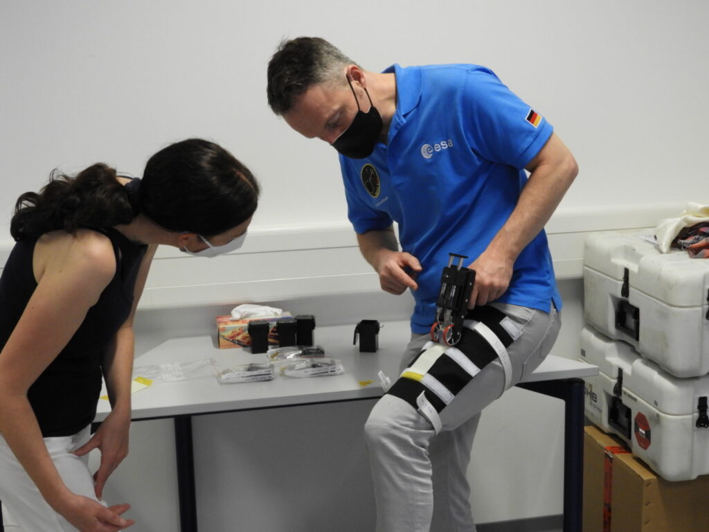
ESA astronaut Matthias Maurer is shown during preflight training for the BioPrint First Aid investigation, which tests a bioprinted tissue patch for enhanced wound healing.
Credit: ESA

A suitably advanced piece of wound care technology will be sent into orbit to the space station in the next few days: a prototype for portable bioprinter that can cover a wound area on the skin by applying a tissue-forming bio-ink that acts like a patch, and accelerates the healing process.

While the aim is to provide a effective wound treatment for astronauts millions of kilometres from the nearest hospital, such a personalised wound healing patch would also have a great benefit on Earth. Since the cultured cells are taken from the patient, immune system rejection is unlikely, allowing a safe regenerative and personalised therapy. Other advantages are the possibilities of treatment and greater flexibility regarding wound size and position. In addition, due to its small size and portability, physicians could take the device anywhere to an immobile patient if their cells were cultivated in advance.

“On human space exploration missions, skin injuries need to be treated quickly and effectively,” said project manager Michael Becker from the German Space Agency. “Mobile bioprinting could significantly accelerate the healing process. The personalised and individual bioprinting-based wound treatment could have a great benefit and is an important step for further personalised medicine in space and on Earth.”

The use of bioprinting for skin reconstruction following burns is one growing application for the technology. However, it presently requires large bioprinters that first print the tissue, allow it to mature, before it is implanted onto the patient. By testing it in the gravity-free environment of space, Bioprint FirstAid will help optimise of bioprinting materials and processes. Microgravity-based 3D tissue models are important for greater understanding of the bioengineering and bio-fabrication requirements that are essential to achieve highly viable and functional tissues. Under microgravity conditions, the pressure of different layers containing cells is absent, as well as the potential sedimentation effect of living cell simulants. The stability of the 3D printed tissue patch, and the potentially gravity-dependent (electrolyte to membrane interface) crosslinking process, can be analysed for future applications.

The Bioprint FirstAid prototype contains no cells at this point. The surprisingly simple prototype is a robust, purely mechanical handheld bioprinter consisting of a dosing device in the handle, a print head, support wheels, and an ink cartridge. The cartridge contains a substitution (in total two different substitutions, both without skin cells) and a crosslinker, which serves as a stabilising matrix. To test it out, the simulant will be applied to the arm or leg of a crew member wrapped in foil, or alternatively at any other surface wrapped in foil. On Earth, a printed sample with human cells will be tested, and the distribution pattern will be compared to the cell-free sample that was printed in space.

Source: NASA

Faster 3-D Bioprinting A Step Closer to Printing Whole Organs

With the demonstration of a new type of more rapid 3-D bioprinting, University at Buffalo engineers have taken a step closer to the fabrication of whole organs.

In a video of the process, a hand emerges over a matter of seconds from a vat of liquid almost as if out of a science fiction movie. In reality, the video was sped up from its original duration of 19 minutes, but even this is a quantum leap ahead of the six or so hours such a process previously took. 
“The technology we’ve developed is 10-50 times faster than the industry standard, and it works with large sample sizes that have been very difficult to achieve previously,” said co-lead author Ruogang Zhao, PhD, associate professor of biomedical engineering.

The new method involves a 3-D printing technology called stereolithography and hydrogels. Hydrogels have applications in wound dressings, contact lenses and hygiene products, as well as scaffolds for tissue engineering.

Scaffolds are particularly important in 3-D bioprinting, and the team has spent a great deal of its time and effort on these in order to come up with an optimised solution for its fast, accurate 3-D printing technique.
“Our method allows for the rapid printing of centimeter-sized hydrogel models. It significantly reduces part deformation and cellular injuries caused by the prolonged exposure to the environmental stresses you commonly see in conventional 3-D printing methods,” said the other co-lead author, Chi Zhou, PhD, associate professor of industrial and systems engineering.

This method is readily suited for the printing of cells with embedded networks of blood vessels. It is expected that this emerging technology will be key to producing whole 3-D printed organs and tissue.

Source: Medical Xpress

Journal information: Nanditha Anandakrishnan et al, Fast Stereolithography Printing of Large‐Scale Biocompatible Hydrogel Models, Advanced Healthcare Materials (2021). DOI: 10.1002/adhm.202002103
https://medicalxpress.com/news/2021-03-rapid-3d-method-3d-printed.html