Category: Cancer

Cancer Surgery Patients Have a Reduced Hospital Stay with ‘Prehabilitation’

Photo by Martha Dominguez de Gouveia on Unsplash


A new approach to improve their fitness for surgery reduced the length of hospital stay for cancer patients, according to a new study.

Termed ‘prehabilitation’, the study’s approach includes exercise, nutrition and psychological and social interventions to bolster physical and mental health before surgery.

The study, published in the Annals of Surgery, found that prehabilitation interventions of between one and four weeks reduced cancer patients’ stay in hospital by 1.8 days compared with usual care.

Study author Dr Chris Gaffney from Lancaster Medical School said: “Surgery is like a marathon in terms of stressing the body, and you wouldn’t run a marathon without training.”

The researchers found that as little as one week can still benefit patient outcomes, indicating that prehabilitation should be recommended to accelerate recovery from cancer surgery, as shown by a reduced hospital length of stay.

Study author Dr Joel Lambert, now a postgraduate student at Lancaster Medical School and a surgeon at East Lancashire Teaching Hospitals NHS Trust, said: “We think that it may also confer a survival advantage for cancer patients as they can get to follow up treatments like chemotherapy more quickly.

“We think that the patient groups most likely to benefit are the ones with lower levels of fitness at baseline. In the Northwest we have some of the most socioeconomically deprived populations in the UK. This subset tend to have more co-morbid conditions hence less fit.”  

The patients studied were those with liver, colorectal, and upper gastrointestinal cancer, and who are often less fit than other cancer patients.

The study interventions were grouped into three types

  • Multimodal prehabilitation: exercise, which included both nutrition and psychosocial support,
  • Bimodal prehabilitation: exercise and nutrition or psychosocial support
  • Unimodal prehabilitation: exercise or nutrition alone

The exercise interventions included aerobic, resistance, and both aerobic and resistance exercises at all levels of intensity, some supervised by a kinesiologist or physiotherapist, while others were home-based exercise regimes. These ranged from one to four weeks and all interventions were within the current NHS surgery targets for cancer surgery.

The researchers concluded: “Future studies should focus on identifying patients who would benefit most from prehabilitation and the mechanistic underpinning of any improvement in clinical outcomes. Studies should closely monitor nutrition intake to determine if the response to exercise prehabilitation is dependent upon nutritional status. Lastly, mortality should be monitored for 12 months post surgery to determine if prehabilitation has any effect beyond 30 or 90 days.”

Source: Lancaster University

Fewer Side Effects in New Breast Cancer Therapy

Breast cancer cells. Image source: National Cancer Institute on Unsplash

Researchers have shown that a new treatment for breast cancer using an antibody linked to a cytotoxic drug is as effective as the previous combination, but with less side effects. The study was published in JAMA Oncology.

The development of treatment with antibodies directed towards HER2 positive breast cancer trastuzumab and pertuzumab, has proven the possibility of improved treatment and cure of this cancer type if these antibodies are combined with chemotherapy, often taxaner. Preoperative (neoadjuvant) treatment with this combination confers complete response in a high proportion of cases. Trastuzumab emtansine (T-DM1) consists of trastuzumab (T) and the cytotoxic substance emtansine (DM1) which clinical trials have shown have good efficacy and low toxicity.

The randomised phase II study PREDIX HER2 was conducted at 9 Swedish clinics to investigate the effect (the proportion of complete response after neoadjuvant treatment) of this treatment in relation to frequency and degree of side effects. The standard treatment was a combination of docetaxel, trastuzumab and pertuzumab which was compared with T-DM1 as experimental treatment, and patients received 6 treatments every 3rd week. The treatment effect was monitored with mammography and PET-CT with 18- fluorodeoxyglucose (18F-FDG). Tissue and blood samples were regularly taken from the patients for later analysis.

The study showed that the effect measured as pathologic complete response was similar in both treatment groups. With T-DM1 treatment, the frequency and degree of side effects was significantly lower and quality of life was higher during treatment. After a median follow up time of 40,4 months no difference was observed between the treatment groups.

In conclusion, the study showed that both treatments were equally efficient, but with fewer side effects in patients treated with T-DM1. A phase III study will allow definitive conclusions to be drawn regarding the efficiency. Meanwhile, the collected samples are being analysed to investigate factors which can explain the response in individuals.

Source: Karolinska Institute

How do Patients Who Exit Clinical Trials Early Feel?

Source: JD Mason on Unsplash

A new study has helped researchers understand the experiences of people who withdraw from clinical cancer trials.

Cancer clinical trials (CCTs) provide patients with an opportunity to receive experimental drugs, tests, and/or procedures that may lead to remissions. Such opportunities can be a great benefit for those who took part, but there is little known of the experiences of participants who withdraw from CCTs.

To address this, a first-of-its-kind study from the University of Pennsylvania School of Nursing (Penn Nursing) was conducted to better understand the post-trial needs of these patients and define responsible transitions when patients exit CCTs.

“Understanding the post-trial needs of patients with cancer and their families represents a measure of ethical respect of the many contributions that patients with cancer make to advancing our scientific knowledge and finding treatments that save lives,” said the study’s lead researcher, Connie M Ulrich, the Lillian S Brunner Chair in Medical and Surgical Nursing, professor of nursing, professor of medical ethics and health policy.

The study revealed three important areas:

  • Patients exiting CCTs feel intense symptoms, emotions, and awareness that their life spans are short and options seem limited.
  • The limited discussions with patients who are exiting on their immediate post-trial care needs can result in many feeling that there is no clear path forward.
  • Good communication that deliberately includes attention to post-trial needs throughout the CCT is needed to help scared and disappointed patients navigate their next steps.

The study is set for publication on the JAMA Network.

Source: University of Pennsylvania

New Test Makes Prostate Cancer Screening More Affordable

Photo by mari lezhava on Unsplash

Researchers have found that, coupled with MRI, the novel Stockholm3 blood test, could greatly cut overdiagnoses and thereby improve prostate cancer screening. The same research group previously showed that Magnetic resonance imaging (MRI) could also reduce overdiagnoses, and the Stockholm3 test can reduce the number of MRIs performed by a third while further preventing the detection of minor, low-risk tumours.

The research group published the findings of their study in The Lancet Oncology,

“Overall, our studies show that we have identified the tools needed to be able to carry out effective and safe screening for prostate cancer. After many years of debate and research, it feels fantastic to be able to present knowledge that can improve healthcare for men,” said Tobias Nordström, associate professor of urology at the Department of Clinical Sciences, Danderyd Hospital at Karolinska Institutet, who is responsible for the STHLM3MRI study.

The disease is currently screened for by using PSA (prostate-specific antigen) tests combined with traditional biopsies, result in unnecessary biopsies and overdiagnosis from detection of numerous minor, low-risk tumours. As a result of these costs outweighing benefits, no country save Lithuania has implemented nationwide screening programmes.

Results from the STHLM3MRI study published in NEJM indicated that overdiagnosis could be reduced by substituting traditional prostate biopsies with magnetic resonance imaging (MRI) and targeted biopsies. The new results, now published in y, show that the addition of the Stockholm3 test, which was developed by researchers at Karolinska Institutet, can be an important complement. It is a blood test that uses an algorithm to analyse a combination of protein markers, genetic markers and clinical data.

Fewer biopsies needed
“The availability of MRI in healthcare will be a limiting factor. We now show that a novel blood test as adjunct to MRI can reduce the number of MRIs performed by a third. Compared with traditional screening, overdiagnosis is reduced by as much as 69 percent. At the same time, the number of biopsies is halved, while we can find just as many clinically significant tumours,” said Martin Eklund, associate professor at the Department of Medical Epidemiology and Biostatistics, Karolinska Institutet.

In the STHLM3MRI study, 12 750 male participants provided an initial blood sample for PSA analysis and analysis using the new Stockholm3 test. Men with test results showing elevated PSA levels were then randomly selected for traditional biopsies or MRI. In the MRI group, biopsies were conducted strictly on suspected tumours identified by MRI.

“Separate use of the Stockholm3 test and MRI has previously been shown to be cost-effective. We have now analysed the cost-effectiveness when these tools are combined and will shortly report exciting results from that analysis,” Tobias Nordström concluded.

Source: Karolinska Institute

Cervical Cancer Rates Falling Worldwide

Photo by Becca Tapert on Unsplash

Cervical cancer incidence and mortality rates are holding steady or falling in most countries across the globe, according to a new analysis. Each country’s socioeconomic development level, cervical cancer screening use, and human papillomavirus (HPV) vaccination rates affect the rates. The findings are published online in CANCER.

Cervical cancer can be potentially prevented through screening for and treatment of precancerous lesions and through HPV vaccination. A team led by Mingjuan Jin, PhD, of the Zhejiang University School of Medicine in China, examined information on 31 countries released by the International Agency for Research on Cancer.

The analysis found that cervical cancer incidence and mortality rates were lower in more socioeconomically developed countries. Also, both past and predicted trends appear to be stable or decreasing in most countries, especially in those with effective cervical cancer screening and HPV vaccination programs.

Over the past decade, 12 countries had stable cervical cancer incidence rates, while rates fell in 14 and rose in five. Twelve countries had stable mortality rates from cervical cancer, with falling rates in 18 and only one had an increased rate.

The researchers predicted that for 27 countries, most are expected to have stable or decreasing trends over the next 15 years. Ten of the 27 countries are predicted to have stable incidence rates, nine to have decreasing rates, and eight to have increasing rates. Mortality rates are predicted to be stable in 16, decreasing in 10 and one to have an increasing rate.

“Effective cervical cancer screening programs and HPV vaccination should be further popularized to increase their coverage and ultimately decrease cervical cancer’s short-, mid-, and long-term burden,” said Dr Jin.

Source: Wiley Online

How Cancer Cells Develop Resistance to Chemotherapy

Source: National Cancer Institute on Unsplash

Researchers have found some answers as to why cancer cells can develop resistance to the cytotoxic drugs used in chemotherapy.

“We haven’t understood very much about how this resistance to chemotherapy develops and even less about how the microenvironment in cancer can affect the process,” said Kaisa Lehti, a professor at the Norwegian University of Science and Technology’s (NTNU) Department of Biomedical Laboratory Science.

Lehti has led this study into how cancerous tissues develop resistance to a particular form of chemotherapy, the results of which appear in Nature Communications.

If ovarian cancer is picked up early, almost all patients survive the first five years, while chances of survival are much worse if detected later. Finding effective treatment is therefore very important.

Platinum chemotherapy is one of the standard treatments for ovarian cancer, but cancer cells often develop resistance to this particular treatment. The reason lies in how the platinum-based cytotoxin itself can change the cancer cells and their environment.

Cytotoxin influences cancer cells and their environment
Lehti summed up the process: “The cytotoxin can change the way the cancer cells send and perceive signals and can modify the microenvironment around the cells.”

This change allows the cancer cells to withstand the damage caused by the cytotoxin—and can thus survive the chemotherapeutic attack. The researchers have found this key to the puzzle in a layer of tissue that often surrounds cancer cells.

“A fibrotic network of proteins, known as the extracellular matrix or ECM, surrounds the cancer cells, particularly the most aggressive ones,” said Lehti.

The fibrotic tissue, with the ECM network around the cancer cells, is mainly produced by normal connective tissue cells. But the cancer cells and connective tissue cells in the network can alter this tissue themselves.

“Previously, we haven’t known how the communication between the cancer cells and the extracellular matrix is affected by, or even itself influences, the development of cancer and its response to chemotherapy,” said Prof Lehti.

But it is now known that chemical and mechanical signals in the surrounding ECM tissue help cancer develop its ability to spread and to resist treatment.

“Certain signals from the ECM can critically change the cancer cells’ resistance to platinum-based cytotoxic drugs,” Prof Lehti explained.

In this way, the cytotoxin itself helps change both the microenvironment around the cancer cells and the ability of the cancer cells to sense their environment, and so resist the cytotoxin. By understanding this process, better therapies can be developed.

Source: MedicalXpress

Powering Up NK Cells With Magnetism

NK cells attack a cancer cell (centre). Source: NIH

Powering up natural killer cells with magnetic nanoparticles could enhance cancer immunotherapy, according to a Northwestern Medicine study published in ACS Nano.

This method could allow for the unleashing of natural killer (NK) cells on a variety of solid tumours, according to senior author Dong-Hyun Kim, PhD, associate professor at Northwestern.

“People have had trouble applying NK cells to solid tumours,” said Prof Kim. “If we can provide an easy path to modulate NK cells, perhaps this can become a useful therapy.”

Most cell-based immunotherapies involve T-cells, but these chimeric antigen receptor (CAR) T-cell therapies are costly and have a long incubation period and strong side effects.

On the other hand, NK cells belong to the innate immune system and are quicker to respond to pathogens. NK cell immunotherapy has been explored, according to Prof Kim, but that too has barriers.

“It’s pretty hard for these cells to penetrate inside the tumours which have thick barrier tissues,” Prof Kim said.

Magnetically activated NK cells
Boosting NK cell function with cytokines have proven unsuccessful and, like CAR T-cell therapy, have a high cost and lengthy manufacturing time. However, Prof Kim’s previous work with nanoparticles inspired a different approach.

Prof Kim and colleagues developed a magnetic nanocomplex that binds with NK cells and, when activated with an alternating magnetic field, exerts force on the exterior of the cell, promoting secretion of cytotoxic compounds. Testing this nanocomplex in animal models of hepatocellular carcinoma, the investigators found that magnetic activation increased the cancer-killing ability of NK cells when injected into solid tumours.

As a bonus, these nanoparticles show up with MRI, allowing for precise monitoring of NK cell distribution during and after injection.

“This creates a stronger NK cell, and can hopefully enhance the efficacy of the treatment,” Prof Kim said.

Source: Northwestern University

Cancer Survivors Experience Accelerated Ageing

Source: JD Mason on Unsplash

A new study published in Journal of the American Geriatrics Society indicates that cancer survivors, especially older ones, are more likely to experience faster functional decline as they age, compared with those without a history of cancer.

For the study, 1728 men and women (aged 22 to 100 years) were evaluated from 2006 to 2019, with 359 of these adults reporting a history of cancer. Among all participants, a history of cancer was associated with a 1.42 greater odds of weak grip strength. Those with a history of cancer and over 65 had a 1.61 greater odds of slow gait speed than those with no cancer history, and also had lower physical performance scores. Additionally, compared with those with no history of cancer, older individuals with a history of cancer experienced steeper declines in grip strength and gait speed. Reduced prefrontal cortex area is one of the factors thought to contribute to slow gait.

“Findings from our study add to the evidence that cancer and its treatment may have adverse effects on aging-related processes, putting cancer survivors at risk for accelerated functional decline,” said senior author Lisa Gallicchio, PhD, of the National Cancer Institute. “Understanding which cancer survivors are at highest risk, and when the accelerated decline in physical functioning is most likely to begin, is important in developing interventions to prevent, mitigate, or reverse the adverse aging-related effects of cancer and its treatment.”

Source: EurekAlert!

Sunlight-fuelled Chemical Changes Drive Melanomas

Photo by Amy Humphries on Unsplash

Mutations in DNA that lead to melanoma result from a sunlight-fuelled chemical conversion in DNA, not just a DNA copying error as previously believed, according to a new study.

The findings of the study, led by the Van Andel Institute scientists and published in Science Advances, upend long-held beliefs about the mechanisms underlying the disease, reinforce the importance of prevention efforts and offer a path forward for investigating the origins of other cancer types.

“Cancers result from DNA mutations that allow defective cells to survive and invade other tissues,” said corresponding author Gerd Pfeifer, PhD, a VAI professor. “However, in most cases, the source of these mutations is not clear, which complicates development of therapies and prevention methods. In melanoma, we’ve now shown that damage from sunlight primes the DNA by creating ‘premutations’ that then give way to full mutations during DNA replication.”

Melanoma begins in pigment-producing skin cells. Although less common than other types of skin cancer, melanoma is more likely to metastasise, significantly reducing patient survival. Previous studies have shown that melanoma has the most DNA mutations of any cancer. Like other skin cancers, melanoma is linked to sun exposure, specifically UVB radiation which damages cells and DNA.

Most cancers are to arise when damaged DNA causes a mutation that is propagated through subsequent cellular generations. In the case of melanoma, however, Pfeifer and his team found a different mechanism that produces disease-causing mutations – the introduction of a chemical base not normally found in DNA that makes it prone to mutation.

In melanoma, the problem occurs when UVB radiation from the sun hits certain sequences of bases: CC, TT, TC and CT, causing them to chemically link together and become unstable. This resulting instability induces a chemical change to cytosine that transforms it into uracil, a chemical base found in the messenger molecule RNA but not in DNA. This change, called a “premutation,” primes the DNA for mutation during normal cell replication and eventually melanoma.

These mutations may lay dormant for years, not causing disease. More mutations can build up throughout a person’s lifetime exposure to sunlight, resulting in a stubborn cancer that evades many therapeutic options.

“Safe sun practices are very important. In our study, 10–15 minutes of exposure to UVB light was equivalent to what a person would experience at high noon, and was sufficient to cause premutations,” Prof Pfeifer said. “While our cells have built-in safeguards to repair DNA damage, this process occasionally lets something slip by. Protecting the skin is generally the best bet when it comes to melanoma prevention.”

The study used a method developed by Prof Pfeifer’s lab called Circle Damage Sequencing, enabling scientists to ‘break’ DNA at each point where damage occurs. DNA is coaxed into circles and replicated with PCR. With enough DNA, next-generation sequencing then identifies which DNA bases are present at the breaks. Pfeifer and colleagues plan to use this technique going forward to examine other types of DNA damage in different kinds of cancer.

Source: Van Andel Institute (VAI)

Journal reference: Jin, S-G., et al. (2021) The major mechanism of melanoma mutations is based on deamination of cytosine in pyrimidine dimers as determined by circle damage sequencing. Science Advances. doi.org/10.1126/sciadv.abi6508.

Unleashing the Immune System to Attack Cancers

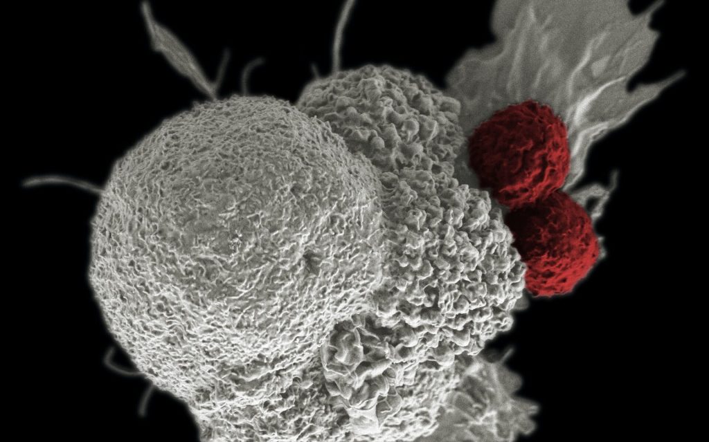
Shown here is a pseudo-colored scanning electron micrograph of an oral squamous cancer cell (white) being attacked by two cytotoxic T cells (red), part of a natural immune response. Photo by National Cancer Institute on Unsplash

A potential treatment has been identified, that could boost the immune system’s ability to find and destroy cancer cells, by impeding certain cells which regulate the immune system, which in turn can unleash other immune cells to attack tumours in cancer patients.

“A patient’s immune system is more than able to detect and remove cancer cells and immunotherapy has recently emerged as a novel therapy for many different types of cancers,” explained study leader Nullin Divecha, Professor of Cell Signalling at the University of Southampton. “However, cancer cells can generate a microenvironment within the tumour that stops the immune system from working thereby limiting the general use and success of immunotherapy,” he continued.

One of a number of types of T cells, Teffector cells (Teffs) carry out the task of detection and removal of cancer cells . How well Teff cells work in detecting and removing cancer cells is partly governed by other T cells called T-regulatory cells, or Tregs for short. Tregs physically interact with the Teff cells, producing molecules which dampen the functioning of the Teff cells.

Prof Divecha added, “Tregs carry out an important function in the human body because without them, the immune system can run out of control and attack normal cells of the body. However, in cancer patients we need to give the Teff cells more freedom to carry out their job.”

Molecules released by tumour cells exacerbate the problem by attracting and gathering Tregs, reducing the activity and function of Teff cells even further. Though there are mechanisms to inhibit Treg cells, since Treg and Teff cells are very similar, Teff cells are also generally inhibited.

In this new study, published in PNAS, scientists from the University of Southampton and the National Institute of Molecular Genetics in Milan showed that inhibition of a family of enzymes in cells called PIP4K could be the answer to how to restrict Tregs without affecting Teffs.

The research team isolated Tregs from healthy donors and used genetic technology to suppress the production of the PIP4K proteins. They saw that loss of PIP4Ks from Treg cells stopped their growth and response to immune signals, in turn stopping them from impeding Teff cell growth and function.

Importantly, the loss of the same enzymes in Teff cells did not limit their activity.

“This was surprising because PIP4Ks are in both types of T cells in similar concentrations but our study shows that they seem to have a more important function for Tregs than Teffectors,” said Dr. Alessandro Poli who carried out the experimental research.

Scientists must next develop molecules in order to inhibition of PIP4K as a potential therapy for patients. “Towards this end we show that treatment with a drug like inhibitor of PIP4K could enable the immune system to function more strongly and be better equipped to destroy tumour cells.”

Source: EurekAlert!