Tag: immune system

Neanderthal Genetic Influences on Human Immune System and Metabolism

Diagram comparing the nose shape of a Neanderthal with that of a modern human by Dr Macarena Fuentes-Guajardo.

Neanderthal genes comprise some 1 to 4% of the genome of present-day humans whose ancestors migrated out of Africa, and new research has shown that their lingering presence shapes the immune systems and metabolism of people of non-African ancestry. Some of these genetics changes are detrimental, but are slowly being replaced by human versions.

A multi-institution research team including Cornell University has developed a new suite of computational genetic tools to address the genetic effects of interbreeding between humans of non-African ancestry and Neanderthals that took place some 50 000 years ago. (The study applies only to descendants of those who migrated from Africa before Neanderthals died out, and in particular, those of European ancestry.)

In a study published in eLife, the researchers reported that some Neanderthal genes are responsible for certain traits in modern humans, including several with a significant influence on the immune system. Overall, however, the study shows that modern human genes are winning out over successive generations.

“Interestingly, we found that several of the identified genes involved in modern human immune, metabolic and developmental systems might have influenced human evolution after the ancestors’ migration out of Africa,” said study co-lead author April (Xinzhu) Wei, an assistant professor of computational biology in the College of Arts and Sciences. “We have made our custom software available for free download and use by anyone interested in further research.”

Using a vast dataset from the UK Biobank consisting of genetic and trait information of nearly 300 000 British people of non-African ancestry, the researchers analysed more than 235 000 genetic variants likely to have originated from Neanderthals. They found that 4303 of those differences in DNA are playing a substantial role in modern humans and influencing 47 distinct genetic traits, such as how fast someone can burn calories or a person’s natural immune resistance to certain diseases.

Unlike previous studies that could not fully exclude genes from modern human variants, the new study leveraged more precise statistical methods to focus on the variants attributable to Neanderthal genes.

While the study used a dataset of almost exclusively white individuals living in the United Kingdom, the new computational methods developed by the team could offer a path forward in gleaning evolutionary insights from other large databases to delve deeper into archaic humans’ genetic influences on modern humans.

“For scientists studying human evolution interested in understanding how interbreeding with archaic humans tens of thousands of years ago still shapes the biology of many present-day humans, this study can fill in some of those blanks,” said senior investigator Sriram Sankararaman, an associate professor at the University of California, Los Angeles. “More broadly, our findings can also provide new insights for evolutionary biologists looking at how the echoes of these types of events may have both beneficial and detrimental consequences.”

Source: Cornell University

Pseudomonas Aeruginosa Locks out Immune Cells

Pseudomonas
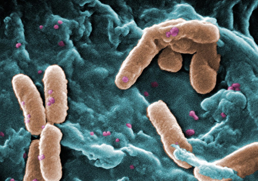
Scanning Electron Micrograph of Pseudomonas aeruginosa. Credit: CDC/Janice Carr

Pseudomonas aeruginosa bacteria are a common menace in hospital wards, causing life-threatening infections, and are often resistant to antibiotics. Researchers have discovered a mechanism that likely contributes to the severity of P. aeruginosa infections, which could also be a target for future treatments. The results were recently appeared in the journal EMBO Reports.

Many bacterial species use sugar-binding molecules called lectins to attach to and invade host cells. Lectins can also influence the immune response to bacterial infections. However, these functions have hardly been researched so far. A research consortium led by Prof Dr Winfried Römer at the University of Freiburg and Prof Dr Christopher G. Mueller at the CNRS/University of Strasbourg has investigated the effect of the lectin LecB from P. aeruginosa on the immune system. It found that isolated LecB can render immune cells ineffective: The cells are then no longer able to migrate through the body and trigger an immune response. The administration of a substance directed against LecB prevented this effect and led to the immune cells being able to move unhindered again.

LecB blockades immune cells

As soon as they perceive an infection, cells of the innate immune system migrate to a nearby lymph node, where they activate T and B cells, triggering a targeted immune response. LecB, according to the current study, prevents this migration. “We assume that LecB not only acts on the immune cells themselves in this process, but also has an unexpected effect on the cells lining the inside of the blood and lymph vessels,” Römer explains. “When LecB binds to these cells, it triggers extensive changes in them.” Indeed, the researchers observed that important structural molecules were relocated to the interior of the cells and degraded. At the same time, the cell skeleton became more rigid. “The cell layer thus becomes an impenetrable barrier for the immune cells,” Römer said.

An effective agent against LecB

Can this effect be prevented? To find out, the researchers tested a specific LecB inhibitor that resembles the sugar building blocks to which LecB otherwise binds. “The inhibitor prevented the changes in the cells, and T-cell activation was possible again,” Mueller said. The inhibitor was developed by Prof Dr Alexander Titz, who conducts research at the Helmholtz Institute for Pharmaceutical Research Saarland and Saarland University.

Further studies are needed to determine how clinically relevant the inhibition of the immune system by LecB is to the spread of P. aeruginosa infection and whether the LecB inhibitor has potential for therapeutic application. “The current results provide further evidence that lectins are a useful target for the development of new therapies, especially for antibiotic-resistant pathogens such as P. aeruginosa,” the authors conclude.

Source: University of Freiburg

How Shigella Suppresses the Immune System

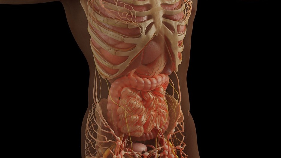
Anatomy of the gut
Source: Pixabay CC0

Researchers report in the journal Cell on insights they have made into the molecular process by which bacteria such as the highly contagious Shigella suppresses the immune system, preventing it from recognising impending infection.

Interferons are the first line of the immune system’s defence against infection. These warn neighbouring cells and prepare them to fight off an incoming infection. Many viruses – including SARS-CoV2 – have evolved proteins which inhibit normal interferon functions in order to increase infectivity.

Whether bacteria such as Shigella, which causes dysentery, are also able to interfere with the immune system and its capacity to fight the infection was previously unknown. Shigella is highly contagious, requiring only a small inoculum (10 to 200 organisms) to cause an infection.

The study found that Shigella inject a protein called “OspCs” into cells, which blocks the host’s interferon response, allowing the bacteria to successfully infect the host.

Interestingly, OspCs blocked interferon signalling by preventing cells from adapting to changing concentrations of calcium – a molecular signal that usually alerts a cell to infection and damage.

This newly identified strategy tricks the immune system, preventing the body from mounting an effective immune response to infection by decoding host calcium signals.

“This study was another perfect example of how studying pathogens can not only lead to a better understanding of infectious processes, but can also reveal the complexity of host responses to infection,” said Dr Charlotte Odendall.

These findings may be the first steps towards new bacterial treatments in the future, the researchers said.

Source: Kings College London

Hypoxia can Trigger Immune System Reaction

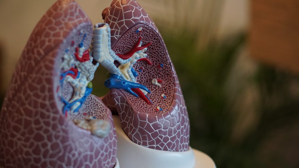
Anatomical model of lungs
Photo by Robina Weermeijer on Unsplash

New research from scientists at La Jolla Institute for Immunology (LJI), shows that hypoxia can activate the same group of immune cells that cause inflammation during asthma attacks. During hypoxia, these cells flood the airways with lung-damaging molecules.

Hypoxia is a known trigger for developing and worsening lung conditions such as severe asthma, chronic obstructive pulmonary disease (COPD), and fibrosis. To treat and prevent these diseases, researchers need to understand why a lack of oxygen would affect the immune system.

“We show how lack of oxygen can be part of a feedback loop that can contribute to even worse inflammation,” says LJI Professor and Chief Scientific Officer Mitchell Kronenberg, PhD, a member of the LJI Center for Autoimmunity and Inflammation. “This work gives us insight into the causes of fibrosis of the lung and severe asthma.”

Prof Kronenberg and colleagues worked with a genetically altered mouse model to mimic the signals of hypoxia in the airway’s epithelial cells, which line the paths to the lungs. They discovered that combining the hypoxia signals with inflammatory signals stimulated the “innate,” or rapidly responding immunity, and an immune cell type called an ILC2.

An ILC2’s job is to make signaling molecules – cytokines – that quickly alert other immune cells to react to a pathogen. Unfortunately, ILC2s sometimes over-react and respond to harmless environmental allergens. In these cases, ILC2s churn out cytokines that drive mucus production and inflammation in the lungs. All this swelling and mucus leads to hypoxia.

As they report in Journal of Experimental Medicine, ILC2s respond to hypoxia as well, adding to the lung damage already caused during an asthma attack.

“That hypoxia may then contribute further to inflammation,” said Prof Kronenberg.

The next step was to figure out exactly how epithelial cells activate ILC2 during hypoxia. LJI Postdoctoral Fellow Jihye Han, PhD, led the work to uncover an unexpected culprit: adrenomedullin (ADM). ADM is known for its role in helping blood vessels dilate, but until now it had no known role in immune function.

Prof Kronenberg was surprised to see ADM involved — but not shocked. “We’re finding that many molecules with no previously known role in the immune system can also be important for immune function,” said Prof Kronenberg. “We need to understand that more generally.”

The researchers showed that human lung epithelial cells exposed to hypoxia also produced ADM. This means ADM or its receptor could be targets for treating inflammatory and allergic lung diseases.

The challenge is to find a balance between dampening the harmful immune response without leaving the body vulnerable to infections. Prof Kronenberg points out that the epithelial cell-ADM-ILC2 connection protected mice from hookworm infections, which damage the lungs and gut.

“ADM is a new target for lung diseases and has been implicated in bacterial pneumonia as well,” said Prof Kronenberg. “But blocking it would have to be done carefully.”

Source: La Jolla Institute for Immunology

Long COVID May be Due to Suppressed Immune System

Man wearing mask with headache
Source: Usman Yousaf on Unsplash

Scientists studying the effect of the monoclonal antibody Leronlimab on long COVID may have found a surprising clue to the baffling syndrome, one that contradicts their initial hypothesis. The cause may be down to an abnormally suppressed immune system, and not a persistently hyperactive one as they initially suspected.

The study was published in Clinical Infectious Diseases.

“While this was a small pilot study, it does suggest that some people with long COVID may actually have under-active immune systems after recovering from COVID, which means that boosting immunity in those individuals could be a treatment,” said senior author Professor Otto Yang.

COVID is known to be caused by hyperactive immune responses against SARS-CoV-2 resulting in damage to lungs and other organs, and sometimes a cytokine storm that overwhelms the individual, which could lead to severe illness and death.

For some who recover from COVID, various symptoms can persist for months, such as fatigue, mental haziness, and shortness of breath. Classified as long COVID, a limited understanding of the causes makes it difficult to develop treatments.

One suggested possibility is that persistence of immune hyperactivity after COVID is a major contributor. The researchers therefore ran a small exploratory trial of Leronlimab, an antibody that attaches to an immune receptor called CCR5 that is involved in inflammation, on 55 people with the syndrome. Leronlimab was originally being developed as an HIV treatment.

Participants were randomised to receiving either weekly injections of the antibody or a saline placebo for eight weeks, and changes in 24 symptoms associated with long COVID were tracked, including loss of smell and taste, muscle and joint pain, and brain fog.

Originally, the researchers believed that blocking CCR5 would calm an overactive immune system after COVID infection. Indeed, preliminary results from an earlier trial appeared to show an improvement with Leronlimab.

“But we found just the opposite,” Prof Yang said. “Patients who improved were those who started with low CCR5 on their T cells, suggesting their immune system was less active than normal, and levels of CCR5 actually increased in people who improved. This leads to the new hypothesis that long COVID in some persons is related to the immune system being suppressed and not hyperactive, and that while blocking its activity, the antibody can stabilize CCR5 expression on the cell surface leading to upregulation of other immune receptors or functions.”

The findings, the researchers wrote, “suggests a complex role for CCR5 in balancing inflammatory and anti-inflammatory effects, eg through T regulatory cells,” although the results need to be confirmed in a larger, more definitive study.

Source: University of California – Los Angeles Health Sciences

New Insights Into Atopic Dermatitis Yield Possible Therapy

Source: CC0

Atopic dermatitis (AD) is often thought of as an inflammatory disease that arises from a breakdown in the barrier function of the skin. Now a new study pinpoints a cascade of inflammatory signalling that precedes the appearance of skin ulcers, shedding light on the early stages of the condition and possible new drug targets.

The work, published in the journal Science Translational Medicine, was the result of a cross-school and cross-institutional collaboration among researchers.

“You have researchers in the dental school noticing a skin condition, broadening their work to the medical school and experts on computational systems biology,” said Professor Dana Graves, a co-corresponding author on the paper. “Without this interdisciplinary collaboration, that initial finding would not have gone anywhere.”

John Seykora, a co-corresponding author and professor of dermatology, agreed. “This shows one of the benefits of being part of a university with experts across fields,” he says. “Our dental school colleagues developed a mouse that manifested a particular skin phenotype, and the question was, What was this and did it resemble any disease we might know? And in the end it did, and it’s providing some novel insights into a very common skin condition in humans.”

The work began with an exploration of the role of inflammatory signalling in bone fracture healing in diabetes. A focus was on nuclear factor kappa-B (NF-kB), a master regulator of inflammatory responses. As part of that work, researchers developed a mouse model lacking an activator of NF-kB signalling, IKKB. The researchers noticed that these animals developed skin lesions as they became young adults.

“That was interesting to us because these ulcerations looked like an inflammatory event, but we had effectively turned off the activity of NF-kB, which should reduce inflammation,” said Prof Graves. “So this was a paradox.”

To better understand what was driving this response, they sought expertise in skin diseases from Prof Seykora. When they examined the mice, they noted several features quite similar to AD, “albeit the mouse version” said Prof Graves.

In particular, they noted skin thickening and an infiltration of certain types of white blood cells that are also seen in human AD. Delving deeper into how the loss of IKKB was driving these effects, the team performed single-celled RNA analysis combined with a new analysis method. The team learned that fibroblasts were the culprit, a major component of the skin’s dermis layer and typically thought to support the structural integrity of skin.

Though NF-kB typically promotes inflammation, here, decreased NF-kB activity was paradoxically leading to recruitment of immune cells and associated inflammation. Data from the team’s single-cell RNA analysis pointed to high activities of a protein transcription factor called CEBPB, as well as a signalling molecule, CCL11, “We worked out the mechanism in the mouse,” Prof Sekora said, “then showed that much of it applied in human tissue as well.”

When the researchers compared what they had seen in the mouse cells to skin samples from people with AD, they found similar patterns; CCL11 and CEBPB were both found at higher levels in the affected skin than in unaffected skin.

Testing a monoclonal antibody against CCL11 in mice tamped down the inflammatory response they had initially seen in animals lacking IKKB, suggesting that this could be a target to reduce AD-associated inflammation.

The researchers say the work also highlights a developing appreciation that fibroblasts play important roles in immune processes in the skin, indicating that they are important regulators of white blood cells.

AD  typically emerges in childhood, often manifesting along with asthma. Indeed, in the mice, too, the signalling abnormalities the researchers observed occurred in a period corresponding to the animals “childhood.” The group’s findings suggest that fibroblasts may be involved during this period in helping to establish appropriate immune signalling in the skin.

“We have viewed NF-kB as a factor that stimulates inflammation, but it could be that, during development, its activation might be important for maintaining homeostasis,” said Prof Graves.

The team’s next steps are to further explore NF-kB signalling in fibroblasts.

Source: University of Pennsylvania

New Coating Makes the Nanomedicine Go Down

Upon injection into the blood, nanomedicines (blue spheres) are immediately attacked by proteins of the immune system called complement proteins (orange). Complement proteins cause rapid destruction of the nanomedicine, and also induce an anaphylaxis-like reaction. By attaching complement-degrading proteins (yellow ninjas made of protein) to the surface of nanomedicines, Penn researchers have largely solved this problem, potentially allowing more diseases to be safely treated by nanomedicine. Credit: University of Pennsylvania

In nanomedicine, immune reactions against the nanoparticles that contain the medicine or vaccine, reducing its effectiveness. Researchers have now come up with a new method to prevent the body from treating nanomedicines like foreign invaders, by covering those nanoparticles with a coating to suppress the immune response.

As soon as they are injected into the bloodstream, unmodified nanoparticles are swarmed by complement proteins, triggering an inflammatory response and preventing the nanoparticles from reaching their treatment targets. Penn Medicine researchers, whose findings are published in Advanced Materials, have devised a coating for nanoparticles that suppresses complement activation.

Nanoparticles are tiny capsules, typically made from proteins or fat-related molecules, that contain certain types of treatment or vaccine. The best-known examples of nanoparticle-delivered medicines are mRNA COVID vaccines.

“It turned out to be one of those technologies that just works right away and better than anticipated,” said study co-senior author Jacob Brenner, MD, PhD.

RNA- or DNA-based therapies generally need delivery systems to get them through the bloodstream into target organs. Harmless viruses often have been used as carriers or “vectors” of these therapies, but nanoparticles are increasingly considered safer alternatives. Nanoparticles also can be tagged with antibodies or other molecules that make them hone in precisely on targeted tissues.

The complement attack problem has been a serious impediment to nanomedicine. Circulating complement proteins treat nanoparticles as if they were bacteria, immediately coating nanoparticle surfaces and summoning macrophages to engulf them. Researchers have attempted to reduce the problem by pre-coating nanoparticles with camouflaging molecules, such as forming a watery, protective shell around nanoparticles using polyethylene glycol (PEG).

But nanoparticles camouflaged with substances like PEG still draw at least some complement attack. In general, nanoparticle-based medicines that move through the bloodstream (mRNA COVID vaccines are injected into muscle, not the bloodstream) have had a very low efficiency in getting to their target organs, usually under 1%.

In the study, the researchers came up with a new approach to protect nanoparticles, based on natural complement-inhibitor proteins that circulate in the blood, attaching to human cells to help protect them from complement attack.

In vitro tests using standard PEG-protected nanoparticles with one of these complement inhibitors, called Factor I, provided dramatically better protection from complement attack. In mice, the same strategy prolonged the half-life of standard nanoparticles in the bloodstream, allowing a much larger fraction of them to reach their targets.

“Many bacteria also coat themselves with these factors to protect against complement attack, so we decided to borrow that strategy for nanoparticles,” said co-senior author Jacob Myerson, PhD, a senior research scientist in the Department of Systems Pharmacology and Translational Therapeutics at Penn.

In a set of experiments in mouse models of severe inflammatory illness, the researchers also showed that attaching Factor I to nanoparticles prevents the hyper-allergic reaction that otherwise could be fatal.

Further testing will be needed before nanomedicines incorporating Factor I can be used in people, but in principle, the researchers said, attaching the complement-suppressing protein could make nanoparticles safer and more efficient as therapeutic delivery vehicles so that they could be used even in severely ill patients.

The researchers now plan other protective strategies for medical devices, such as catheters, stents and dialysis tubing, which are similarly susceptible to complement attack. They also plan to investigate other protective proteins.

“We’re recognising now that there’s a whole world of proteins that we can put on the surface of nanoparticles to defend them from immune attack,” Dr Brenner said.

Source: University of Pennsylvania School of Medicine

How Immune Cells Fight Infection Using Body Fat

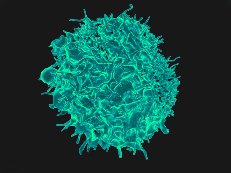
T lymphocyte. Credit: NIH/NIAID

A new study from the University of East Anglia and Quadram Institute sheds light on how our immune cells make use of body fat to fight infection. The research, published today in Nature Communications, could lead to new approaches to treating people with bacterial infections.

The work could one day help treat infections in vulnerable and older people, the researchers said. The team studied Salmonella bacteria and tracked fatty acid movement and consumption in live stem cells. They then examined the immune response to Salmonella bacterial infection, by analysing liver damage.

They uncovered how blood stem cells respond to infection, by acquiring high energy fatty acids from the body’s fat stores. In the bone marrow where blood stem cells are resident, infection signals drive adipocytes to release their fat stores as fatty acids into the blood.

And they identified that these high energy fatty acids are then taken up by blood stem cells, effectively feeding the stem cells and enabling them to make millions of Salmonella-fighting white blood cells. The researchers also identified the mechanism by which the fatty acids are transferred and discusses the potential impact this new knowledge could have on future treatment of infection.

Dr Stuart Rushworth, from UEA’s Norwich Medical School, said: “Our results provide insight into how the blood and immune system is able to respond to infection.

“Fighting infection takes a lot of energy and fat stores are huge energy deposits, which provide the fuel for the blood stem cells to power up the immune response.

“Working out the mechanism through which this ‘fuel boost’ works gives us new ideas on how to strengthen the body’s fight against infection in the future.”

Dr Naiara Beraza, from the Quadram institute, said: “Our results allow us to understand how our immune system uses fat to fuel the response to infection. Defining these mechanisms will enable us to develop new therapeutics to treat infections in the liver.”

Source: University of East Anglia

Differences in Influenza Responses According to Genetic Ancestries

Photo by Andrea Piacquadio on Unsplash

Researchers have uncovered differences in immune pathway activation to influenza infection between individuals of European and African genetic ancestry, according to a study published in Science. Many of the genes that were associated with these immune response differences to influenza are also enriched among genes associated with COVID disease severity. 

“The lab has been interested in understanding how individuals from diverse populations respond differently to infectious diseases,” said first author Haley Randolph, a graduate student at the University of Chicago. “In this study, we wanted to look at the differences in how various cell types respond to viral infection.”

The researchers examined gene expression patterns in peripheral mononuclear blood cells, a diverse set of specialised immune cells that play important roles in the body’s response to infection. These cells were gathered from men of European and African ancestry and then exposed the cells to flu in a laboratory setting. This let the team examine the gene signatures of a variety of immune cell types, and observe how the flu virus affected each cell type’s gene expression.

The results showed that individuals of European ancestry showed an increase in type I interferon pathway activity during early influenza infection.

“Interferons are proteins that are critical for fighting viral infections,” said senior author Luis Barreiro, PhD, Associate Professor of Medicine at UChicago. “In COVID-19, for example, the type I interferon response has been associated with differences in the severity of the disease.”

This increased pathway activation hindered the replication of the virus more and limited viral replication later on. “Inducing a strong type I interferon pathway response early upon infection stops the virus from replicating and may therefore have a direct impact on the body’s ability to control the virus,” said Barreiro. “Unexpectedly, this central pathway to our defense against viruses appears to be amongst the most divergent between individuals from African and European ancestry.”

The researchers saw a variety of differences in gene expression across different cell types, suggesting a constellation of cells that work together to fight disease.

Such a difference in immune pathway activation could explain influenza outcome disparities between different racial groups; Non-Hispanic Black Americans are more likely to be hospitalised due to the flu than any other racial group.

However, these results are not evidence for genetic differences in disease susceptibility, the researchers point out. Rather, possible differences in environmental and lifestyle between racial groups could be influencing gene expression, and affecting the immune response.

“There’s a strong relationship between the interferon response and the proportion of the genome that is of African ancestry, which might make you think it’s genetic, but it’s not that simple,” said Barreiro. “Genetic ancestry also correlates with environmental differences. A lot of what we’re capturing could be the result of other disparities in our society, such as systemic racism and healthcare inequities. Although some of the differences we show in the paper can be linked to specific genetic variation, showing that genetics do play some role, such genetic differences are not enough to fully explain the differences in the interferon response.”

These differences in viral susceptibility may not be confined to just influenza. Comparing a list of genes associated with differences in COVID severity, the researchers found that many of the same genes showed significant differences in their expression after flu infection between individuals of African and European ancestry.

“We didn’t study COVID patient samples as part of this study, but the overlap between these gene sets suggests that there may be some underlying biological differences, influenced by genetic ancestry and environmental effects, that might explain the disparities we see in COVID outcomes,” said Barreiro.

As they explore this further, the researchers hope to figure out which factors contribute to the differences in the interferon response, and immune responses more broadly, to better predict individual disease risk.

Source: EurekAlert!