Tag: natural killer cells

Natural Killer Cells are Suppressed by Anxiety and Insomnia

Photo by Cottonbro on Pexels

Anxiety and insomnia have been shown to weaken the immune system and make us more prone to disease. Now, researchers found that this may be because experiencing symptoms of either can reduce the number of natural killer cells, our bodies’ machinery for defence. Their findings showed that in young women who experience insomnia symptoms, the number of total NK cells was lower. If they experienced anxiety symptoms, the number of NK cells that circulate through the body was lower. These findings could inform the development of novel strategies to raise awareness about the physiological consequences of anxiety and insomnia and help in the prevention of immune-related disorders and cancers, the team said.

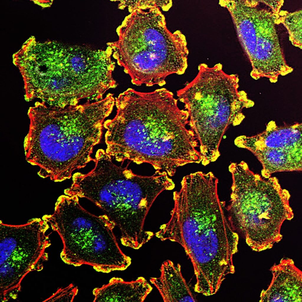
Natural killer (NK) cells are the bodyguards of our immune system. As a first line of defense, they destroy invading pathogens, foreign bodies, and infected cells in early stages, thereby preventing them from spreading. NK cells can circulate within the blood stream (circulatory) or reside in tissue and organs. Having too few NK cells can lead to immune system dysfunction and increase susceptibility to disease.

Anxiety disorder and insomnia are two conditions that can disrupt the normal functioning of the immune system. Given these disorders are on the rise, researchers in Saudi Arabia have now examined the association between anxiety, insomnia, and NK cells in young, female students. They published their results in Frontiers in Immunology.

“We found that in students with insomnia symptoms, count and percentage of total NK cells and their sub-populations were declined,” said first author Dr Renad Alhamawi, an assistant professor of immunology and immunotherapy at Taibah University. “Students with general anxiety symptoms, on the other hand, had a lower percentage and number of circulatory NK cells and their sub-populations, compared to symptom-free students.”

Decimated defence

60 female students, aged between 17 and 23 years old, participated in the study. They filled out three questionnaires about sociodemographics as well as anxiety and insomnia symptoms. The symptoms of the latter two were self-reported. The surveys showed that around 53% of the participants reported sleeping disturbance suggestive of insomnia, and 75% reported anxiety symptoms, with around 17% and 13% reporting moderate and severe symptoms, respectively.

Participants also provided blood samples through which percentages of NK cells and their subtypes were determined. NK cells have two subtypes: CD16+CD56dim cells make up the majority of NK cells in the nervous system that connects the central nervous system to the rest of the body (peripheral NK cells). Cells belonging to this subtype also exhibit cytotoxicity, which means they can damage or kills cells that invade the body. The other subtype, CD16+CD56high cells, are less frequent and involved in the production of proteins that function as chemical messengers and in immunoregulation. Both subtypes are circulatory NK cells.

The results showed that students with anxiety symptoms had a lower percentage and number of circulatory NK cells and their sub-populations, compared to students who did not report symptoms. Severity of symptoms also played a role as students with moderate and severe anxiety symptoms had a significant lower percentage of circulatory NK cells compared to students without them. Among students with minimal or mild anxiety symptoms, only a statistically insignificant decline in NK cell percentage was observed. In students with insomnia symptoms, higher anxiety scores were negatively associated with the proportion of total peripheral NK cells.


Read and download original article


Stressed immune systems

A reduction of these cells can lead to the impairment of the immune system, which may result in diseases, cancers, and mental disease, including depression. “Understanding how these psychological stressors influence the distribution and activity of immune cells, especially peripheral NK cells, may provide valuable insights into the mechanisms underlying inflammation and tumorigenesis,” Alhamawi explained.

The study is limited in some respects, the team pointed out. It only included young females – the group amongst whom anxiety and sleeping disorders have been rising disproportionally, limiting the generalizability of the results. The researchers said that future studies that include different age groups, sexes, and people from different regions, are necessary to gain a better overall view of the hidden effects of anxiety and insomnia on the proportion and function of these immune cells.

Previous studies have suggested healthy lifestyles with regular physical activity, stress reduction, and a healthy and balanced diet can boost the number and function of NK cells. However, the impact of anxiety and insomnia can disrupt the normal functioning of various body systems, including the immune system, thereby contributing to the development of chronic and inflammatory diseases. “Such impacts ultimately compromise overall health and quality of life,” concluded Alhamawi.

Source: Frontiers

Elevated NK Cells Found in Children with Severe RSV

Photo by Andrea Piacquadio on Unsplash

Respiratory syncytial virus (RSV) is the leading cause of hospitalisation in young children due to respiratory complications such as bronchiolitis and pneumonia. Yet little is understood about why some children develop only mild symptoms while others develop severe disease.

To better understand what happens in these cases, clinician-scientists from Brigham and Women’s Hospital, and Boston Children’s Hospital analysed samples from patients’ airways and blood, finding distinct changes in children with severe cases of RSV, including an increase in the number of natural killer (NK) cells in their airways.

The descriptive study, which focuses on understanding the underpinnings of severe disease, may help to lay groundwork for identifying new targets for future treatments. Results are published in Science Translational Medicine.

“As a physician, I help to care for children who have the most severe symptoms, and as a researcher, I’m driven to understand why they become so sick,” said corresponding author Melody G. Duvall, MD, PhD, of the Division of Pulmonary and Critical Care Medicine at Brigham and Women’s Hospital (BWH) and the Division of Critical Care Medicine at Boston Children’s Hospital. “NK cells are important first responders during viral infection – but they can also contribute to lung inflammation. Interestingly, our findings fit with data from some studies in COVID-19, which reported that patients with the most severe symptoms also had increased NK cells in their airways. Together with previous studies, our data link NK cells with serious viral illness, suggesting that these cellular pathways merit additional investigation.”

Duvall and colleagues, including lead author Roisin B. Reilly of the Division of Pulmonary and Critical Care Medicine at BWH, looked at samples from 47 children critically ill with RSV, analysing immune cells found in their airways and peripheral blood. Compared to uninfected children, those with severe illness had elevated levels of NK cells in their airways and decreased NK cells in their blood. In addition, they found that the cells themselves were altered, both in appearance and in their ability to perform their immunological function of killing diseased cells.

Duvall and co-authors have previously described a post-pandemic surge in paediatric RSV infections. While clinicians can only provide supportive care to the most severely sick children, vaccines to prevent RSV are now available for children 19 months and younger, adults 60 years and over, and people who are pregnant.

Source: Brigham and Women’s Hospital

New Research Explains Differences in Men’s and Women’s Immune Systems

Photo by Daniil Onischenko on Unsplash

By analysing the immune system of female-to-male transgender individuals, Swedish researchers demonstrate the role of sex hormones in regulating the immune system. This newfound knowledge, published in Nature, explains differences between men and women, particularly in terms of immune signalling, and can be used to develop new immunological medications according to researchers.

Sex differences in the immune system are regulated both by genetics and by sex hormones. However, immunological comparisons between men and women can never fully distinguish the significance of genetic versus hormonal variations.

Now, three Swedish research groups led by Karolinska Institutet and Uppsala University has conducted a unique study analysing the regulation and adaptation of the immune system over time in 23 trans men who have undergone gender-affirming testosterone treatment, starting at the age of 18–37 years.

“We have followed individuals who were assigned female sex at birth and later received testosterone treatment in adulthood. Their genetic profile remains unchanged, while their hormone profile shifts entirely from typically female to male hormone levels,” says Petter Brodin, paediatrician and professor of paediatric immunology at the Department of Women’s and Children’s Health, Karolinska Institutet, who led the study together with Nils Landegren, assistant professor at Uppsala University, and Olle Kämpe, Professor at the Department of Medicine, Solna, Karolinska Institutet. “This unique change allows us, for the first time, to identify which parts of a person’s immune system are directly regulated by sex hormones rather than genetic sex differences.” 

The researchers can now demonstrate that increased testosterone levels and the accompanying reduction in oestrogen particularly affect the balance between two crucial immune signalling systems: antiviral interferon type 1 (IFN-1) and proinflammatory signals such as tumour necrosis factor alpha (TNFα).  

Specifically, they found that testosterone modulates a cross-regulated axis between type-I interferon and tumour necrosis factor. This is mediated by functional attenuation of type-I interferon responses in both plasmacytoid dendritic cells and monocytes. Conversely, testosterone potentiates monocyte responses leading to increased tumour necrosis factor, interleukin-6 and interleukin-15 production and downstream activation of nuclear factor kappa B-regulated genes and potentiation of interferon-γ responses, primarily in natural killer cells. 

The immune system changes throughout life

They also have a hypothesis about why the immune system needs to be dynamically regulated by hormones throughout life. 

“All individuals must be able to adjust their immune systems over the course of their lives to be optimally regulated for the conditions and challenges we face. During puberty and sexual maturation, new demands arise, and the immune system must be regulated differently to enable pregnancy in women and muscle growth in men,” says Petter Brodin. 

By regulating these key functions via sex hormones, this can be achieved, and in women, it is dynamically controlled even during a menstrual cycle,” he adds. 

The results of the study open an entirely new field of research, according to Nils Landegren. 

“The newfound knowledge will help us better influence people’s immune systems even without using sex hormones. For example, new drugs can be developed to impact these regulatory mechanisms and thus rebalance the immune response, especially for women with the autoimmune rheumatic disease SLE,” he explains. 

However, the results also have a more direct implications for transgender individuals. 

“This research is also of crucial for transgender individuals undergoing gender-affirming hormone therapy, and I believe that this group deserves significantly more scientific attention and follow-up to ensure their long-term health,” says Petter Brodin. 

Source: Karolinska Institutet

Neuroblastomas: ‘New’ Immune System Responds Better to Therapy

Credit: National Cancer Institute

Cancer researchers have shown that immunotherapy after stem cell transplantation effectively combats neuroblastomas in children. Crucially, stem cells from a parent provide children with a new immune system that responds much better to immunotherapies. These results of an early clinical trial were published in the Journal of Clinical Oncology.

Tumours of the nervous system, neuroblastomas are associated with an unfavourable prognosis if the tumour is classified as a high-risk type. and particularly poor for patients in the relapsed stage. In this study by scientists at St. Anna Children’s Cancer Research Institute and the Eberhard Karls University of Tübingen, immunotherapy following stem cell transplantation is now associated with long-term survival in a substantial proportion of the patients. Compared to an earlier study the survival rate was increased.

“After the transplantation of stem cells from a parent, the patients are equipped with a new immune system. This enables a better immune response to the subsequent immunotherapy and clearly improves the outcome,” explains Prof Ruth Ladenstein, MD, co-first author.

Five-year survival exceeds 50%

“After a median follow-up of about eight years, we see that more than half of the study patients live five years or longer with their disease,” Prof Ladenstein reports (5-year overall survival: 53%). In comparison, the 5-year overall survival in an earlier study, in which stem cell transplantation was not followed by immunotherapy, was only 23%. Those patients who showed a complete or partial response to prior treatment had significantly better survival.

“In summary, immunotherapy with dinutuximab beta following transplantation of stem cells from matched family donors resulted in remarkable outcomes when patients had at least a partial response to prior treatment,” says Prof Ladenstein. “In our study, there were no unexpected side effects and the frequency of graft-versus-host-disease was low.”

Restoring natural killer cell potency

Dinutuximab beta is a monoclonal antibody that binds to a molecule, GD2, on the surface of tumour cells, marking them for destruction by natural killer cells. But prior chemotherapies may impair natural killer cells“Therefore, a transplantation of intact natural killer cells from matched family donors seems reasonable before immunotherapy is administered. The transplanted, new natural killer cells are now able to target the tumour cells more efficiently – by means of an antibody-dependent reaction,” explains Prof Ladenstein.

According to the authors, further studies are needed to determine the individual components of the therapeutic approaches. Recently, conventional chemotherapy has also been combined with immunotherapy early in the treatment strategy, resulting in similarly improved response rates. The hope is that a renewed immune system through a healthy parent in combination with the described transplantation procedure could further increase survival rates: “Our approach could thus result in stronger and longer lasting tumour control. A randomised study would be necessary to scientifically substantiate the additional potential benefit of a new immune system in the context of relapse therapy,” Prof Ladenstein adds.

Source: St. Anna Children’s Cancer Research Institute

Memory Killer Cells can Improve Melanoma Survival

Melanoma Cells. Credit: National Cancer Institute

In a study published in the journal Immunity, Researchers at Karolinska Institutet and the University of Copenhagen shown that high levels of memory killer cells in cancer tissue correlate with a better survival rate in people with melanoma.

Certain immune T cells called tissue-resident memory cells are formed locally in the skin and other tissue, and protect against infections that they have encountered before. Some of them express proteins that enable them to kill infected cells. These “memory killer cells” can also contribute to the inflammatory skin disorders vitiligo and psoriasis. Recent research has shown that they are also involved in the body’s immune response to various cancers.

Varying responses to treatment

The memory killer cells have been shown to respond to immunotherapy, a Nobel Prize-winning cancer therapy involving the tweaking/activation of the immune system. Immunotherapy is normally administered as a complement to other cancer treatments, and there is considerable variation in how patients respond to it.

“We don’t know so much about how and why memory killer cells are formed in the skin and what it means for cancer patients,” says Professor Yenan Bryceson at Karolinska Institutet. “Finding out how these cells develop enables us to contribute to the development of more efficacious immunotherapy for diseases like melanoma.”

The study charted the development of memory killer cells in human skin, performed as a collaborative effort between KI researchers Beatrice Zitti and Elena Hoffer. The researchers isolated T cells from the skin and blood of healthy volunteers and used advanced techniques to examine gene activity and expression of different proteins. This allowed them to identify T cells in the blood with the potential to develop into memory killer cells in skin or other tissues. After knocking out specific genes, they could also demonstrate which genes are required for the maturation of memory killer cells in tissue.

More effective immunotherapy

The researchers then went on to study tumour samples from melanoma patients and found that those with a higher rate of survival also had a larger accumulation of epidermal memory killer cells.

We’ve been able to identify several factors that control the formation of memory killer cells, which play an important part in maintaining a healthy skin,” says dermatologist Liv Eidsmo, professor at the University of Copenhagen in Denmark and researcher at Karolinska Institutet, who led the study with Professor Bryceson. “There’s a fine balance between effective protection against tumours and infections in the skin and contribution to inflammatory diseases like vitiligo and psoriasis.”

The researchers now aim to harness their findings to optimise the immunotherapy-induced T-cell response to make it even better at eliminating cancer cells in tissues.

Source: Karolinska Institutet

After a Pregnancy, Natural Killer Cells Suppress Tumours

Source: Anna Hecker on Unsplash

After a pregnancy, breast cells call in Natural Killer T (NKT) cells as reinforcements to prevent tumours from arising, according to a study published in Cell Reports. This finding from the lab of Associate Professor Camila dos Santos at Cold Spring Harbor Laboratory (CSHL) illuminates a new way in which pregnancy reduces the risk of breast cancer.

Two lines of defence exist in the immune system: the innate response, which involves immune cells that attack any foreign molecule they encounter, and the adaptive response, which consists of immune cells that respond specifically to calls for help. NKT cells are a unique subset of cells that are present throughout the body which can participate in both responses. 

CSHL graduate student Amritha Varshini Hanasoge Somasundara said that after a pregnancy: “There is an increase in this specific [NKT] cell type, and only in the mammary gland. We don’t see the expansion everywhere else in the body, even though NKT cells are present everywhere else in the body.”

The team sought to uncover the reason behind the larger number of NKT cells were doing in the breast tissue. Hanasoge discovered that in mice, breast epithelial cells, which line lactation ducts, produce a specific protein called CD1d after pregnancy. If the cells did not present CD1d, no increase in NKT cells was seen in the tissue; the epithelial cells became cancerous and grew into tumours. Hanasoge and dos Santos think that CD1d molecules are calling in NKT cells to monitor the epithelial cells in the breast tissue after pregnancy. If they become cancerous, the NKT cells can quickly kill them to prevent tumour growth.

The team’s findings establish a novel link between pregnancy and the immune system in preventing breast cancer. They want to know how these findings can be translated into humans and what other factors may influence an abundance of NKT cells in breast tissue, such as aging and menopause, which are both associated with increased breast cancer risk.

Discussing the results, Associate Professor dos Santos said: “One of the hypotheses that we are working on now is: do pregnancies later on in life bring in the same expansion of the same subtypes of immune cells as pregnancies that took place early in life?”

Source: Cold Spring Harbor Laboratory

Powering Up NK Cells With Magnetism

NK cells attack a cancer cell (centre). Source: NIH

Powering up natural killer cells with magnetic nanoparticles could enhance cancer immunotherapy, according to a Northwestern Medicine study published in ACS Nano.

This method could allow for the unleashing of natural killer (NK) cells on a variety of solid tumours, according to senior author Dong-Hyun Kim, PhD, associate professor at Northwestern.

“People have had trouble applying NK cells to solid tumours,” said Prof Kim. “If we can provide an easy path to modulate NK cells, perhaps this can become a useful therapy.”

Most cell-based immunotherapies involve T-cells, but these chimeric antigen receptor (CAR) T-cell therapies are costly and have a long incubation period and strong side effects.

On the other hand, NK cells belong to the innate immune system and are quicker to respond to pathogens. NK cell immunotherapy has been explored, according to Prof Kim, but that too has barriers.

“It’s pretty hard for these cells to penetrate inside the tumours which have thick barrier tissues,” Prof Kim said.

Magnetically activated NK cells
Boosting NK cell function with cytokines have proven unsuccessful and, like CAR T-cell therapy, have a high cost and lengthy manufacturing time. However, Prof Kim’s previous work with nanoparticles inspired a different approach.

Prof Kim and colleagues developed a magnetic nanocomplex that binds with NK cells and, when activated with an alternating magnetic field, exerts force on the exterior of the cell, promoting secretion of cytotoxic compounds. Testing this nanocomplex in animal models of hepatocellular carcinoma, the investigators found that magnetic activation increased the cancer-killing ability of NK cells when injected into solid tumours.

As a bonus, these nanoparticles show up with MRI, allowing for precise monitoring of NK cell distribution during and after injection.

“This creates a stronger NK cell, and can hopefully enhance the efficacy of the treatment,” Prof Kim said.

Source: Northwestern University