Category: Cancer

New Genes, Natural Toxins Offer Hope for Patients with Head and Neck Cancer – and Maybe Others
Photo by National Cancer Institute on Unsplash

Research led by Queen Mary University of London and published in Molecular Cancer has revealed two new genes that cause head and neck cancer patients to be resistant to chemotherapy. The study also shows that silencing either gene can make cancer cells that were previously unresponsive chemotherapy subsequently respond to it.

The two genes discovered actively ‘work’ in most human cancer types, meaning the findings could potentially extend to other cancers with elevated levels of the genes.

The researchers also looked through a chemical library, commonly used for drug discovery, and found two substances that could target the two genes specifically and make resistant cancer cells almost 30 times more sensitive to a common chemotherapy drug called cisplatin. They do this by reducing the levels of the two genes and could be given alongside existing chemotherapy treatment such as cisplatin. One of these substances is a fungal toxin – Sirodesmin A – and the other – Carfilzomib – comes from a bacterium. This shows that there may be existing drugs that can be repurposed to target new causes of disease, which can be cheaper than having to develop and produce new ones.

The research is the first evidence for the genes NEK2 and INHBA causing chemoresistance in head and neck squamous cell carcinoma (HNSCC) and gene silencing of either gene overturning chemoresistance to multiple drugs.

The scientists first used a method known as data mining to identify genes that may be affecting tumour responsiveness to drug therapy. They tested 28 genes on 12 strains of chemoresistant cancer cell lines, finding 4 ‘significant’ genes that were particularly responsive that they then investigated further and tested multidrug-resistance.

Senior study author Dr Muy-Teck Teh, from Queen Mary University of London, said: “These results are a promising step towards cancer patients in the future receiving personalised treatment based on their genes and tumour type that give them a better survival rate and treatment outcome.

“Unfortunately, there are lots of people out there who do not respond to chemotherapy or radiation. But our study has shown that in head and neck cancers at least it is these two particular genes that could be behind this, which can then be targeted to fight against chemoresistance.

“Treatment that doesn’t work is damaging both for the NHS and patients themselves. There can be costs associated with prolonged treatment and hospital stays, and it’s naturally extremely difficult for people with cancer when their treatment doesn’t have the results they are hoping for.”

90% of all head and neck cancers are caused by HNSCCs, with tobacco and alcohol use being key associations. In the UK, there are 12 422 new cases of head and neck cancer each year, and the overall 5-year survival rate of patients with advanced HNSCC is less than 25%. A major cause of poor survival rates of HNSCC is because of treatment failure that stems from resistance to chemotherapy and/or radiotherapy.

Unlike lung and breast cancer patients, all HNSCC patients are treated with almost the same combinations of treatment irrespective of the genetic makeup of their cancer.

Source: Queen Mary University of London

Statins Might Reduce the Risk of Colorectal Cancer in Those with Ulcerative Colitis

Photo by Towfiqu Barbhuiya on Unsplash

New research published in eClinicalMedicine suggests that statins might protect patients with ulcerative colitis from developing and dying from colorectal cancer. The study, by Karolinska Insitut researchers, also found that statin treatment was associated with a lower risk of death regardless of cause in patients with ulcerative colitis or Crohn’s disease.

First author Jiangwei Sun notes that previous studies have shown that the risk of colorectal cancer in patients with IBD, such as ulcerative colitis and Crohn’s disease, is 50% higher than in the general population. This is likely to be because of the chronic gut inflammation that these patients have. Researchers have long sought drugs that can reduce the inflammation-related cancer risk.

“Even though more studies are needed to confirm our results, our study suggests that statins can prevent colorectal cancer in patients with inflammatory bowel disease (IBD), which is a high-risk group for this kind of cancer,” says Dr Sun.

The observational study conducted by Dr Sun and his colleagues compared over 10 500 IBD patients from around the country, of whom half were statin users; the other half of the group, who were matched with the first, were not. After a follow-up period of, on average, 5.6 years, 70 of the statin group and 90 of the non-statin group had been diagnosed with colorectal cancer.

The effect increased over time

The protective effect was directly proportional to the length of time the patient had been on statins and could be demonstrated after two years’ treatment.

There were also fewer deaths from colorectal cancer in the statin group (20) than in the non-statin group (37) during the study period, and deaths regardless of cause (529 versus 719).

The study shows that some 200 IBD patients need to be treated with statins to avoid one case of colorectal cancer or death from the cancer within ten years of treatment onset. The protective effect was only statistically valid for patients with ulcerative colitis.

“We think this is because the study contained fewer patients with Crohn’s disease,” explains Dr Sun. “More and larger studies compiling data from patient populations in many countries will probably be needed to achieve statistical significance for Crohn’s disease.”

Significantly fewer deaths

To avoid death regardless of cause during the same ten-year period, the number of treated patients dropped to 20, on account of how statins also protect against more common conditions, such as cardiovascular disease. Statins were linked to fewer deaths in both ulcerative colitis and Crohn’s disease patients.

The study was based on the ESPRESSO-cohort, which is run by its initiative-taker Jonas F Ludvigsson, paediatrician at Örebro University Hospital and professor at Karolinska Institutet, and the study’s last author.

“In that we can combine tissue data from patients with colorectal cancer with data from Swedish health registries, we’re uniquely placed to study the long-term effects of drugs for IBD,” he says. “Our hope is that these studies will improve the care of IBD patients.”

The most solid evidence so far

According to the researchers, the new results provide the most solid evidence so far that statins could be an effective prophylactic for colorectal cancer among people with IBD. However, more knowledge must be gathered before the treatment can be recommended in general guidelines.

“More studies are needed to ascertain if there is a causal relationship, at what point of the pathological process statins should be administered, what a reasonable dose would be and how long treatment needs to last if it’s to be of benefit,” says Dr Sun.

Source: Karolinska Institut


How High-fat Diets Affect Gut Bacteria and Increase Colorectal Cancer Risk

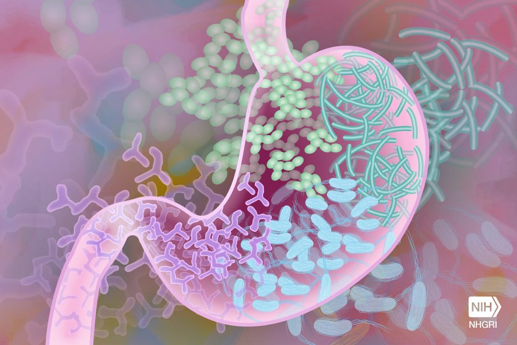
Gut Microbiome. Credit Darryl Leja National Human Genome Research Institute National Institutes Of Health

The increasing rate of obesity and high-fat diets are suspected to be behind the growing rates of colorectal cancers in people aged under 50. Now, in a study published in Cell Reports, researchers at have discovered how high-fat diets can change gut bacteria and alter digestive molecules called bile acids that are modified by those bacteria, predisposing mice to colorectal cancer.

In the study, researchers from the Salk Institute and UC San Diego found increased levels of specific gut bacteria in mice fed high-fat diets. They showed that those gut bacteria alter the composition of the bile acid pool in ways that cause inflammation and affect the replenishment rate of intestinal stem cells replenish.

“The balance of microbes in the gut is shaped by diet, and we are discovering how alterations in the gut microbial population (the gut microbiome) can create problems that lead to cancer,” says co-senior author and Professor Ronald Evans, director of Salk’s Gene Expression Laboratory. “This paves the way toward interventions that decrease cancer risk.”

In 2019, Evans and his colleagues showed in mice how high-fat diets boosted the overall bile acid levels. The shift in bile acids, they found, shut down a key protein in the gut, the Farnesoid X receptor (FXR). and increased the prevalence of cancer.

However, there were still missing links in the story, including how the gut microbiome and bile acids are changed by high-fat diets.

In the new work, Evans’ group teamed up with the labs of Rob Knight and Pieter Dorrestein at UC San Diego to examine the microbiomes and metabolomes (collections of dietary and microbially derived small molecules) in the digestive tracks of animals on high-fat diets. They studied mice genetically more susceptible to colorectal tumours.

The scientists discovered that although mice fed high-fat diets had more bile acids in their guts, it was a less diverse collection with a higher prevalence of certain bile acids that had been changed by gut bacteria. They also showed that these modified bile acids affected the proliferation of stem cells in the intestines. Without frequent replenishment, they accumulate mutations – a key step toward encouraging the growth of cancers, which often arise from these stem cells.

“We are only just beginning to understand these bacterially-conjugated bile acids and their roles in health and disease,” says co-author Michael Downes, a staff scientist at Salk.

There were also striking differences in the microbiomes of the mice on high-fat diets: the collections of gut bacteria in these mice’s digestive tracts were less diverse and contained different bacteria than the microbiomes of mice not on high-fat diets. Two of these bacteria – Ileibacterium valens and Ruminococcus gnavus – were able to produce these modified bile acids.

The scientists were surprised to discover that a high-fat diet actually had a greater impact on the microbiome and modified bile acids than a genetic mutation that increases cancer susceptibility in the animals.

“We’ve pinpointed how high-fat diet influences the gut microbiome and reshapes the bile acids pool, pushing the gut into an inflamed, disease-associated state,” says co-first author Ting Fu, a former postdoctoral fellow in the Evans lab.

The researchers believe high-fat diets change the composition of the microbiome, encouraging the growth of bacteria like I. valens and R. gnavus. In turn, that boosts levels of modified bile acids. In a vicious cycle, those bile acids create a more inflammatory environment that can further change the makeup of gut bacteria.

“We’ve deconstructed why high-fat diets aren’t good for you, and identified specific strains of microbes that flare with high-fat diets,” says Evans, March of Dimes Chair in Molecular and Developmental Biology. “By knowing what the problem is, we have a much better idea of how to prevent and reverse it.”

In the future, the team will study how quickly the microbiome and bile acids change after an animal begins eating a high-fat diet. They also plan to study ways to reverse the cancer-associated effects of a high-fat diet by targeting FXR – the protein that they previously discovered to be associated with bile acid changes.

Source: Salk Institute

Suicide in Cancer not Affected by the Option of Assisted Suicide

Source: Pixabay CC0

In countries which allow the practice, assisted suicide would seem to be an alternative to conventional suicide – but new research shows that this is not a simple relationship. An analysis published in Cancer Medicine reveals the trends of self-initiated deaths – including assisted suicide (AS) and conventional suicide (CS) – in Switzerland over a 20-year period, focusing on people who suffered from cancer. While cancer-related AS rose, CS fell but then stabilised – suggesting that cancer-related CS has more complex reasons behind it.

Although supporters of assisted dying state that access to AS should lead to a reduction in violent CS, the study’s findings do not confirm this assumption. The situations and motivations for cancer-associated CS seem to be clearly different from those for cancer-related AS.

In Switzerland, assisting in a suicide is not punishable as long as it does not serve selfish motives. In this analysis of data from 1999–2018, investigators found that cancer was the most often listed principal disease for AS: 3580 people with cancer died by AS, representing 41.0% of AS cases. Cancer was listed in only a small minority of CS cases (832 people, representing 3.8% of CS cases).

There was approximately a doubling of AS cases among patients with cancer every 5 years. Also, the percentage of cancer-associated AS in relationship with all cancer-associated deaths increased over time to 2.3% in 2014–2018. The numbers of cancer-associated CS showed a downward trend in 1999–2003 and were stable through 2009–2018.

“Obviously, the situations and motivations for cancer-associated CS seem to be clearly different from those for cancer-related AS,” said corresponding author Uwe Güth, MD, of the University of Basel.

Source: Wiley

Neuroblastomas: ‘New’ Immune System Responds Better to Therapy

Credit: National Cancer Institute

Cancer researchers have shown that immunotherapy after stem cell transplantation effectively combats neuroblastomas in children. Crucially, stem cells from a parent provide children with a new immune system that responds much better to immunotherapies. These results of an early clinical trial were published in the Journal of Clinical Oncology.

Tumours of the nervous system, neuroblastomas are associated with an unfavourable prognosis if the tumour is classified as a high-risk type. and particularly poor for patients in the relapsed stage. In this study by scientists at St. Anna Children’s Cancer Research Institute and the Eberhard Karls University of Tübingen, immunotherapy following stem cell transplantation is now associated with long-term survival in a substantial proportion of the patients. Compared to an earlier study the survival rate was increased.

“After the transplantation of stem cells from a parent, the patients are equipped with a new immune system. This enables a better immune response to the subsequent immunotherapy and clearly improves the outcome,” explains Prof Ruth Ladenstein, MD, co-first author.

Five-year survival exceeds 50%

“After a median follow-up of about eight years, we see that more than half of the study patients live five years or longer with their disease,” Prof Ladenstein reports (5-year overall survival: 53%). In comparison, the 5-year overall survival in an earlier study, in which stem cell transplantation was not followed by immunotherapy, was only 23%. Those patients who showed a complete or partial response to prior treatment had significantly better survival.

“In summary, immunotherapy with dinutuximab beta following transplantation of stem cells from matched family donors resulted in remarkable outcomes when patients had at least a partial response to prior treatment,” says Prof Ladenstein. “In our study, there were no unexpected side effects and the frequency of graft-versus-host-disease was low.”

Restoring natural killer cell potency

Dinutuximab beta is a monoclonal antibody that binds to a molecule, GD2, on the surface of tumour cells, marking them for destruction by natural killer cells. But prior chemotherapies may impair natural killer cells. “Therefore, a transplantation of intact natural killer cells from matched family donors seems reasonable before immunotherapy is administered. The transplanted, new natural killer cells are now able to target the tumour cells more efficiently – by means of an antibody-dependent reaction,” explains Prof Ladenstein.

According to the authors, further studies are needed to determine the individual components of the therapeutic approaches. Recently, conventional chemotherapy has also been combined with immunotherapy early in the treatment strategy, resulting in similarly improved response rates. The hope is that a renewed immune system through a healthy parent in combination with the described transplantation procedure could further increase survival rates: “Our approach could thus result in stronger and longer lasting tumour control. A randomised study would be necessary to scientifically substantiate the additional potential benefit of a new immune system in the context of relapse therapy,” Prof Ladenstein adds.

Source: St. Anna Children’s Cancer Research Institute

Research Identifies Metformin for Potential Treatment of Metastatic Prostate Cancer

Credit: Darryl Leja National Human Genome Research Institute National Institutes Of Health

While treatment for localised prostate cancer has a relatively high success rate, mortality is high in advanced metastatic cancer. The precise mechanism behind the spread of the tumour has not been fully explained. Researchers have now decoded the underlying cellular signal pathway and have demonstrated that the common diabetes drug metformin could provide a new treatment option. The study has just been published in the journal Molecular Cancer.

Using a complex mouse model, the research team under Lukas Kenner at the Medical University of Vienna examined prostate cancer cells and identified the key factors in the regulation of tumour cell growth and the way they interact with each other. The protein signal transducer and activator of transcription 3 (STAT3) plays the leading role – its activation by another protein called interleukin 6 (IL6) has been a focus for cancer researchers in connection with tumour progression for some time now.

“Interestingly, our study showed for the first time that permanent activation of STAT3 prevents the development of prostate cancer as well as the development and spread of metastases. Conversely, we discovered that the loss of the signal pathway between STAT3 and IL6 in the prostate can lead to massive tumour growth and metastasis, which significantly increases the aggressiveness of the cancer and the mortality rate,” explained Kenner.

Metformin as a potential medication

In the course of the study, the researchers also found that activation of STAT3 in the prostate leads to increased levels of cell components (LKB1/pAMPK) that are responsible for the regulation of glucose metabolism and are linked to type 2 diabetes mellitus. The proteins LKB1/pAMPK block certain cancer molecules (mTOR and CREB) and as a result also stop the tumour growing. “In light of this finding, we used a common diabetes drug in our research,” said Kenner. Kenner and his team discovered that the active ingredient metformin, which is used in the treatment of type 2 diabetes to regulate glucose levels, can significantly slow the progression of STAT3-positive prostate cancer, a condition with a metabolism that is very similar to type 2 diabetes. “As metformin is already available, our research findings could be useful in developing new treatment options for patients with STAT3-positive prostate cancer in the foreseeable future,” Kenner pointed out, looking ahead to further research into the newly discovered approach.

Source: Medical University of Vienna

In A First, Immunotherapy for Glioblastoma Successfully Tested in Mice

Photo by Kanashi ZD on Unsplash

Immunotherapy has dramatically improved survival against many cancers but efforts to use it against glioblastomas have to date proven fruitless. Now, Salk scientists have found the immunotherapy treatment anti-CTLA-4 leads to considerably greater survival of mice with glioblastoma. Furthermore, they discovered that this therapy was dependent on immune cells called CD4+ T cells infiltrating the brain and triggering the tumour-destructive activities of other immune cells called microglia, which permanently reside in the brain.

The findings, published in the journal Immunity, show the benefit of harnessing the body’s own immune cells to fight brain cancer and could lead to more effective immunotherapies for treating brain cancer in humans.

Glioblastoma, the most common and deadly form of brain cancer, grows rapidly to invade and destroy healthy brain tissue. The tumour sends out cancerous tendrils into the brain that make surgical tumour removal extremely difficult or impossible.

“There are currently no effective treatments for glioblastoma – a diagnosis today is basically a death sentence,” says Professor Susan Kaech, senior author and director of the NOMIS Center for Immunobiology and Microbial Pathogenesis. “We’re extremely excited to find an immunotherapy regimen that uses the mouse’s own immune cells to fight the brain cancer and leads to considerable shrinkage, and in some cases elimination, of the tumour.”

For some tumours, immunotherapy can be used, in which the body’s own immune cells to seek and destroy cancer cells, leading to strong, lasting anti-cancer responses for many patients. Kaech sought new ways of harnessing the immune system to develop more safe and durable treatments for brain cancer.

Her team found three cancer-fighting tools that have been somewhat overlooked in brain cancer research that may cooperate and effectively attack glioblastoma: an immunotherapy drug called anti-CTLA-4 and specialized immune cells called CD4+ T cells and microglia.

Anti-CTLA-4 immunotherapy works by blocking cells from making the CTLA-4 protein, which, if not blocked, inhibits T cell activity. It was the first immunotherapy drug designed to stimulate our immune system to fight cancer, but it was quickly followed by another, anti-PD-1, that was less toxic and became more widely used. Whether anti-CTLA-4 is an effective treatment for glioblastoma remains unknown since anti-PD-1 took precedence in clinical trials. Unfortunately, anti-PD-1 was found to be ineffective in multiple clinical trials for glioblastoma – a failure that inspired Kaech to see whether anti-CTLA-4 would be any different.

As for the specialized immune cells, CD4+ T cells are often overlooked in cancer research in favour of a similar immune cell, the CD8+ T cell, because CD8+ T cells are known to directly kill cancer cells. Microglia live in the brain full time, where they patrol for invaders and respond to damage – whether they play any role in tumour death was not clear. When treated with anti-CLA-4, mice with glioblastoma had longer lifespans than those receiving anti-PD-1.

Upon investigation, they found that after anti-CTLA-4 treatment, CD4+ T cells secreted a protein called interferon gamma that caused the tumour to throw up “stress flags” while simultaneously alerting microglia to start eating up those stressed tumour cells. As they gobbled up the tumour cells, the microglia would present scraps of tumour on their surface to keep the CD4+ T cells attentive and producing more interferon gamma, creating a cycle that lasts until the tumour is destroyed.

“Our study demonstrates the promise of anti-CTLA-4 and outlines a novel process where CD4+ T cells and other brain-resident immune cells team up to kill cancerous cells,” says co-first author Dan Chen, a postdoctoral researcher in Kaech’s lab.

To understand the role of microglia in this cycle, the researchers collaborated with co-author and Salk Professor Greg Lemke. For decades, Lemke has investigated critical molecules, called TAM receptors, used by microglia to send and receive crucial messages. The researchers found that TAM receptors told microglia to gobble up cancer cells in this novel cycle.

“We were stunned by this novel codependency between microglia and CD4+ T cells,” says co-first author Siva Karthik Varanasi, a postdoctoral researcher in Kaech’s lab. “We are already excited about so many new biological questions and therapeutic solutions that could radically change treatment for deadly cancers like glioblastoma.”

Connecting the pieces of this cancer-killing puzzle brings researchers closer than ever to understanding and treating glioblastoma.

“We can now reimagine glioblastoma treatment by trying to turn the local microglia that surround brain tumours into tumour killers,” says Kaech. “Developing a partnership between CD4+ T cells and microglia is creating a new type of productive immune response that we have not previously known about.”

Next, the researchers will examine whether this cancer-killing cell cycle is present in human glioblastoma cases. Additionally, they aim to look at other animal models with differing glioblastoma subtypes, expanding their understanding of the disease and optimal treatments.

Source: Salk Institute

Study Shows a Link Between Sugar-sweetened Beverages and Liver Cancer

Photo by Breakingpic on Pexels

One of the first studies to look at the association between intake of sugar-sweetened beverages, artificially sweetened beverages, and incidence of liver cancer and chronic liver disease mortality, has found an 85% increase in liver cancer incidence between postmenopausal women who consume one sweetened drink per day and those who consume them rarely. Results from the study, which was led by Brigham and Women’s Hospital, are published in JAMA.

“To our knowledge, this is the first study to report an association between sugar sweetened beverage intake and chronic liver disease mortality,” said first author Longgang Zhao, PhD, of the Brigham’s Channing Division of Network Medicine. Zhao is a postdoctoral researcher who works with senior author Xuehong Zhang, MBBS, ScD, in the Channing Division. “Our findings, if confirmed, may pave the way to a public health strategy to reduce risk of liver disease based on data from a large and geographically diverse cohort.”

This observational study included nearly 100 000 postmenopausal women from the large, prospective Women’s Health Initiative study. Participants reported their usual soft drink, fruit drink (not including fruit juice) consumption, and then reported artificially sweetened beverage consumption after three years. Participants were followed for a median of more than 20 years. Researchers looked at self-reported liver cancer incidence and death due to chronic liver disease such as fibrosis, cirrhosis, or chronic hepatitis, which were further verified by medical records or the National Death Index.

A total of 98 786 postmenopausal women were included in the final analyses. The 6.8% of women who consumed one or more sugar-sweetened beverages daily had an 85% higher risk of liver cancer and 68% higher risk of chronic liver disease mortality compared to those who had fewer than three sugar sweetened beverages per month. No such increase was observed for consumption of artificially-sweetened beverages.

The authors note that the study was observational, and causality cannot be inferred, and relied on self-reported responses about intake, sugar content and outcomes. More studies are needed to validate this risk association and determine why the sugary drinks appeared to increase risk of liver cancer and disease. Furthermore, more research is needed to elucidate the potential mechanisms by integrating genetics, preclinical and experimental studies, and -omics data.

Source: Mass General Brigham

Robust Analysis Challenges the Link Between Cancer and Anxiety and Depression

Photo by Karolina Grabowska on Pexels

Depression and anxiety are thought to increase a person’s risk of developing cancer, but research results have been inconclusive. In an analysis of multiple studies from the Netherlands, the UK, Norway, and Canada, investigators found that depression and anxiety are not linked to higher risks for most types of cancer among this population. The analysis is published in the journal CANCER.

Experts have suspected that depression and anxiety may increase cancer risk by affecting a person’s health-related behaviours or by having biological effects on the body that support cancer development. Some research has supported an association between depression, anxiety, and cancer incidence, while other investigations have found no or negligible associations.

To provide additional insights, Lonneke A. van Tuijl, PhD, of the University Medical Center Groningen, and her colleagues examined data from the international Psychosocial Factors and Cancer Incidence consortium, which includes information from 18 prospective study groups with more than 300 000 adults from the Netherlands, the United Kingdom, Norway, and Canada.

The team found no associations between depression or anxiety and overall, breast, prostate, colorectal, and alcohol-related cancers during a follow-up of up to 26 years. The presence of depression or anxiety was linked with a 6% higher risk of developing lung cancer and smoking-related cancers, but this risk was substantially reduced after adjusting for other cancer-related risk factors including smoking, alcohol use, and body mass index. Therefore, this analysis supports the importance of addressing tobacco smoking and other unhealthy behaviours including those that may develop as a result of anxiety or depression.

“Our results may come as a relief to many patients with cancer who believe their diagnosis is attributed to previous anxiety or depression,” said Dr van Tuijl. “However, further research is needed to understand exactly how depression, anxiety, health behaviours, and lung cancer are related.”

Source: Wiley

Study Finds No Evidence of Fungus Link to Pancreatic Cancer

Source: CC0

Four years ago, a report that a common species of fungus might fuel pancreatic cancer offered a promising new view of the deadly disease. But in working to validate the finding, Duke Health researchers have found no such association. In a study published in the journal Nature, the researchers conducted a multi-pronged analysis of data from the earlier study and found no link between the pancreatic microbiome and the development of pancreatic cancer.

“We were intrigued by the original finding, as were many research teams,” said senior author Peter Allen, MD, professor in the Department of Surgery and chief of the Division of Surgical Oncology at Duke University School of Medicine.

“There is a growing body of literature connecting the human microbiome to disease, and this was particularly compelling for pancreatic cancer,” Allen said. “But our findings did not support an association between fungi and the development of pancreatic cancer in humans.”

Allen and colleagues worked to recreate the 2019 findings published in Nature by a different research team. The original study raised hopes that there might be a possible method of preventing pancreatic cancer with the use of antifungals or some other approach to protect from infection.

Focusing on the research team’s original raw sequencing data, the Duke researchers were unable to reproduce the findings. Additional studies, using pancreatic cancer tissue in Duke repositories, also failed to produce the original results.

“We believe our findings highlight the challenges of using low biomass samples for microbiome sequencing studies,” Allen said. “The inclusion of appropriate negative controls and efforts to identify and remove sequencing contaminants is critical to the interpretation of microbiome data.”

Source: Duke University Medical Center