Category: Cancer

The Urgent Need for Early Detection Emphasised this Bone Marrow & Leukaemia Awareness Month

Credit: National Cancer Institute

Leukaemia has been identified as the most prevalent cancer among the country’s youth, according to the latest report from the National Cancer Registry (NCR) of South Africa (2021). However, approximately half of children with cancer remain undiagnosed, with the majority of cases only being detected during the advanced stages of the illness. This is partly attributed to a lack of awareness of the early warning signs of childhood cancer.

As the world observes Bone Marrow & Leukaemia Awareness Month until the 15th of October, Dr Candice Hendricks, Paediatric Haematologist and Medical Spokesperson for DKMS Africa shares that leukaemia can be categorised as acute leukaemias or chronic leukaemias, each with varying symptoms. “Acute leukaemias are far more common in children and can further be divided into acute lymphoblastic- (ALL) and acute myeloid leukaemia (AML). Among children, especially those aged between two and 10, Acute Lymphoblastic Leukaemia (ALL) is the most common blood cancer in this age group.

“This disease arises from genetic mutations in immature lymphocytes called lymphoblasts which are located in the bone marrow. The mutations lead to uncontrolled growth of these lymphoblasts,” she explains. “Lymphoblasts are abnormal blood stem cells that lose the ability to make mature blood cells. The uncontrolled growth of these cells in the bone marrow displaces normal blood cell development and leads to a decrease in properly functioning red blood cells, white blood cells, and platelets. Patients may potentially present with an elevated white blood cell count on blood results, however, their impaired function leaves the body vulnerable to infections.”

Aligned with the NCR, Dr Hendricks emphasises that early symptoms often go unnoticed, as they mimic common, mild conditions, causing many patients and those who care for them to overlook them. “However, the severity of these symptoms escalates rapidly with acute leukaemias and persist even after standard treatment for infections. A high index of suspicion is required in diagnosing patients, and if any symptom persists, an immediate full blood count test is necessary, followed by additional tests if irregularities are detected.”

Prominent symptoms indicating the disease include:

  • Blood clotting disorders or blood diathesis characterised by easy bruising from minor impacts and the appearance of small reddish spots on the skin. Other signs encompass blood in urine, as well as uncontrollable gum and nose bleeding.
  • Muscle and joint pain, particularly in the limbs, along with frequent limb numbness.
  • Fever and night sweats.
  • Anaemia caused by a deficiency of red blood cells, leading to constant fatigue, reduced exercise capacity, lethargy, sleepiness, and pale skin.
  • Recurrent infections that persist despite antibiotic treatment due to cancer cells impairing the immune system. Pathological cancer cells displace healthy leukocytes, rendering the body susceptible to various viral, bacterial, and fungal infections.
  • Loss of appetite and weight loss.
  • Enlarged lymph nodes.
  • Stomach pain resulting from spleen and/or liver enlargement.

In support of Bone Marrow & Leukaemia Awareness Month, DKMS Africa continues to raise awareness and funds to cover the registration costs for as many potential stem cell donors as possible. Stem cell donations offer the best chance of survival for children afflicted by high-risk leukaemia which does not respond to or recurs after standard treatment. Answer the call! If you’re aged between 17 and 55 and in good general health, please register at https://www.dkms-africa.org/register-now. Registration is entirely free and takes less than five minutes.

For further information, get in touch with DKMS Africa at 0800 12 10 82.

About DKMS
DKMS is an international non-profit organisation dedicated to saving the lives of patients with blood cancer and blood disorders. Founded in Germany in 1991 by Dr. Peter Harf, DKMS and organisations of over 1,000 employees have since relentlessly pursued the aim of giving as many patients as possible a second chance at life. With over 11 million registered donors, DKMS has succeeded in doing this 100,000 times to date by providing blood stem cell donations to those in need. This accomplishment has led to DKMS becoming the global leader in the facilitation of unrelated blood stem cell transplants. The organisation has offices in Germany, the US, Poland, the UK, Chile and South Africa. In India, DKMS has founded the joint venture DKMS-BMST together with the Bangalore Medical Services Trust. International expansion and collaboration are key to helping patients worldwide because, like the organisation itself, blood cancer knows no borders.

DKMS is also heavily involved in the fields of medicine and science, with its own research unit focused on continually improving the survival and recovery rate of patients. In its high-performance laboratory, the DKMS Life Science Lab, the organisation sets worldwide standards in the typing of potential blood stem cell donors.


New Treatment Combination could Prevent Cystectomy in Invasive Bladder Cancer

Photo by cottonbro studio

Mount Sinai investigators have developed a new approach for treating invasive bladder cancer without the need for surgical removal of the bladder, they report in their study published in Nature Medicine. At present, cystectomy (removal of the bladder) is currently a standard approach when cancer has invaded the muscle layer of the bladder.

In a phase 2 clinical trial that was the first of its kind, doctors found that some patients could be treated with a combination of chemotherapy and immunotherapy without the need to remove their bladder. Radical cystectomy can be curative in muscle-invasive bladder cancer, but the procedure is a life-changing operation due to the need for urinary diversion and is associated with a 90 day mortality risk of up to 6–8%.

“Treatment for muscle-invasive bladder cancer is in need of major improvements from both a quality-of-life and an effectiveness standpoint,” said Matthew Galsky, MD, Co-Director of the Center of Excellence for Bladder Cancer at The Tisch Cancer Institute, a part of the Tisch Cancer Center at Mount Sinai. “If additional research confirms our findings, this may lead to a new paradigm in the treatment of muscle-invasive bladder cancer.”

The 76 patients received four cycles of gemcitabine, cisplatin, plus nivolumab followed by clinical restaging. Approximately 43% (33 patients) achieved a complete response (no detectable cancer) when treated with this combination of chemotherapy and immunotherapy. Patients with a clinical complete response were offered the opportunity to proceed with additional immunotherapy, without surgical removal of the bladder. Among patients opting to proceed without surgical removal of the bladder, about 70% had no evidence of recurrent cancer after two years.

The most common adverse events were fatigue, anaemia, neutropenia and nausea. Somatic alterations in pre-specified genes or increased tumour mutational burden did not improve the positive predictive value of complete response.

Based on the results of this trial, two follow-up studies were launched to build on this approach; one is ongoing, and another will open in the next six months.

Source: The Mount Sinai Hospital / Mount Sinai School of Medicine

High Rates of Hyperglycaemia with Alpelisib Treatment for Breast Cancer

Photo by National Cancer Institute on Unsplash

New research has uncovered high rates of hyperglycaemia among breast cancer patients being treated with the oral medication alpelisib. The researchers say that patients receiving this medication should have their blood sugar levels monitored and managed well before treatment with alpelisib. The results are published by Wiley online in CANCER, a peer-reviewed journal of the American Cancer Society.

Alpelisib targets the phosphoinositide 3-kinase (PI3K) protein that is involved in cell growth and when mutated can fuel cancer. In 2019, the US Food and Drug Administration approved the use of this drug in combination with fulvestrant, an oestrogen receptor blocker, for certain cases of metastatic breast cancer that have mutations in the gene that codes for a PI3K subunit.

Unfortunately, targeting PI3K can lead to hyperglycaemia as a side effect which, if severe, can result in dehydration or kidney damage and can require hospitalisation. Sherry Shen, MD, of Memorial Sloan Kettering Cancer Center, and her colleagues set out to describe the incidence, risk factors, and treatment patterns of alpelisib-associated hyperglycaemia in patients with metastatic breast cancer treated in a clinical trial or as standard care at their institution.

Among 147 patients treated with alpelisib as standard care, the rate of hyperglycaemia was 80.3%, and the rate of serious hyperglycaemia was 40.2%. Among 100 patients who were treated during a clinical trial, rates were lower (34.0% any grade and 13.0% serious hyperglycaemia). The median time to onset of hyperglycaemia after initiating alpelisib was 16 days. An initially elevated haemoglobin A1c, an indicator of high blood sugar such as in prediabetes or diabetes, was a risk factor for later developing hyperglycaemia.

Among patients who developed hyperglycemia, 66.4% received treatment, most commonly with the diabetes drug metformin.

“If a patient is identified to have a PI3KCA mutation and thus eligible for treatment with alpelisib, we should be checking haemoglobin A1c level and partnering with the patient’s primary care physician and/or endocrinologist to optimise their blood sugar levels,” said Dr Shen. “This needs to be done months before initiating alpelisib, because once alpelisib is started, hyperglycaemia usually develops within the first two weeks of treatment. Being pre-emptive about improving glycaemic status and treating prediabetes/diabetes will hopefully lower the patient’s risk of developing hyperglycaemia and thus, lower their risk of needing to discontinue a drug that could be effective for their cancer.”

Senior author Neil M. Iyengar, MD noted that optimising a patient’s blood sugar levels often involves changes to dietary and exercise patterns, and potentially introducing certain medications. “Improving metabolic risk factors through lifestyle interventions may also improve dose delivery of alpelisib, and ongoing clinical trials by our group and other groups are testing whether metabolic interventions such as the ketogenic diet or newer medications used to treat diabetes could also improve the treatment efficacy of cancer therapies that target the PI3K pathway,” he said.

Source: Wiley

First-in-human Treatment with Tumour Vaccine Shows Positive Results against Gliomas

Photo by Anna Shvets on Pexels

Tumour vaccines alert a cancer patient’s immune system to proteins that are carrying cancer-typical alterations, and may prove useful in gliomas, which are resistant to most forms of treatment including immune checkpoint inhibitors. Reporting in Nature Medicine, physicians and cancer researchers have now performed a first-in human treatment of eight adult patients with advanced midline gliomas with a peptide vaccine. The vaccine, which mimicked a histone protein mutation typical of this type of cancer, proved to be safe and induced the desired immune responses against the brain tumour, with one patient experiencing remission for more than 31 months.

Cancer vaccinations depend on distinct protein structures on cancer cells by which immune cells can differentiate them from healthy one. Mutations in the tumour genome often lead to protein structures that are altered in a way typical of cancer.

Diffuse midline gliomas are among the most aggressive brain tumours. They usually occur in children and young adults near the brain stem and are therefore difficult to access surgically. Chemotherapy or radiotherapy also have limited effectiveness. In this type of cancer, mutations characteristically occur in the gene encoding histone H3 (H3K27M), a packaging protein of DNA. The mutation gives rise to a novel protein structure (a neoepitope) that can be recognised as foreign by the patient’s immune system.

“Such mutations, which occur in identical form in many patients, are rare in cancer. They literally lend themselves to the development of tumor vaccines because they occur in all cancer cells, since the mutated histone is causative for the development of midline gliomas. This means that vaccination against the mutated protein gets to the root of the problem,” explains Michael Platten, Director of the Department of Neurology at the University Medical Center Mannheim and Head of Department at the German Cancer Research Center (DKFZ).

The researchers led by Katharina Sahm and Michael Platten synthetically reconstructed the section of the histone H3 protein with the characteristic mutation. Using this peptide, they were able to curb the growth of H3K27M-mutated tumours in a mouse model. Encouraged by the results, the team decided to test the mutation-specific vaccine produced at the University of Tübingen in patients in a phase I-trial*, which is still ongoing.

In parallel, the physicians, together with colleagues from Munich, Berlin, Bonn and Münster, treated eight adult patients with the peptide vaccine in time-limited individual curative trials. These patients, who could not be enrolled in the trial protocol, suffered from diffuse midline gliomas with H3K27M mutation that progressed after standard therapy. Some of the affected individuals received therapy with immune checkpoint inhibitors in addition to tumor vaccination.

No serious side effects were observed in any of the vaccinated patients. Five of the eight treated patients developed specific immune responses against the mutant protein, which were dominated by CD4 T-helper cells. In one of the patients who had shown a strong immune response, the tumour regressed completely and she remained tumour-free for 31 months.

The vaccine peptide, which is comparatively long at 27 amino acids, worked in patients with different HLA variants. HLA proteins are responsible for the presentation of the mutant peptide on the cell surface and differ from person to person depending on their genetic background. Supported by the HI-TRON Mainz — Helmholtz Institute of the DKFZ, the researchers also observed that immune responses decreased over time, so repeated administration of the vaccine could support a sustained effect.

“We cannot make any further statements about the efficacy of the vaccination based on these treatments. In any case, the current study has given us valuable information that will help us to further optimise the development of brain tumour vaccines in the future,” explains the study’s senior author Katharina Sahm, senior physician at the Neurological University Hospital Mannheim and DKFZ researcher. A phase I-trial is currently underway to test the vaccine against the H3K27M mutation in patients with newly diagnosed midline gliomas. Evaluation is expected to begin around 2025.

Source: German Cancer Research Center (Deutsches Krebsforschungszentrum, DKFZ)

New Online Tool for Hard-to-identify Breast Cancer

A new diagnostic scoring system, developed by renowned breast cancer experts, is now available as an easy-to-use online tool through Susan G. Komen®, the world’s leading breast cancer organisation. This tool will help health care providers recognise and effectively diagnose a rare and aggressive breast cancer, inflammatory breast cancer.

The new Inflammatory Breast Cancer (IBC) Scoring System online tool is available at https://www.komen.org/ibc and may help to increase diagnostic accuracy, predict outcomes, guide treatment decisions and inclusion in clinical trials.  

Before the development of the proposed IBC Scoring System, IBC lacked a formal, objective medical definition and diagnosis was often delayed, misdiagnosed or missed altogether. The new online tool is intended to provide the proposed IBC diagnostic criteria in a convenient tool to help more quickly and effectively recognize IBC in the clinic. 

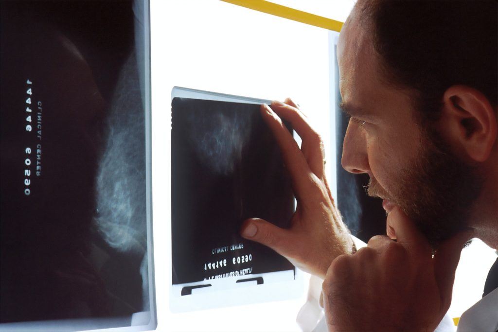
IBC often develops rapidly and can easily be confused with a breast infection because of symptoms such as redness and swelling, and the frequent lack of a breast lump. IBC can be hard to see on a mammogram as it may only show up as skin thickening. This results in approximately 30% of IBC patients being first diagnosed at stage IV (de novo metastatic breast cancer), meaning their breast cancer has already spread to other parts of the body. 

“IBC has historically been difficult to diagnose and no changes to diagnostic approach have been made since the 1960s,” said Dr Reshma Jagsi, Komen Scholar and Lawrence W. Davis Professor and Chair of the Department of Radiation Oncology at Emory University School of Medicine. “This first-of-its-kind tool may help health care providers recognise and diagnose IBC and may also enable researchers to study the biology of IBC, making discoveries to advance progress toward personalized care for all IBC patients in the future.”

The proposed IBC Scoring System was developed through a collaborative effort between Susan G. Komen, the Inflammatory Breast Cancer Research Foundation (IBCRF) and the Milburn Foundation, which brought together a team of leading breast cancer experts including clinicians, researchers, and IBC patients.  It is now being validated by a team of researchers at two of the largest IBC centres in the world led by Dr Filipa Lynce at Dana-Farber Cancer Institute, and Dr Wendy A. Woodward at MD Anderson Cancer Center. This work validating the scoring system is supported by a grant awarded by Susan G. Komen and is part of the groups’ collaborative efforts to advance IBC research and care through innovative approaches.

“I encourage my fellow health providers to use the IBC Scoring System when addressing patients having concerns about changes in their breast, such as swelling and redness. Using this tool may accelerate the diagnosis of IBC and start treatment at an earlier stage for those who have a confirmed diagnosis of invasive breast cancer,” said Dr Lynce.

“The creation of this tool reflects the deep commitment of Komen, the Inflammatory Breast Cancer Research Foundation and the Milburn Foundation to accelerate progress in detecting and treating inflammatory breast cancer. With the help of leading scientists and medical providers across the US, we will help thousands of patients receive an earlier and more accurate diagnosis of this aggressive disease and get the high-quality care they need to survive,” said Senior Vice President of Mission for Susan G. Komen, Victoria Wolodzko Smart. “I have no doubt this tool will improve outcomes for all IBC patients in the future.”

Source: EurekAlert!

Gastric Reflux without Oesophagitis does not Increase Cancer Risk

Source: Pixabay cc0

Reflux disease manifests as acid regurgitation and heartburn and is a known risk factor for oesophageal cancer. However, a new study published in The BMJ by researchers at Karolinska Institutet now reports that the majority of patients do not have a higher risk of cancer. A large-scale study from three Nordic countries shows that the cancer risk is only elevated in patients whom gastroscopy reveals to have changes in the oesophageal mucosa.

“This is a gratifying result since reflux disease is a very common condition and most patients are found to have a completely normal mucus membrane on gastroscopic examination,” says the study’s first author Dag Holmberg, Karolinska Institutet researcher and resident doctor of surgery at Karolinska University Hospital in Sweden.

In reflux disease, acidic stomach contents leak into the oesophagus. This can sometimes cause oesophagitis, inflammation in the oesophageal mucus membrane, which is diagnosed via gastroscopy. It is common knowledge that reflux disease increases the risk of oesophageal cancer, but what the cancer risk is for patients with normal mucosa has remained unknown.

Symptoms generally persist

The symptoms of reflux disease can come and go but generally persist, which means that many patients frequently seek medical attention and often undergo repeated gastroscopies to detect mucosal lesions or prodromal cancer.

“Our study suggests that these repeated gastroscopies are probably unnecessary for people with reflux disease who have a normal oesophageal mucosa,” says Dr Holmberg. “These findings should be reassuring for this large patient group and can guide GPs who often treat them.”

The present study is based on national health data registries in Sweden, Denmark and Finland, and included over 285 000 individuals with reflux disease and no gastroscopic evidence of oesophagitis. The patients were followed for up to 31 years and the researchers registered all cases of oesophageal cancer.

The cancer risk was then compared with that for individuals from the general population matched by age and sex and at the same period in the three countries. No increased risk of oesophageal cancer was observed in patients with reflux disease and a normal mucus membrane.

Increased risk in patients with oesophagitis

By way of comparison, the researchers also analysed the cancer risk in over 200 000 individuals with reflux disease and oesophagitis. These people were at a clearly increased relative risk of developing oesophageal cancer.

“We now intend to examine what factors other than oesophagitis can be linked to tumour growth in people with reflux disease,” says the study’s last author Jesper Lagergren, professor of surgery at Karolinska Institutet.

Source: Karolinska Institutet

An Electrifying Solution to Destroy Glioblastomas

Photo by Zoltan Tasi on Unsplash

In Nature Nanotechnology, researchers report a new way to target and kill cancer cells in glioblastomas, hard-to-treat brain tumours, using electrically charged molecules to trigger self-destruction, that could be developed into a spray treatment used during surgery.

Researchers from the University of Nottingham found a new way to harness the extraordinary capabilities of bio-nanoantennae: gold nanoparticles intricately coated with specialised redox active molecules to induce apoptosis, in cancer cells on electrical stimulation.

The research focuses on patient-derived glioblastoma cells, an elusive and formidable form of brain cancer that has long evaded effective treatment. The five-year survival rate for glioblastoma is only 6.8% and the estimated average length of survival is about only 8 months from diagnosis.

The bio-nanoantennae were able to specifically target glioblastoma cells, leaving healthy cells unscathed. This unprecedented level of precision opens up new possibilities for developing treatment for glioblastoma during surgical resection of the tumour, when the bio-nanoantennae would be sprayed or injected.

The researchers, which included experts from the Schools of Engineering, Physics and Medicine have now established what is thought to be the first ‘quantum therapeutic’, which taps into the potential of quantum signalling to combat cancer.

Dr Frankie Rawson led the research and explains: “The team showed that cancer cells succumb to the intricate dance of electrons, orchestrated by the enchanting world of quantum biology. With the advent of bio-nanoantennae, this vision of real-world quantum therapies edge closer to reality. By precisely modulating quantum biological electron tunnelling, these ingenious nanoparticles create a symphony of electrical signals that trigger the cancer cells’ natural self-destruction mechanism.”

The team has now secured MRC impact acceleratory funding, have filed patent, to begin translating the technology to this eventual clinical application. Further rigorous research and validation are essential to ensure the safety and effectiveness of bio-nanoantennae for human use.

Dr Ruman Rahman from the School of Medicine and co-author of the study, adds: “Treating glioblastoma tumours has long presented challenges for clinicians and prognosis for patients is still poor, which is why any research showing the promise of a new effective treatment is hugely exciting. This research has shown the possibilities presented by quantum therapeutics as a new technology to communicate with biology. The fusion of quantum bioelectronics and medicine brings us one step closer to a new treatment paradigm for disease.”

Source: University of Nottingham

Why Tumours so Often Metastasise to the Spine

The vertebral bones that constitute the spine are derived from a distinct type of stem cell that secretes a protein favouring tumour metastases, according to a study led by researchers at Weill Cornell Medicine. The discovery, published in Nature, opens up a new line of research on spinal disorders and helps explain why solid tumours so often spread to the spine, and could lead to new orthopaedic and cancer treatments.

Vertebral bone was found to be derived from a stem cell that is different from other bone-making stem cells. Using bone-like “organoids” made from vertebral stem cells, they showed that the known tendency of tumours to spread to the spine rather than long bones is due largely to a protein called MFGE8, secreted by these stem cells.

“We suspect that many bone diseases preferentially involving the spine are attributable to the distinct properties of vertebral bone stem cells,” said study senior author Dr Matthew Greenblatt.

In recent years, Dr Greenblatt and other scientists have found that different types of bone are derived from different types of bone stem cells. Since vertebrae develop along a different pathway early in life, and also appear to have had a distinct evolutionary trajectory, Dr Greenblatt and his team hypothesised that a distinct vertebral stem cell probably exists.

The researchers started out by isolating what are broadly known as skeletal stem cells, which give rise to all bone and cartilage, from different bones in lab mice based on known surface protein markers of such cells. They then analysed gene activity in these cells to see if they could find a distinct pattern for the ones associated with vertebral bone.

This effort yielded two key findings. The first was a new and more accurate surface-marker-based definition of skeletal stem cells as a whole. This new definition excluded a set of cells that are not stem cells that had been included in the old stem cell definition, thus clouding some prior research in this area.

The second finding was that skeletal stem cells from different bones do indeed vary systematically in their gene activity. From this analysis, the team identified a distinct set of markers for vertebral stem cells, and confirmed these cells’ functional roles to form spinal bone in further experiments in mice and in lab-dish cell culture systems.

The researchers next investigated the phenomenon of the spine’s relative attraction for tumour metastases, including breast, prostate and lung tumours, compared to other types of bone. The traditional theory, dating to the 1940s, is that this “spinal tropism” relates to patterns of blood flow that preferentially convey metastases to the spine versus long bones. But when the researchers reproduced the spinal tropism phenomenon in animal models, they found evidence that blood flow isn’t the explanation, finding instead a clue pointing to vertebral stem cells as the possible culprits.

“We observed that the site of initial seeding of metastatic tumour cells was predominantly in an area of marrow where vertebral stem cells and their progeny cells would be located,” said study first author Dr Jun Sun, a postdoctoral researcher in the Greenblatt laboratory.

Subsequently, the team found that removing vertebral stem cells eliminated the difference in metastasis rates between spine bones and long bones. Ultimately, they determined that MFGE8, a protein secreted in higher amounts by vertebral compared to long bone stem cells, is a major contributor to spinal tropism. To confirm the relevance of the findings in humans, the team collaborated with investigators at Hospital for Special Surgery to identify the human counterparts of the mouse vertebral stem cells and characterise their properties.

The researchers are now exploring methods for blocking MFGE8 to reduce the risk of spinal metastasis in cancer patients. More generally, said Dr Greenblatt, they are studying how the distinctive properties of vertebral stem cells contribute to spinal disorders.

“There’s a subdiscipline in orthopaedics called spinal orthopaedics, and we think that most of the conditions in that clinical category have to do with this stem cell we’ve just identified,” Dr Greenblatt said.

Source: Weill Cornell Medicine

Studies Point to New, Better Ways to Monitor Head and Neck Cancer Recurrence

Photo by National Cancer Institute on Unsplash

Early findings of a pair of studies from the University of Michigan Rogel Cancer Center shed light on new ways to anticipate recurrence in HPV-positive head and neck cancer sooner. The papers, published in Cancer and Oral Oncology, offer clinical and technological perspectives on how to measure if recurrence is happening earlier than current blood tests allow, and provide a framework for a new, more sensitive blood test that could help in this monitoring.

“When metastatic head and neck cancer returns, it impacts their quality of life and can be disfiguring, interfering with the ability to talk, swallow, and even breathe,” said Paul Swiecicki, MD, associate medical director for the Oncology Clinical Trials Support Unit at Rogel. “As of now, there’s no test to monitor for its recurrence except watching for symptoms or potentially using a blood test which may not detect cancer until shortly before it clinically recurs.”

The paper in Cancer aims to identify different clinical ways that providers can more strategically track for recurrence. To do this, Swiecicki and his team needed to first understand what patient population was at the highest risk to then figure out an appropriate monitoring pattern.

The team examined 450 patients with metastatic head and neck cancer, including people with HPV-positive and HPV-negative cancer. HPV-positive cancer is caused by the human papillomavirus and is increasingly more common in head and neck cancer patients. The team identified some predictors of when recurrences would happen, and to what organs the recurrent cancer would most commonly spread. Patients with HPV-positive cancers were found to develop recurrent disease significantly later than those that were HPV-negative, and also were more likely to spread to the lungs. Taken together, these characteristics may help create a “surveillance” method in the future that combines routine blood testing and imaging to hopefully catch these recurrences and intervene before it’s incurable.

Swiecicki is quick to mention that, at this point, the results of this study are largely theoretical and provide a helpful framework to direct further research. That’s where the newly developed blood test, highlighted in Oral Oncology, comes into play.

Current blood biomarker tests which test for pieces of tumour-shed DNA, may not be sensitive enough to detect a recurrence significantly earlier than clinical surveillance, though several studies with multiple types of tests are ongoing. A research team, led by Muneesh Tewari, MD, PhD, Swiecicki and Chad Brenner, PhD, aimed to create a highly sensitive blood test to detect cancer even when a smaller number of DNA fragments were present, with the intention of providing a better option for detecting cancer earlier in patients.

Not only is this test more sensitive and able to detect a smaller number of DNA fragments in blood, but it’s innovative in other ways too, says first author Chandan Bhambhani, PhD: “We achieved this level of sensitivity by looking for nine different pieces of the HPV genome DNA all at once,” Bhambhani says.

Tewari says this is a step towards a more proactive approach to tackling recurrence in head and neck cancer. “As of now, we only have the tools to react to symptoms when they recur. We want to find a way to be able to detect what’s causing the symptoms much, much sooner, even before the symptoms appear.”

As a clinician, Swiecicki agrees. “It’s exciting to have the ability to potentially detect cancer before it’s incurable and offer us a window for clinical trials to see if we could intervene on cancer to help give people both a better quality of life and perhaps longer quality of life, and even convert their disease from incurable to curable. We don’t know if that’s the case yet, but this is the first tool needed for that to develop.”

Source: Michigan Medicine – University of Michigan

Exercise Stress Tests Pick up More than Just Cardiovascular Problems

Photo by Stephen Andrews

The exercise stress test, which involves treadmill exercise test with electrocardiogram (ECG), is one of the most familiar tests in medicine. While exercise testing typically is focused on diagnosing coronary artery disease, a recent study from Mayo Clinic finds that exercise test abnormalities, such as low functional aerobic capacity, predicted non-cardiovascular causes of death such as cancer in addition to cardiovascular-related deaths. These new findings are published in Mayo Clinic Proceedings.

The exercise stress test is noninvasive, easily available and provides important diagnostic information. In addition to the ECG itself, the test produces data on functional aerobic capacity, heart rate recovery and chronotropic index, the standardised measure of heart rate during exercise that reflects age, resting heart rate and fitness.

“In our exercise testing cohort, non-cardiovascular deaths were more frequently observed than cardiovascular deaths,” says Thomas Allison, PhD, MPH, director of Mayo Clinic’s Integrated Stress Testing Center and the study’s senior author. “Though this was a cardiac stress test, we found that cancer was the leading cause of death, at 38%, whereas only 19% of deaths were cardiovascular. Exercise test results including low exercise capacity, low peak heart rate, and a slow recovery of the heart rate after exercise test were associated with increased mortality.”

The study looked at 13 382 patients who had no baseline cardiovascular issues or other serious diseases and who had completed exercise tests at Mayo Clinic between 1993 and 2010, then were followed closely for a median period of 12.7 years.

The findings suggest that clinicians should focus not only on ECG results but on data in the exercise test results such as low functional aerobic capacity, low chronotropic index and abnormal heart rate recovery. Patients should be encouraged to increase their physical activity if these results are atypical, even if the ECG results show no significant cardiovascular-related risk, Dr Allison says.

Source: Mayo Clinic