Category: Cancer

Doing the Impossible: New Drug Kills 100% of Solid Tumours by Hitting ‘Undruggable’ Target

Assembled human PCNA (PDB ID 1AXC), a sliding DNA clamp protein that is part of the DNA replication complex and serves as a processivity factor for DNA polymerase. The three individual polypeptide chains that make up the trimer are shown. Source: Wikimedia CC0

A ‘cure for cancer’ has long been something of a holy grail for medical research – but experience has shown that cancers are highly individualised and respond differently to therapy, adapting to resist them. Now, in an early study, researchers have tested a cancer drug that kills all solid cancer tumours while leaving other cells unharmed and resulting in no toxicity. The new molecule targets a common key cancer cell protein, the proliferating cell nuclear antigen (PCNA), that is key to helping them grow and metastasise – a target previously believed to be ‘undruggable’.

The new drug, AOH1996, was tested in vitro against 70 different cancer cell lines, including breast, prostate, brain, ovarian, cervical, skin, and lung cancer. It proved effective against all of them, as well as sparing healthy cells. What’s more, developing resistance against the drug is unlikely due to the nature of PCNA as a mistranslation rather than a mutation. The results were published in Cell Chemical Biology. Instructions for synthesis were included in supplementary material.

The last great breakthrough in cancer treatment was immunotherapy, and since then cancer research has looked for the next big leap. A search of journal articles in the Pubmed database showed that “cancer” has grown from 6% of all results in 1950 to 16% by 2016. More recent development in cancer therapies has included gene-based approaches, naked nucleic acids based therapy, targeting micro RNAs, oncolytic virotherapy, suicide gene based therapy, targeting telomerase, cell mediated gene therapy, and CRISPR/Cas9 based therapy.

Shutting down the hub

The research was led by Dr Linda Malkas, a professor at City of Hope Hospital, who said that the molecule selectively disrupts DNA replication and repair in cancer cells, leaving healthy cells unaffected. Animal models also showed a reduction of tumour burden with no apparent adverse effects, with the no observed adverse effect level (NOAEL) calculated being six times higher than the administered dose.

She explained the drug in simple terms to the Daily Mail: “Most targeted therapies focus on a single pathway, which enables wily cancer to mutate and eventually become resistant,” she said. “PCNA is like a major airline terminal hub containing multiple plane gates.

“Data suggests PCNA is uniquely altered in cancer cells, and this fact allowed us to design a drug that targeted only the form of PCNA in cancer cells. Our cancer-killing pill is like a snowstorm that closes a key airline hub, shutting down all flights in and out only in planes carrying cancer cells.”

Dr Malkas said results so far have been ‘promising’ as the molecule can suppress tumour growth on its own or in combination with other cancer treatments without resulting in toxicity.

The development of AOH1996 is the culmination of nearly two decades of work by City of Hope Hospital in Lose Angles.

Decades in the making

PCNA in breast cancer was identified as a potential target in 2006 since it is an isomer, allowing antibodies to target it. The researchers’ first attempts with antibodies to target PCNA were unsuccessful as these were too big to penetrate into solid tumours. Next, they tried a small molecule, which appeared to work in vitro but in vivo proved to have a half-life of only 30 minutes. But they were able to tweak that molecule and arrive at the current drug, AOH1996. It was named after Anna Olivia Healy who died in 2005 from neuroblastoma, and she became the inspiration for the research.

“She died when she was only 9 years old from neuroblastoma, a children’s cancer that affects only 600 kids in America each year,” Malkas said. “I met Anna’s father when she was at her end stages. I sat him down for two hours in my office and showed him all of my data on this protein I had been studying in cancer cells.”

At the time, Dr Malkas was researching breast cancer, studying a protein found in cancer cells but not normal cells. Dr Malkas eventually took Anna’s father, Steve, and his wife, Barbara, to see her lab.

“[Steve] asked if I could do something about neuroblastoma and he wrote my lab a cheque for $25 000,” Dr Malkas said. “That was the moment that changed my life – my fork in the road. I knew I wanted to do something special for that little girl.”

MRI-guided Radiation Therapy Reduces Side Effects from Prostate Cancer Radiotherapy

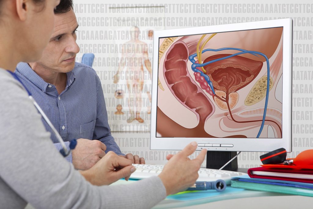
A technique that uses MRI as a guide can make radiotherapy safer for prostate cancer patients by better aiming beams at the prostate while sparing nearby tissue in the bladder, urethra, and rectum. That is the finding of a thorough analysis of all published clinical trials of the technique, called magnetic resonance–guided daily adaptive stereotactic body radiotherapy (MRg-A-SBRT). The analysis is published in CANCER.

By providing detailed images, MRg-A-SBRT can be used to adjust a patient’s radiation plan every day to account for anatomical changes and to monitor the position of the prostate in real time while the radiation beam is on to ensure that treatment is being directed accurately to the prostate. Although MRg-A-SBRT is becoming more popular and multiple clinical trials have tested it, it is unclear whether the technique, which requires more time and resources than standard procedures, has an impact on clinical outcomes and side effects compared with other ways of delivering radiation.

To investigate, Jonathan E. Leeman, MD, of the Dana-Farber Cancer Institute and Brigham and Women’s Hospital, and his colleagues combined data from 29 clinical trials testing MRg-A-SBRT versus conventional CT-guided treatment, with a total of 2457 patients.

MRg-A-SBRT was associated with significantly fewer urinary and bowel side effects in the short term following radiation. Specifically, there was a 44% reduction in urinary side effects and a 60% reduction in bowel side effects.

“The study is the first to directly evaluate the benefits of MR-guided adaptive prostate radiation in comparison to another more standard and conventional form of radiation, and it provides support for use of this treatment in the management of prostate cancer,” said Dr Leeman.

Dr Leeman noted that the study also raises further questions regarding this type of treatment. For example, will the short-term benefits lead to long-term benefits, which are more impactful for patients? Longer follow-up will help answer this question because MRg-A-SBRT is a relatively new treatment. Also, which aspect of the technology is responsible for the improved outcomes seen in clinical trials? “It could potentially be the capability for imaging-based monitoring during the treatment or it could be related to the adaptive planning component. Further studies will be needed to disentangle this,” said Dr Leeman.

An accompanying editorial discusses the analysis’ findings, weighs the potential benefits and shortcomings of adopting this treatment strategy for patients, and questions the value of broad adoption.

Source: Wiley

Scientists Create Protein that Blocks Breast Cancer Metastasis

In a paper published in the journal Biomolecules, UK and Chinese researchers report their creation of a biomedical compound that has the potential to stop breast cancer metastasis.

The scientists from the Chemistry and Biochemistry Departments at the University of Liverpool and Nanjing Medical School in China have discovered a possible way to block proteins produced by cancer cells that promote metastasis – the chief impediment to successful cancer treatment.

Prof Philip Rudland from the University of Liverpool explained: “As a general rule, cancer that has spread is treated with chemotherapy, but this treatment can rarely be given without severely harming or becoming toxic to the patient. The importance of our work was to identify a specific and important target to attack, without toxic side effects.”

The University’s research team have in the past discovered that specific proteins are involved in the metastatic process; these proteins are different from those involved in the production of the primary tumour. One such example is a protein called ‘S100A4’, and is the protein chosen by the research team to target for the identification of chemical inhibitors of metastasis, using model systems of cells from the highly metastatic and incurable hormone receptor-free breast cancer.

Using these model systems, researchers at the University’s Department of Biochemistry discovered a novel compound that can specifically block the interaction of this metastasis-inducing protein S100A4 with its target inside the cell. Researchers in the Department of Chemistry then synthesised a simpler chemical and connected it to a warhead which stimulates cells’ normal protein-degrading machinery. This compound now works at very low doses to inhibit properties associated with metastasis, an improvement of over 20 000-fold on the original unarmed inhibitor, with virtually no toxic side effects. Moreover, in collaboration with Chinese researchers at Nanjing Medical School, they have shown that this compound inhibits metastasis in similar metastatic tumours in mice, suggesting a potential therapeutic role.

Dr Gemma Nixon, Senior Lecturer in Medicinal Chemistry at the University of Liverpool said: “This is an exciting breakthrough in our research. We now hope to take the next steps, and repeat this study in a large group of animals with similar metastatic cancers so that the efficacy and stability of the compounds can be thoroughly investigated and if necessary improved by further design and syntheses, prior to any clinical trials.”

“Significantly, this particular protein we’re investigating occurs in many different cancers, which could mean this approach may be valid for many other commonly occurring human cancers.”

Source: University of Liverpool

Green Tea Extract may Reduce Uterine Fibroids

Photo by Andrea Piacquadio on Pexels

In a pre-clinical, proof-of-concept study from Johns Hopkins Medicine, researchers found that epigallocatechin gallate (EGCG), a green tea compound with powerful antioxidant properties, could be promising for both treating and preventing uterine fibroids. Results of the study, appearing in Scientific Reports, add to growing evidence that EGCG may reduce fibroid cell growth, though the study is still at an early stage. The study was specifically designed to identify the biochemical mechanisms responsible for EGCG action in fibroid cells.

The investigators emphasise that their study involves in vitro human fibroid cells treated with EGCG extract to explore the possibility of oral EGCG supplementation as a therapy, rather than just drinking cups of green tea as a preventative measure for uterine fibroids.

“The purpose of this study was to examine how EGCG works to treat and prevent uterine fibroids,” says James Segars Jr., MD, professor of gynaecology and obstetrics. “There is no standard protocol for uterine fibroid disease management or prevention, no tools to prevent their growth, so finding a safe nonsurgical therapy is important.”

Uterine fibroids are the most common benign tumours of the uterus. Made up of smooth muscle cells and a large matrix of connective tissue, the fibroids range in size from nearly microscopic to bulky masses that can enlarge and distort the uterus.

An estimated 77% of women will develop fibroids in their lifetime, most of them by age 50. Black and Hispanic women develop them at 1.5 to two times the rate of white women.

While many people with uterine fibroids are symptom-free, about 25% experience significant symptoms including heavy uterine bleeding, pelvic pain and infertility. In addition to complete removal of the uterus, surgical treatment may include various means of removing fibroid tumours from the uterine wall.

For the new study, researchers used laboratory cultures of uterine fibroids collected from living patients. Because uterine fibroid cells have a large extracellular matrix compared to normal cells, researchers designed their experiments to see if treatment of cells with EGCG affects protein expression associated with this matrix. Specifically, they studied fibronectin, a matrix protein; cyclin D1, a protein involved with cell division; and connective tissue growth factor (CTGF) protein.

Cells were dosed with 100mmol/L of EGCG in growth media for 24 hours, and then a Western blot was performed. In this study, researchers looked for levels of cyclin D1 and CTGF proteins in EGCG-treated fibroid cells compared to untreated cell.

They found that EGCG reduced protein levels of fibronectin by 46% to 52%, compared with an untreated controls. They also found that EGCG disrupted pathways involved in fibroid tumour cell growth, movement, signalling and metabolism, and they saw up to an 86% decrease in CTGF proteins compared with the control group.

“The results from this study show that EGCG targets many signalling pathways involved in fibroid growth, particularly the extracellular matrix,” says study lead author Md Soriful Islam, PhD, MSc. “EGCG supplements could be an easily accessible and natural way to relieve symptoms and slow fibroid growth.”

These results lend support to the FRIEND study, an ongoing clinical trial of EGCG in women with fibroids who are seeking pregnancy. While results from this study show promise, researchers caution that more studies need to be done, and consumers should not try to self-dose with green tea supplements. Future research on EGCG will include clinical trials with large and diverse patient groups to determine optimal doses as well as possible side effects of EGCG supplementation.

Source: John Hopkins Medicine

Scientists Find a Protein That Keeps Melanoma Hidden from the Immune System

3D structure of a melanoma cell derived by ion abrasion scanning electron microscopy. Credit: Sriram Subramaniam/ National Cancer Institute

New research has helped explain how melanoma evades the immune system and may guide the discovery of future therapies for the disease. The study found that a protein known to be active in immune cells is also active inside melanoma cells, helping promote tumour growth. The findings, published in the journal Science Advances, suggest that targeting this protein with new drugs may deliver a powerful double hit to melanoma tumours.

“The immune system’s control of a tumour is influenced by both internal factors within tumour cells, as well as factors from the tumour’s surroundings,” says first author Hyungsoo Kim, PhD, a research assistant professor at Sanford Burnham Prebys in the lab of senior author Ze’ev Ronai, PhD. “We found that the protein we’re studying is involved in both, which makes it an ideal target for new cancer therapies.”

“Immunotherapy is the first-line therapy for several cancers now, but the success of immunotherapy is limited because many cancers either don’t respond to it or become resistant over time,” says Kim. “An important goal remains to improve the effectiveness of immunotherapy.”

To find ways to boost immunotherapy in melanoma, the research team analysed data from patient tumours to identify genes that may coincide with patients’ responsiveness to immunotherapy. This led to the identification of a protein that helps tumours evade the immune system – called NR2F6 – which was found not only in tumour cells, but also in the surrounding noncancerous cells.

“Often we find that a protein has the opposite effect outside of tumours compared to what it does within a tumour, which is less effective for therapy,” says Kim. “In the case of NR2F6, we found that it elicits the same change in the tumour and in its surrounding tissues, pointing to a synergistic effect. This means that treatments that block this protein’s activity could be twice as effective.”

In a mouse model, the researchers then deleted the NR2F6 protein in both melanoma tumours and in the tumours’ environment. This inhibited melanoma growth more strongly, compared to when this effect occurs in either the tumour or its microenvironment alone. The cancer’s response to immunotherapy was also enhanced upon loss of NR2F6 in both tumours and their microenvironment.

“This tells us that NR2F6 helps melanoma evade the immune system, and without it, the immune system can more readily suppress tumour growth,” adds Kim.

To help advance their discovery further, the team is working with the Institute’s Conrad Prebys Center for Chemical Genomics to identify new drugs that can target NR2F6.

“Discovering drugs that can target this protein are expected to offer a new way to treat melanomas, and possibly other tumours, that would otherwise resist immunotherapy,” says Kim.

Source: Sanford Burnham Prebys

An Existing Cancer Drug Could Have a New Target: Cancer Cells’ ‘Fountain of Youth’

A team of scientists has found that ponatinib, an existing cancer drug could be repurposed to target a subset of cancers that currently lack targeted treatment options and is often associated with poor outcomes. Their findings are published in Nature Communications.

Cancerous cells have a ‘fountain of youth’ by continually lengthening telomeres, but some use a different mechanism called the alternative lengthening of telomeres (ALT). This subset makes up 15% of all cancers and is especially prevalent in aggressive tumours such as osteosarcoma and glioblastoma. The team, led by Nanyang Technological University, Singapore (NTU Singapore), showed that ponatinib, a cancer drug approved by the US Food and Drug Administration, blocks key steps in the ALT mechanism that leads it to fail.

The scientists found that ponatinib helped to shrink bone tumours (a type of ALT cancer) without causing weight loss, a common side effect associated with cancer drugs. In mice with tumours treated with ponatinib, they found a reduction in a biomarker for ALT cancer as compared to untreated mice.

The researchers say that the findings move them a step closer to developing a targeted therapeutic option for ALT cancers, which lack clinically approved targeted treatments to date.

Dr Maya Jeitany and a team of researchers from the NTU School of Biological Sciences, together with collaborators are seeking to address this unmet need.

Dr Jeitany, study lead and senior research fellow at NTU’s School of Biological Sciences, said: “A prominent feature of cancer is its ability to evade cell death and acquire indefinite replication – to stay immortal, in other words – which it can do through the alternative lengthening of telomeres (ALT) mechanism. While a sizeable portion of cancer cells depend on this mechanism, there is no clinically approved targeted therapy available.

“Through our study, we identified a novel signalling pathway in the ALT mechanism and showed that the FDA-approved drug ponatinib inhibits this pathway and holds exceptional promise in stopping the growth of ALT cancer cells. Our findings may provide a new direction for the treatment of ALT cancers by repurposing an FDA-approved drug for these types of tumours.”

Commenting as an independent expert, Assistant Professor Valerie Yang, medical oncologist with the Department of Lymphoma and Sarcoma at the National Cancer Centre Singapore, said: “Sarcomas and glioblastomas are both highly complex cancers that are more prevalent in young people and currently have limited treatment options. The identification of a drug that is FDA-approved which can be repurposed to target ALT, an Achilles heel in these cancers, is very exciting.”

To date, there is no clinically approved targeted treatment for ALT cancers. Furthermore, many ALT cancers, such as osteosarcoma and glioblastoma, show resistance to chemotherapy, highlighting the need for a more targeted form of treatment.

Drug affects telomeres in ALT cancer cells

Through high-throughput drug screening and subsequent testing of shortlisted compounds, the scientists discovered that ponatinib, a drug approved by the FDA for a type of bone marrow cancer, can kill ALT cancer cells effectively.

When osteosarcoma and liposarcoma cells were treated with ponatinib, the scientists found that the drug led to DNA damage, dysfunctional telomeres, and triggered senescence. Importantly, the synthesis of telomeres in the cells also dropped after 18 to 20 hours of treatment with the drug.

Pre-clinical studies conducted on mice that had received transplants of human bone cancer cells further validated the potential of ponatinib. The drug reduced the tumour sizes without affecting the mice’s body weight, a common side effect associated with cancer treatments.

In mice with tumours treated with ponatinib, there was also a reduction in a biomarker for ALT cancer as compared to untreated mice – an indicator that the drug was effective in inhibiting ALT cancer growth.

The scientists ran further tests to identify ponatinib’s mode of action on telomeres in ALT cancer cells and identified a signalling pathway (a series of chemical reactions in which a group of molecules in a cell work together to control a cell function) that could be responsible for the drug’s effect on ALT.

The researchers are now studying further how ponatinib affects telomeres to understand in more detail the signalling pathway they have identified. They are also assessing potential ponatinib-based combinatorial drug treatments for ALT cancers.

Source: Nanyang Technological University

Faecal Microbiota Transplants Could Boost Melanoma Immunotherapy

3D structure of a melanoma cell derived by ion abrasion scanning electron microscopy. Credit: Sriram Subramaniam/ National Cancer Institute

In a world-first clinical trial published in the journal Nature Medicine, a multi-centre study has found faecal microbiota transplants (FMT) from healthy donors are safe and show promise in improving response to immunotherapy in patients with advanced melanoma.

While immunotherapy drugs can significantly improve survival outcomes in those with melanoma, they are only effective in 40–50% of patients. Preliminary research has suggested that the human microbiome may play a role in whether or not a patient responds.

“In this study, we aimed to improve melanoma patients’ response to immunotherapy by improving the health of their microbiome through faecal transplants,” says Dr John Lenehan, Medical Oncologist at London Health Sciences Centre’s (LHSC).

A faecal transplant involves collecting stool from a healthy donor, screening and preparing it in a lab, and transplanting it to the patient. The goal is to transplant the donor’s microbiome so that healthy bacteria will prosper in the patient’s gut.

“The connection between the microbiome, the immune system and cancer treatment is a growing field in science,” explains Dr Saman Maleki, senior investigator on the study. “This study aimed to harness microbes to improve outcomes for patients with melanoma.”

The phase I trial included 20 melanoma patients recruited from LHSC, CHUM and Jewish General Hospital. Patients were administered approximately 40 faecal transplant capsules orally during a single session, one week before they started immunotherapy treatment.

The trial found that combining faecal transplants with immunotherapy is safe for patients. The study also found 65% of patients who retained the donors’ microbiome had a clinical response to the combination treatment. Five patients experienced adverse events sometimes associated with immunotherapy and had their treatment discontinued.

“We have reached a plateau in treating melanoma with immunotherapy, but the microbiome has the potential to be a paradigm shift,” says oncologist Dr Bertrand Routy.

The study is unique due to its administration of faecal transplants (from healthy donors) in capsule form to cancer patients – a technique pioneered in London by Dr Michael Silverman.

“Our group has been doing faecal transplants for 20 years, initially finding success treating C. difficile infections. This has enabled us to refine our methods and provide an exceptionally high rate of the donor microbes surviving in the recipient’s gut with just a single dose,” says Dr Silverman. “Our data suggests at least some of the success we are seeing in melanoma patients is related to the efficacy of the capsules.”

The team has already started a larger phase II trial involving centres in Ontario and Quebec.

Source: Lawson Health Research Institute

Defeating Cancer Cells by Knocking out their Extra Chromosomes

Chromosomes. Credit: NIH

Most cancer cells are aneuploid, having extra chromosomes, and they depend on those chromosomes for tumour growth, a new study in the journal Science reveals. Eliminating them prevents the cells from forming tumours, which suggests that selectively targeting extra chromosomes may lead to a new form of cancer treatment which could spare healthy tissue which has the typical 23 pairs.

“If you look at normal skin or normal lung tissue, for example, 99.9% of the cells will have the right number of chromosomes,” said senior study author Jason Sheltzer, assistant professor of surgery at Yale School of Medicine. “But we’ve known for over 100 years that nearly all cancers are aneuploid.”

However, it was unclear what role extra chromosomes played in cancer, such as whether they cause cancer or are caused by it.

“For a long time, we could observe aneuploidy but not manipulate it. We just didn’t have the right tools,” said Sheltzer. “But in this study, we used the gene-engineering technique CRISPR to develop a new approach to eliminate entire chromosomes from cancer cells, which is an important technical advance. Being able to manipulate aneuploid chromosomes in this way will lead to a greater understanding of how they function.”

Using their newly developed approach, which they dubbed Restoring Disomy in Aneuploid cells using CRISPR Targeting (ReDACT), the researchers targeted aneuploidy in melanoma, gastric cancer, and ovarian cell lines. Specifically, they removed an aberrant third copy of the long portion, or ‘q arm’, of chromosome 1, which is found in several types of cancer, is linked to disease progression, and occurs early in cancer development.

“When we eliminated aneuploidy from the genomes of these cancer cells, it compromised the malignant potential of those cells and they lost their ability to form tumours,” said Sheltzer.

Based on this finding, the researchers proposed cancer cells may have an ‘aneuploidy addiction’ – a discovery that eliminating oncogenes, which can turn a cell into a cancer cell, disrupts cancers’ tumour-forming abilities. This finding led to a model of cancer growth called ‘oncogene addiction’.

When investigating how an extra copy of chromosome 1q might promote cancer, the researchers found that multiple genes stimulated cancer cell growth when they were overrepresented – because they were encoded on three chromosomes instead of the typical two.

This overexpression of certain genes also pointed the researchers to a vulnerability that might be exploited to target cancers with aneuploidy.

Previous research has shown that a gene encoded on chromosome 1, known as UCK2, is required to activate certain drugs. In the new study, Sheltzer and his colleagues found that cells with an extra copy of chromosome 1 were more sensitive to those drugs than were cells with just two copies, because of the overexpression of UCK2.

Further, they observed that this sensitivity meant that the drugs could redirect cellular evolution away from aneuploidy, allowing for a cell population with normal chromosome numbers and, therefore, less potential to become cancerous. When researchers created a mixture with 20% aneuploid cells and 80% normal cells, aneuploid cells took over: after 9 days, they made up 75% of the mixture. But when the researchers exposed the 20% aneuploid mixture to one of the UCK2-dependent drugs, the aneuploid cells comprised just 4% of the mix nine days later.

“This told us that aneuploidy can potentially function as a therapeutic target for cancer,” said Sheltzer. “Almost all cancers are aneuploid, so if you have some way of selectively targeting those aneuploid cells, that could, theoretically, be a good way to target cancer while having minimal effect on normal, non-cancerous tissue.”

More research needs to be done before this approach can be tested in a clinical trial. But Sheltzer aims to move this work into animal models, evaluate additional drugs and other aneuploidies, and team up with pharmaceutical companies to advance toward clinical trials.

“We’re very interested in clinical translation,” said Sheltzer. “So we’re thinking about how to expand our discoveries in a therapeutic direction.”

Source: Yale University

mRNA ‘Trojan Horse’ Tricks Cancer Cells into Self-destruction

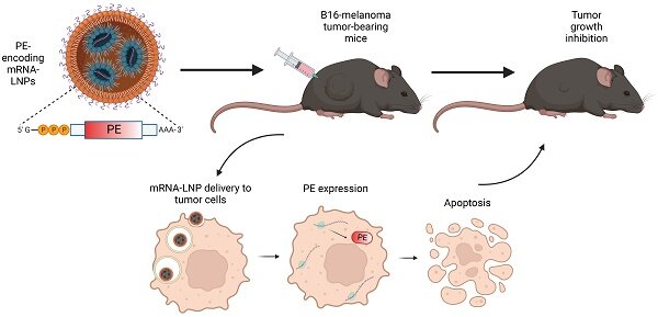
Graphical abstract. Credit: Theranostics (2023). DOI: 10.7150/thno.82228

Tel Aviv University researchers have hit upon a novel method of cancer treatment by creating an mRNA ‘Trojan horse’ that instructed cancer cells to produce a toxin lethal to themselves, eventually killing them with a success rate of about 50%. This ground-breaking study was led by PhD student Yasmin Granot-Matok and Prof Dan Peer, a pioneer in the development of RNA therapeutics. The study’s results were published in Theranostics.

Prof Peer explains: “Many bacteria secrete toxins. The most famous of these is probably the botulinum toxin injected in Botox treatments. Another classic treatment technique is chemotherapy, involving the delivery of small molecules through the bloodstream to effectively kill cancer cells. However, chemotherapy has a major downside: it is not selective, and also kills healthy cells. Our idea was to deliver safe mRNA molecules encoded for a bacterial toxin directly to the cancer cells – inducing these cells to actually produce the toxic protein that would later kill them. It’s like placing a Trojan horse inside the cancer cell.”

First, the research team encoded the genetic info of the toxic protein produced by bacteria of the pseudomonas family into mRNA molecules (resembling the procedure in which genetic info of COVID-19’s ‘spike’ protein was encoded into mRNA molecules to create the vaccine). The mRNA molecules were then packaged in lipid nanoparticles developed in Prof Peer’s laboratory and coated with antibodies to ensure they would reach their target, the cancer cells. These particles were injected into the tumours of animal models with melanoma skin cancer. After a single injection, 44–60% of the cancer cells vanished.  

“In our study, the cancer cell produced the toxic protein that eventually killed it,” says Prof Peer. “We used pseudomonas bacteria and the melanoma cancer, but this was only a matter of convenience. Many anaerobic bacteria, especially those that live in the ground, secrete toxins, and most of these toxins can probably be used with our method. This is our ‘recipe’, and we know how to deliver it directly to the target cells with our nanoparticles. When the cancer cell reads the ‘recipe’ at the other end it starts to produce the toxin as if it were the bacteria itself and this self-produced toxin eventually kills it. Thus, with a simple injection to the tumour bed, we can cause cancer cells to ‘commit suicide’, without damaging healthy cells. Moreover, cancer cells cannot develop resistance to our technology as often happens with chemotherapy – because we can always use a different natural toxin.”

Source: Tel Aviv University

Study Improves Accuracy for Breast Cancer Genetic Markers in Ashkenazi Jewish Women

Photo by Robert Thiemann on Unsplash

A new study by researchers in the UK and Israel has investigated how to improve breast cancer genetic tests for Ashkenazi Jewish women. By analysing genetic samples from women from Ashkenazi Jewish backgrounds, they were able to correctly adjust the risk estimates from commercially available genetic screening tests, giving a more accurate result. They detail their technique in the journal Genetics in Medicine.

New forms of genetic tests can tell women their personal risk of developing breast cancer. However, previous research has shown they are not accurate for many Black, Asian or Ashkenazi Jewish women, or women with a mixed ethnic background.

The study focuses on tiny genetic variations called Single Nucleotide Polymorphisms (SNPs) – which, depending on the unique combination of them, can increase or decrease the risk of breast cancer.

This information is used to produce a Polygenic Risk Score (PRS), which can inform women whether they are at low, average, or high risk of developing breast cancer in the next 10 years. PRS are becoming more widely available through commercial companies and research studies on the NHS breast screening programme.

Current PRSs were developed from large-scale genome studies which predominantly collected genetic data from mainstream White European populations. As a result, the accuracy of a PRS for an individual will depend on how closely their genetic material resemble those of the people whose data was used to develop the risk score.

This means that while commercially available PRS can accurately predict breast cancer risk for mainstream White European women, they often exaggerate this risk for Black, Asian or Ashkenazi Jewish women, or women with a mixed ethnic background.

In this new study, researchers compared two available PRS based on two SNPs – SNP142 and the commercial SNP78 – and analysed their accuracy for women of Ashkenazi Jewish ancestry.

The findings showed that these PRS tests inaccurately predicted Ashkenazi Jewish women to be at higher risk of developing breast cancer.

After adjusting the test for Ashkenazi Jewish ancestry, the researchers were able to generate a more accurate prediction of breast cancer risk for these women.

The research team used genetic information from Ashkenazi Jewish women in both Manchester and Israel, with data from the Predicting the Risk of Cancer at Screening (PROCAS) study conducted in Greater Manchester, a Manchester regional genetics database, and the Breast Cancer in Northern Israel (BCINIS) study.

This research was led by Professor Gareth Evans, a leading expert in breast cancer genetics and SNPs testing and NIHR Manchester BRC Cancer Prevention and Early Detection Theme Lead.

Professor Evans said: “Polygenic Risk Scores (PRS) are a major component of accurate breast cancer risk prediction and have great potential to improve personalised screening methods. However, it is clear from our findings that you cannot simply apply current PRS developed using genetic data from individuals of white European ancestry to those from Ashkenazi Jewish backgrounds.

“A test result which exaggerates a woman’s risk of the disease could lead to undue stress or concern and unnecessary screening and preventative measures that they don’t need. Future PRS for Ashkenazi Jewish women should be based on their genetic data to provide a more accurate risk prediction.

“This study is an important step forward in our continued research into breast cancer genetic testing for people of different ethnic backgrounds to improve equity. More accurate and personalised PRS are required to avoid further increasing health inequalities and so patients can receive high-quality screening, care, and treatments.”

Source: University of Manchester