The journey a cell makes from healthy to metastatic cancer is mostly driven by epigenetic changes, according to a new computational study that has been recently published in the journal Nature.
Every cell makes its own proteins by accessing its genetic information, but genetic mutations may ruin the function of the affected proteins. In oncology, this is regarded as the genetics of cancer. The last decades, however, have seen the rise of a new field: the epigenetics of cancer.
Epigenetic modifications do not change the information but temporarily modify the cell’s ability to read some of its own genes and produce the associated proteins instead. This forms a vast epigenetic program that controls general cell functions and, when altered, it may put it at the starting line of malignant transformation. Is there a way to track these changes and understand the epigenetics of cancer transition?
Now an international team of researchers has made headway towards this goal. They analysed 1.7 million cells from 225 samples from primary and metastatic origin, from 205 patients of 11 different cancer types. For each cell, the team obtained the full transcriptome, exome and epigenome. This covers virtually all gene mutations, gene accessibility and its consequences. With the help of enormous computational resources, they were able deduce the whole functional status of each analysed cell and link it to its particular cancer type.
The results of the work demonstrate that many regions in the DNA are differentially activated or inactivated in a cancer-specific manner, creating a signature for each tumour. These differences are relevant for cancer progression and many correspond to already identified hallmarks of cancer, the steps a cell must undergo to become malignant. Dr Eduard Porta, group leader at the Josep Carreras Leukaemia Research Institute (IJC-CERCA), is part of the team and contributed with his experience in the analysis of large amounts of biological data.
Epigenetic changes at the DNA level stand out as an underlying cause of cancer, according to the new publication. Particularly, the accessibility of enhancer regions, a kind of master regulator acting upon many genes at once. Taken together, the results converges into a short list of genes that can be used as markers for good or poor prognosis, valuable information for the clinical management of patients.
The analysis has also identified the cellular pathways of these important genes, making it possible to track their distant interactions. Sometimes, the affected genes are so fundamental that is impossible to drug them directly without side effects but, knowing the full pathway, researchers may develop strategies to target the weakest link in the chain, maximising the therapeutic benefits while minimising undesirable effects.
In an analysis of information on adults with colorectal cancer, patients who also had diabetes, particularly those with diabetic complications, faced a higher risk of early death. The results are published in CANCER, a peer-reviewed journal of the American Cancer Society.
For the study, Kuo‐Liong Chien, MD, PhD, of National Taiwan University, and his colleagues examined data registered between 2007 and 2015 in the Taiwan Cancer Registry Database, which is linked to health insurance and death records. Their analysis included 59 202 individuals with stage I–III colorectal cancer who underwent potentially curative surgery to remove their tumours. Among these patients, 9448 experienced a cancer recurrence and 21 031 died from any cause during the study period.
Compared with individuals without diabetes, those with uncomplicated diabetes were at a minimally or insignificantly higher risk of all‐cause and cancer‐specific death, whereas those with complicated diabetes had 85% higher odds of death from any cause and 41% higher odds of death from cancer. These associations were more pronounced in women and in patients with early‐stage colorectal cancer.
Also, compared to patients without diabetes, patients with uncomplicated or complicated diabetes had a 10–11% higher risk of colorectal cancer recurrence.
The mechanisms behind the relationship between diabetic severity and poor colorectal cancer prognosis could involve various pathways and responses triggered by high insulin and glucose levels in the blood, as well as elevated inflammatory states, which are characteristic of type 2 diabetes.
“While a higher diabetes prevalence was noted in patients with colorectal cancer, the study suggests that coordinated medical care involving multiple specialists can help prevent diabetes complications, potentially improving long-term colorectal cancer oncological outcomes, particularly in women and patients with early-stage cancer,” said Dr Chien.
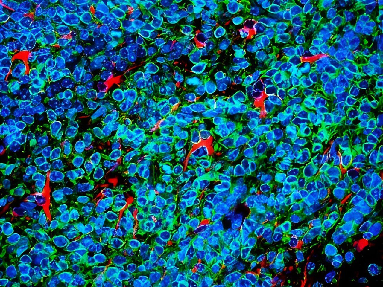
Small cell lung cancer cells (green and blue) that metastasised to the brain in a laboratory mouse recruit brain cells called astrocytes (red) for their protection. Credit: Fangfei Qu
Lung cancer cells that metastasise to the brain survive by convincing brain cells called astrocytes that they are baby neurons in need of protection, according to a study by researchers at Stanford Medicinepublished in Nature Cell Biology.
The cancer cells carry out their subterfuge by secreting a chemical signal prevalent in the developing human brain, the researchers found. This signal draws astrocytes to the tumour, encouraging them to secrete other factors that promote the cancer cells’ survival. Blocking that signal may be one way to slow or stop the growth of brain metastases of small cell lung cancer, which account for about 10% to 15% of all lung cancers, the researchers believe.
In the adult brain, astrocytes play a critical role in maintaining nerve function and connectivity. They are also important during brain development, when they facilitate connections between developing neurons.
The researchers studied laboratory mice, human tissue samples and human mini-brains, or organoids, grown in a lab dish to dissect the unique relationship between the cancer cells and their ‘big sister’ astrocytes, which hover nearby and shower them with protective factors.
“Small cell lung cancers are known for their ability to metastasise to the brain and thrive in an environment that is not normally conducive to tumour growth,” said professor of paediatrics and of genetics Julien Sage, PhD. “Our study suggests that these cancer cells reprogram the brain microenvironment by recruiting astrocytes for their protection.”
Professor Sage is the senior author of the study, while postdoctoral scholar Fangfei Qu, PhD, is the lead author.
Invasion of the brain
Small cell lung cancer excels at metastasising to the brain – about 15% to 20% of people already have clusters of cancer cells in their brains when their lung tumours are first diagnosed. As the cancer progresses, about 40% to 50% of patients will develop brain metastases. The problem is so prevalent, and the clinical outcome so dire, that clinicians recommend cranial radiation even before brain metastases have been found.
How and why small cell lung cancer has such an affinity for the brain has been something of a mystery. Brain metastases are rarely biopsied or removed because doing so has not been shown to affect a patient’s survival, and brain surgery is so invasive. Using laboratory mice is also of little help since small cell lung cancers in those animals rarely develop metastases in the brain, perhaps due to subtle biological differences between species.
Small cell lung cancers have another distinguishing feature – they are neuroendocrine cancers, meaning they arise from cells with similarities to both neurons and hormone-producing cells. Neuroendocrine cells link the nervous system with the endocrine system throughout the body, including in the lung.
Sage and his colleagues wondered whether neuronal-associated proteins on the surface of small cell lung cancer cells give them a leg up when the cells first begin to infiltrate the brain.
“We know the brain is full of neurons,” Sage said. “Maybe that’s why these cancer cells with some neuronal traits are happy in the brain and are accepted into that environment.”
Qu and Sage developed a way to inject mouse small cell lung cancer cells grown in the laboratory into the brains of mice to spark the development of brain tumours. They saw that astrocytes, a subtype of glial cell, flocked to the infant tumours and began to churn out proteins critical during brain development, including factors that stimulate nerve growth.
A plethora of astrocytes
A similar call happens in human brains, they noted: Brain tissue samples from people who had died of metastatic small cell lung cancer, shared by professor of pathology and paper co-author Christina Kong, MD, had many more protective astrocytes in the interior of the tumours than did metastases of melanoma, breast cancer and another type of lung cancer called adenocarcinoma.
Qu worked with assistant professor of paediatrics and co-author Anca Pasca, MD, to fuse aggregates of small cell lung cancer, lung adenocarcinoma or breast cancer cells with what are called cortical organoids – in vitro-grown clumps of brain cells including neurons and astrocytes that begin to mimic the organisation and connectivity of a human cortex. Within 10 days, many more protective astrocytes had infiltrated the small cell lung cancer pseudo-tumours than the adenocarcinoma or breast cancer.
“This showed us that the astrocytes actively move toward the small cell lung cancer cells, rather than simply being engulfed by the growing tumour,” Sage said. “What’s more exciting, though, is that these organoids, or mini-brains, realistically model the developing human brain. So, we’re no longer relying on a mouse model. It’s a perfect system to study brain metastases.”
Further research showed that the small cell lung cancer cells summon protective astrocytes by secreting a protein called Reelin that mediates the migration of neuronal and glial cells during brain development. Triggering Reelin expression in mouse breast cancer cells injected into the brain significantly increased the number of astrocytes in the resulting tumours in the mice, and the tumours were larger than in control animals injected with cells with low Reelin expression.
The apparent reliance of the cancer cells on chemical signals and responses specific to the developing brain may give clues for the development of future therapies, Sage believes.
“Some of these signals may not be as relevant or as highly expressed in the adult brain,” Sage said. “As a result, perhaps they could still be targeted to slow or prevent brain metastases without harming a normal brain. This might be an important window of opportunity for therapy.”
At 18 Spanish hospitals, when a patient with brain metastasis undergoes surgery, they can donate a tiny part of their brain to the first repository of brain metastasis living samples in the world, based at the Spanish National Cancer Research Centre (CNIO). A world-first collection, it was created to accelerate the search for therapies against brain metastasis, a disease that affects up to 30% of patients with systemic cancer.
The creators of this repository, called RENACER (Spanish acronym for the National Brain Metastasis Network), are two CNIO researchers, Manuel Valiente, head of the Brain Metastasis Group, and Eva Ortega-Paíno, director of the Biobank. They explained the advantages of the collection in the journal Trends in Cancer. In just three years RENACER has compiled samples from more than 150 patients.
The truly unique feature of RENACER, which makes it a valuable tool for the international scientific community, is that it contains living samples, conserved in cultures that enable the cells to continue behaving in a similar way as they were in the body.
A living biobank that enables organotypic cultures
“We have built a ‘living’ biobank” write Valiente and Ortega-Paíno. And this characteristic can be “transformative, not only for research but also for clinical trial design, especially when focused on unmet clinical needs, such as brain metastasis”.
The fact that the cells are living allows them, for example, to study their response to specific drugs. RENACER paves the way to create avatars for each patient in order to identify the best therapeutic options in an individualised way.
“Research contracts have been already signed to exploit patient-derived organotypic cultures (PDOCs) as avatars, thus providing the possibility to generate biomarkers of sensitivity or resistance to specific drugs” the authors explain.
The hospitals involved with RENACER work as a network to pass on research findings to patients as quickly as possible. In fact, thanks to this network, there are already two clinical trials underway, which will determine the capacity of two biomarkers to discriminate cases in which radiotherapy – a technique with side effects – will be effective.
From the operating theatre to the biobank in hours
The requirement for cells to be “alive” is not easy to achieve, since it involves a sophisticated logistics chain. The samples are taken from the operating theatre in a special container, in their culture medium, at a temperature of between 4 and 8 degrees centigrade.
They must reach the CNIO Biobank, in Madrid, in less than 24 hours. There, they are processed, organotypic cultures are created, and they are divided into proportional parts that are stored as samples for future investigations. They are also analysed using various techniques and sequenced, to extract as much information as possible from them. All the data are put into a database that is open to the international scientific community.
“It is pivotal to empower patients”
“This is happening just a few years after the project was launched,” said Valiente. “It’s a strategy that helps to improve knowledge as well as diagnosis and treatment options, but also brings all the people involved closer together: patients, core researchers, chemical re searchers, healthcare professionals, and the biobank.”
Patients, “[because they act] as donors during a difficult brain metastasis neurosurgery, play a crucial role and we strongly believe that it is pivotal to empower them,” the researchers explain. GEPAC (Spanish Group of Patients with Cancer) is also involved with RENACER.
University of Bristol researchers have created a robotic hand that could carry out Clinical Breast Examinations (CBE). The device is able to apply very specific forces over a range similar to forces used by human examiners and can detect lumps using sensor technology at larger depths than before.
This could revolutionise how women monitor their breast health by giving them access to safe electronic CBEs, located in easily accessible places, such as pharmacies and health centres, which provide accurate results. The technology is described in the journal Sensors.
Precision, repeatability and accuracy are of paramount importance in these tactile medical examinations to ensure favourable patient outcomes. A range of automatic and semi-automatic devices have been proposed to aid with optimising this task, particularly for difficult to detect and hard to reach situations such as during minimally invasive surgery.
The research team included a mix of postgraduate and undergraduate researchers, supervised by Dr Antonia Tzemanaki from Bristol Robotics Laboratory. Lead author George Jenkinson explained: “There are conflicting ideas about how useful carrying out Clinical Breast Examinations (CBE) are for the health outcomes of the population.
“It’s generally agreed upon that if it is well performed, then it can be a very useful and low risk diagnostic technique.
“There have been a few attempts in the past to use technology to improve the standard to which healthcare professionals can perform a CBE by having a robot or electronic device physically palpate breast tissue. But the last decade or so of technological advances in manipulation and sensor technology mean that we are now in a better position to do this.
“The first question that we want to answer as part of this is whether a specialised manipulator can be demonstrated to have the dexterity necessary to palpate a realistic breast size and shape.”
The team created their manipulator using 3D printing and other Computerised Numerical Control techniques and employed a combination of laboratory experiments and simulated experiments on a fake (silicone) breast and its digital twin, both modelled on a volunteer at the Simulation and Modelling in Medicine and Surgery research group at Imperial College London.
The simulations allowed the team to perform thousands of palpations and test lots of hypothetical scenarios such as calculating the difference in efficiency when using two, three, or four sensors at the same time. In the lab, they were able to carry out the experiments on the silicone breast to demonstrate the simulations were accurate and to experimentally discover the forces for the real equipment.
George added: “We hope that the research can contribute to and complement the arsenal of techniques used to diagnose breast cancer, and to generate a large amount of data associated with it that may be useful in trying to identify large scale trends that could help diagnose breast cancer early.
“One advantage that some doctors have mentioned anecdotally is that this could provide a low-risk way to objectively record health data. This could be used, for example, to compare successive examinations more easily, or as part of the information packet sent to a specialist if a patient is referred for further examination.”
As a next step, the team will combine CBE techniques learned from professionals with AI, and fully equip the manipulator with sensors to determine the effectiveness of the whole system at identifying potential cancer risks.
The ultimate goal is that the device and sensors will have the capability to detect lumps more accurately and deeper than it is possible only from applying human touch. It could also be combined with other existing techniques, such as ultrasound examination.
“So far we have laid all of the groundwork,” said George. “We have shown that our robotic system has the dexterity necessary to carry out a clinical breast examination – we hope that in the future this could be a real help in diagnosing cancers early.”
Pancreatic ductal adenocarcinoma (PDAC) is one of the deadliest cancers worldwide, with a 5-year survival rate of less than 10%. Many PDAC tumours go undetected in early stages since they go undetected by conventional imaging methods such as fluorodeoxyglucose positron emission tomography (PET) scans. To tackle this problem, researchers in Japan are combining diagnostic and therapeutic procedures into a single integrated process: ‘theranostics’.
In an article recently published in the Journal of Nuclear Medicine, the Osaka University-led team has developed a ‘radio-theranostics’ strategy that uses a new radioactive antibody to target glypican-1 (GPC1), a protein highly expressed in PDAC tumours. Theranostics, particularly radio-theranostics, has been receiving increasing attention because, by radio-labelling the compounds used to target certain molecules in cancer cells, diagnosis and treatment can be carried out sequentially.
“We decided to target GPC1 because it is overexpressed in PDAC but is only present in low levels in normal tissues,” explains Tadashi Watabe, lead author of the study.
The team used a monoclonal antibody (mAb) designed to target GPC1. The mAb could be labelled with isotopes of zirconium (89Zr) or astatine (211At). First, they injected the 89Zr-GPC1 mAb into a xenograft mouse model, which has a human pancreatic cancer tumour.
“We monitored 89Zr-GPC1 mAb internalisation over seven days with PET scanning,” explains Kazuya Kabayama, the second author of the article. “There was strong uptake of the mAb into the tumours, suggesting that this method could support tumour visualisation. We confirmed that this was mediated by its binding to GPC1, as the xenograft model that had GPC1 expression knocked out showed significantly less uptake.”
The researchers next tested this model with alpha therapy using 211At-GPC1 mAb, a method that could support radioactive label-based delivery of a therapeutic molecule to its target. Administration of 211At-GPC1 mAb resulted in DNA double-strand break induction in the cancer cells, as well as significantly reduced tumour growth. Control experiments showed that these antitumor effects did not occur when mAb internalisation was blocked. Additionally, non-radiolabelled GPC1 mAb did not induce these effects.
“Both radiolabeled versions of the GPC1 mAb we examined showed promising results in PDAC,” says Watabe. “89Zr-GPC1 mAb showed high humoral uptake, while 211At-GPC1 mAb could be used for targeted alpha therapy to support suppression of PDAC tumour growth.”
These highly impactful data demonstrate the potential for using a theranostics approach in PDAC, a disease in dire need of new diagnostic and therapeutic options. In the future, this could lead to early detection of PDAC with PET imaging and systemic treatment with alpha therapy.
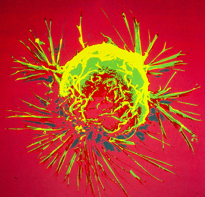
Colourised scanning electron micrograph of a breast cancer cell. Credit: NIH
A novel therapeutic approach that combines human epidermal growth receptor factor 2 (HER2)-targeted therapies with the cholesterol-lowering drug lovastatin can reduce the number of cancer treatments required to prevent tumour growth. Monitored by immuno-PET scans, this combination therapy has the potential to personalise treatment for cancer patients and spare them from harmful side effects. This research was published in The Journal of Nuclear Medicine.
Antibody-drug conjugates (ADCs) have become an eminent cancer treatment because of their ability to precisely target tumours with potent efficacy. HER2-ADC therapies have been effective in treating breast, lung, bladder, and stomach cancers. Although usually well-tolerated, multiple doses of the drugs can result in severe side effects, including low blood counts, liver damage, and lung damage. Strategies that reduce toxic side effects caused by ADCs and predictive biomarkers of ADC toxicity are currently an unmet clinical need.
“In this study, we sought to determine whether a single dose of HER2-ADCs could be administered in combination with lovastatin (which temporarily elevates cell-surface HER2) to achieve therapeutic efficacy similar to that of a multiple dose regime,” said Patricia Pereira, PhD, assistant professor at the Washington University School of Medicine. “We also used HER2-targeted immuno-PET to monitor changes in HER2 expression after ADC therapy.”
Researchers injected mice with cultured gastric cancer cells and patient-derived gastric cancer cells. When tumours grew sufficiently, the mice were divided into groups and received various treatment schedules (no treatment, multiple doses of ADC, multiple doses of ADC with lovastatin, single dose of ADC, or single dose of ADC with lovastatin). Immuno-PET was used to investigate the dosing regimen and the efficacy of the treatment schedules.
A single dose of ADC therapy combined with lovastatin was found to reduce tumour volume at rates similar to those resulting from multiple doses of ADC in a preclinical setting. The study results showed that immuno-PET can noninvasively monitor HER2 tumour levels after treatment with HER2-targeted ADC therapies.
“This preclinical work is significant because it has the potential to improve therapy for patients with HER2-positive cancers,” noted Pereira. “It not only simplifies treatment by exploring single-dose schedules of antibody-drug conjugates but can also reduce side effects by minimizing the number of doses required. Additionally, it personalises therapy using molecular imaging, enhancing treatment efficacy.”
She continued, “The findings suggest a future where molecular imaging techniques play a critical role in guiding drug development and cancer treatment decisions, particularly as various ADCs are being tested and approved for cancer treatment. Currently, there is no perfect way to select tumours or monitor their response to ADCs. This research indicates that molecular imaging can bridge this gap by providing real-time insights into therapy response.”
A study in Frontiers in Immunology has demonstrated that, in animal models, a protein antigen from a childhood vaccine can be delivered into the cells of a malignant tumour to refocus the body’s immune system against the cancer, effectively halting it and preventing its recurrence.
Instead of using vaccines tailored with tumour-specific antigens to prime the immune system to attack a particular cancer, this method makes use of the immune system’s encounter with common vaccines. The bacteria-based intracellular delivering (ID) system uses a non-toxic form of Salmonella that releases a drug, in this case a vaccine antigen, after it’s inside a solid-tumour cancer cell.
“As an off-the-shelf immunotherapy, this bacterial system has the potential to be effective in a broad range of cancer patients,” writes senior author Neil Forbes, professor of chemical engineering, in the recently published article.
The research, carried out in Forbes’s lab, offers promise toward tackling difficult-to-treat cancers, including liver, metastatic breast and pancreatic tumours.
“The idea is that everybody is vaccinated with a whole bunch of things, and if you could take that immunisation and target it towards a cancer, you could use it to eliminate the cancer,” Forbes explains. “But cancers obviously aren’t going to display viral molecules on their surface. So the question was, could we take a molecule inside the cancer cell using Salmonella and then have the immune system attack that cancer cell as if it was an invading virus?”
To test their theory that this immune treatment could work, Forbes and team genetically engineered ID Salmonella to deliver ovalbumin (chicken egg protein) into the pancreatic tumour cells of mice that had been immunised with the ovalbumin ‘vaccine’. The researchers showed that the ovalbumin disperses throughout the cytoplasm of cells in both culture and tumours.
The ovalbumin then triggered an antigen-specific T-cell response in the cytoplasm that attacked the cancer cells. The therapy cleared 43% of established pancreatic tumours, increased survival and prevented tumour re-implantation, the paper states.
“We had complete cure in three out of seven of the pancreatic mice models,” Forbes says. “We’re really excited about that; it dramatically extended survival.”
The team then attempted to re-introduce pancreatic tumours in the immunised mice. The results were exceedingly positive. “None of the tumours grew, meaning that the mice had developed an immunity, not just to the ovalbumin but to the cancer itself,” Forbes says. “The immune system has learned that the tumour is an immunogenic. I’m doing further work to figure out how that’s actually happening.”
In preliminary research, the team previously showed that injecting the modified Salmonella into the bloodstream effectively treated liver tumours in mice. They advanced their findings with the current research on pancreatic tumours.
Before clinical trials can begin, the researchers will repeat the experiments on other animals and refine the ID Salmonella strain to ensure its safety for use in humans. Liver cancer would be the first target, followed by pancreatic cancer.
For the management of children’s cancer symptoms, cannabis products have increased in popularity, but questions remain over their efficacy and safety. A recent review of published studies to date shows a lack of evidence to determine the dosing, safety, and efficacy of medical marijuana or cannabis-containing products for managing symptoms experienced by children with cancer. The analysis is published online in CANCER, a peer-reviewed journal of the American Cancer Society.
Although great strides have been made in treatments for childhood cancer, even leading to cures for many patients, many children still suffer from symptoms such as pain, anxiety, and weight loss related to cancer and its treatment. Over the last decade, cannabis products, both synthetic cannabinoids and natural phytocannabinoids, have gained popularity with patients and families for managing such symptoms, but paediatric oncologists are cautious to authorise cannabis for their patients given the limited data to inform dosing, product selection, and safety monitoring.
To provide insights for clinicians and parents, and to inform an upcoming clinical trial, a team led by Lauren E. Kelly, PhD, associate professor of pharmacology and therapeutics in the Rady Faculty of Health Sciences at the University of Manitoba, searched the medical literature to summarise existing knowledge about the potential benefits and harms of cannabis products in children with cancer.
The investigators identified 19 unique studies with a total of 1927 participants with cancer: eight retrospective chart reviews, seven randomised controlled trials, two open‐label studies, and two case reports. The products studied included medical-grade cannabinoids (such as the prescription drug nabilone), synthetic cannabinoids, and unspecified cannabis herbal extracts. Products were most commonly used to manage chemotherapy‐induced nausea and vomiting.
In the randomised controlled trials, patients who used cannabinoids were more likely to experience drowsiness, feeling high, dizziness, and dry mouth. Also, trial participants who received cannabinoids were almost four times more likely to drop from the study due to adverse events, compared with the control group who received placebo. Across all included studies, no serious cannabis‐related adverse events were reported.
Dr Kelly and her colleagues noted that most studies did not adequately describe the types, dosing, frequencies, and routes of administration of cannabis products, and outcomes were mixed and were reported in different ways. Therefore, researchers should develop standards for reporting cannabis exposures, cannabis‐related effects, and patient outcomes.
“It was difficult to measure benefit across studies, given a range of different outcomes and study designs; however, in interventional studies with active control groups, cannabinoids performed better in managing nausea and vomiting. Data are lacking on cannabinoids’ effects on pain, mood, sleep, and health-related quality of life,” said Dr Kelly. “Given that some children report benefits and some children experience adverse events, it is critical that more rigorous studies evaluating the effects of cannabinoids on children with cancer are conducted and shared with parents, patients, and the health care community.”
This literature review informed the design of a three-arm tolerability trial later this year.
Credit: Darryl Leja National Human Genome Research Institute National Institutes Of Health
Patients with intermediate risk, localised prostate cancer can be treated as effectively using fewer and higher doses of radiation therapy delivered over five treatment sessions as they can with lower doses delivered over several weeks, according to researchers from The Royal Marsden NHS Foundation Trust and The Institute of Cancer Research, London.
Significant reduction in treatment time
Results from the PACE B (Prostate Advances in Comparative Evidence) phase III randomised trial showed that stereotactic body radiotherapy (SBRT) performed as well as standard radiotherapy treatment for people whose prostate cancer had not spread, demonstrating a 96% chance of no disease progression within five years, compared to 95% for conventional radiotherapy.
SBRT, which can be delivered on a CyberKnife or standard radiotherapy machines, allows clinicians to target tumours to sub-millimetre precision. This approach uses advanced imaging and treatment planning techniques to deliver radiation with pinpoint accuracy, minimising damage to surrounding healthy tissue. It delivers five high doses of radiation to patients over one to two weeks, compared to standard radiotherapy, which delivers more moderate doses over a much longer period of time – usually around 20 sessions for patients in the UK, which can take up to one month.
Drawing from 38 centres across the UK, Ireland and Canada, researchers enrolled 874 people who preferred radiation treatment or were unsuitable for surgery. Patients were randomly assigned to receive either SBRT, consisting of five doses over one to two weeks, or standard radiation consisting of 20 doses over four weeks or 39 doses over 7.5 weeks. None of the patients received hormonal therapy.
“For something as serious as a cancer diagnosis, the treatment was incredibly easy”
The trial investigated whether SBRT was non-inferior to conventional radiation for treating people with intermediate risk, localised prostate cancer. Non-inferiority was measured by whether patients remained free of biochemical clinical failure (BCF), defined as an increase in prostate-specific antigen (PSA) levels, distant metastases or other evidence the cancer was returning, or death from prostate cancer.
Five years after treatment, people treated with SBRT had a BCF-event free rate of 95.7% compared to 94.6% for those treated with conventional radiotherapy, demonstrating that SBRT was non-inferior to conventional radiation.
Side effects were low in both groups and at five years not significantly different between treatment arms.
One patient who benefited from the treatment was 64-year-old Alistair Kennedy-Rose, who was diagnosed with prostate cancer in 2014 at his local hospital following a blood test which revealed his prostate-specific antigen levels were raised. Alistair was referred to The Royal Marsden and, after being recruited to the PACE-B trial, was treated with SBRT via CyberKnife.
He said: “When I was diagnosed I had no symptoms at all, so it came as quite a shock. There can be a lot of anxiety following a cancer diagnosis, but just ten days after I was referred to the Royal Marsden, I started SBRT. I still find it unbelievable that five days later I finished my treatment, for something as serious as a cancer diagnosis it was incredibly easy. I haven’t had any side effects and I’ve been able to live my life to the full. I can’t thank The Royal Marsden enough for what they have done for me.”