Category: Cancer

Certain Fatty Acids can ‘Supercharge’ T-Cells’ Antitumour Immunity
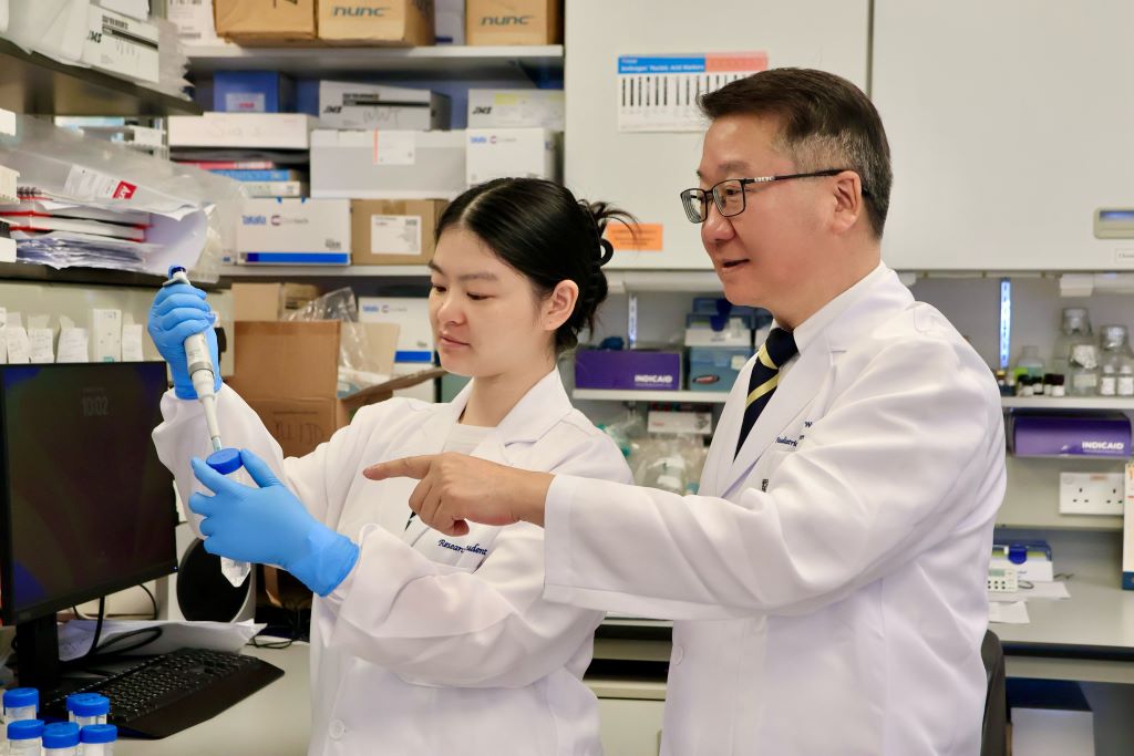
A research team at the LKS Faculty of Medicine of the University of Hong Kong (HKUMed) discovered that certain dietary fatty acids can supercharge the human immune system’s ability to fight cancer. The team found that a healthy fatty acid found in olive oil and nuts, called oleic acid (OA), enhances the power of immune γδ-T cells, specialised cells known for their cancer-fighting properties.

Conversely, they found that another fatty acid, called palmitic acid (PA), commonly found in palm oil and fatty meats, diminishes the ability of these immune cells to attack tumours. This groundbreaking study, published in the academic journal Signal Transduction and Targeted Therapy, offers an innovative approach using dietary OA supplementation to strengthen the antitumour immunity of γδ-T cells.

Dietary fatty acids and cancer immunotherapy

Dietary fatty acids are essential for health, helping with growth and body functions. They may also play a role in cancer prevention and treatment, but understanding how they affect cancer is challenging because of the complexity of people’s diets and the lack of detailed studies. Recently, scientists have learned that fatty acids can influence the immune system, especially in how it fights cancer. Specialised immune cells, called γδ-T cells, are particularly good at attacking tumours. These cells, once activated, have helped some lung and liver cancer patients live longer. However, this therapy is not effective for all patients, partly because the variation of the metabolic status, such as fatty acid metabolism, can influence its efficacy in the patients.

Oleic acid may improve cancer treatment outcomes

The research team identified a correlation between PA and OA levels and the efficacy of cancer therapies. ‘Our research suggests that dietary fatty acid supplementation, particularly with foods rich in OA, such as olive oil and avocados, could enhance γδ-T cell immunosurveillance, leading to more effective cancer treatments,’ said Professor Tu Wenwei from the Department of Paediatrics and Adolescent Medicine, School of Clinical Medicine, HKUMed, who led the study.

The team also discovered that another fatty acid, called PA, can weaken these immune cells and how OA can counteract this. ‘The results indicate that cancer patients should avoid PA and consider OA supplementation in their diets to improve clinical outcomes of γδ-T cell-based cancer therapies,’ added Professor Tu.

Significant impact from simple dietary changes

Professor Tu said, ‘This study is the first to show that the fatty acids we eat can directly affect how well our immune cells fight cancer.’ It reveals how PA can harm these cells and how OA helps them through a specific process involving a protein called IFNγ. By analysing blood samples, the researchers confirmed that the levels of these fatty acids are linked to the outcome of cancer immunotherapy.

‘For cancer patients, this discovery suggests simple changes, like eating more foods rich in OA (such as olive oil, avocados and nuts) and cutting back on PA (found in processed foods, palm oil and fatty meats), could improve the effectiveness of cancer treatments. The study also points to novel strategies, like combining dietary changes with specific drugs to further boost the immune system,’ added Professor Tu.

This study demonstrates that personalised nutrition may serve as an effective strategy to enhance immune function and support cancer treatment. It also suggests that new drugs targeting the processes affected by these fatty acids could enhance the power of γδ-T cell therapies. By integrating nutritional interventions with immunotherapy, this discovery could help more cancer patients achieve better outcomes.

Source: University of Hong Kong

Unique Immunotherapy Could be the ‘Holy Grail’ for a Wide Range of Cancers

Next step is testing ‘holy grail’ therapy’s safety and effectiveness in patients

Squamous cancer cell being attacked by cytotoxic T cells. Image by National Cancer Institute on Unsplash

A new, highly potent class of immunotherapeutics with unique Velcro-like binding properties can kill diverse cancer types without harming normal tissue, University of California, Irvine cancer researchers have demonstrated.

A team led by Michael Demetriou, MD, PhD, reported that by targeting cancer-associated complex carbohydrate chains called glycans with binding proteins, they could penetrate the protective shields of tumor cells and trigger their death without toxicity to surrounding tissue.

Their biologically engineered immunotherapies – glycan-dependent T cell recruiter (GlyTR, pronounced ‘glitter’) compounds, GlyTR1 and GlyTR 2 – proved safe and effective in models for a spectrum of cancers, including those of the breast, colon, lung, ovaries, pancreas and prostate, the researchers reported in the journal Cell.

“It’s the holy grail – one treatment to kill virtually all cancers,” said Demetriou, a professor of neurology, microbiology and molecular genetics at the UC Irvine School of Medicine and the paper’s corresponding author. “GlyTR’s velcro-like sugar-binding technology addresses the two major issues limiting current cancer immunotherapies: distinguishing cancer from normal tissue and cancer’s ability to suppress the immune system.”

The researchers were awarded a Cancer Moonshot Initiative grant from the National Cancer Institute in 2018 for this study.

Landmark research

The study’s publication, the culmination of a decade of research, is a watershed moment and source of pride for UC Irvine and the UCI Health Chao Family Comprehensive Cancer Center.

“This landmark study is a paradigm shift with the very real potential to change how we treat cancer patients,” said Marian Waterman, PhD, former deputy director of research at the cancer centre and champion of the project since Demetriou and his then-postdoctoral fellow, Raymond W. Zhou, the study’s first author, began working on the concept in 2015.

Added Richard A. Van Etten, MD, PhD, director of the cancer centre and also an early supporter of the GlyTR project, “This novel technology may, for the first time, allow the widespread application of targeted T-cell therapy to solid tumours, which is the ‘holy grail’ in the immuno-oncology field.”

Current treatments, such as chimeric antigen receptor (CAR) T therapy, use the body’s white blood cells to attack cancer. They have largely worked only for blood cancers, such as leukaemia. The GlyTR technology also proved effective in targeting leukaemia, the study shows.    

Unorthodox approach

While many cancer researchers have sought protein biomarkers for specific cancers, Demetriou and Zhou aimed at a more abundant target, the unique coating of glycans that surround cancer cells but are found in very low density in normal cells.

These complex sugar chains are the most widespread cancer antigens known, but were generally ignored by researchers because they are inert to the immune system.

To solve this problem, Demetriou and Zhou engineered the GlyTR compounds to attach themselves, Velcro-like, to glycan-dense cancer cells while ignoring low-glycan-density normal cells. Once attached, the GlyTR compounds identify the cancer cells as targets for killing by the body’s immune system.   

In contrast, current cancer  immunotherapies attack cells based on specific proteins regardless of their glycan density and thereby fail to distinguish tumour cells from healthy tissue.

A second impediment to developing broadly active cancer immunotherapies is the shield glycans form around solid tumours.

By targeting glycans and blanketing the tumour cells with the Velcro-like compounds, the GlyTR technology overcomes both obstacles.  

Human trials

The next step will be testing the therapy’s safety and effectiveness in humans. Clinical grade GlyTR1 protein manufacturing is already being developed at the NCI Experimental Therapeutics program labs in Maryland, Demetriou said.

That will enable the launch of a phase 1 clinical trial, which could begin within about two years. It will test the therapy in patients with a range of metastatic solid cancers. The highest glycan density is typically seen in patients with refractory/metastatic disease, a population that also has the greatest unmet need for treatment.

Source: University of California – Irvine

Could Reducing Inflammation Help Combat Fatigue in Early-stage Breast Cancer?

Study links blood levels of inflammatory markers with different aspects of cancer-related fatigue.

Photo by Karolina Grabowska on Pexels

New research reveals that inflammatory responses may play a role in different types of fatigue experienced by many people with cancer. The findings are published by Wiley online in CANCER, a peer-reviewed journal of the American Cancer Society.

Cancer-related fatigue can be a distressing and persistent burden that causes patients to feel physical, emotional, and/or cognitive tiredness or exhaustion. Activation of inflammatory responses by the tumour itself and/or by cancer treatment is thought to be a key biological driver of this symptom, but inflammatory activity across the cancer continuum has not been thoroughly examined.

To investigate, researchers at the University of California, Los Angeles (UCLA) analysed protein markers of inflammation in 192 women with early-stage breast cancer who were examined before radiation or chemotherapy and throughout the 18 months after treatment. At each assessment, women reported on different dimensions of fatigue (general, physical, mental, and emotional) and provided blood that was tested for protein markers of inflammation. These included two pro-inflammatory cytokines (TNF-α and IL-6) and two downstream markers of their activity (sTNF-RII and CRP).

Higher levels of TNF-α, sTNF-RII, and IL-6 were linked with greater general fatigue, which involves feelings of tiredness and exhaustion. These effects remained even after accounting for age, race, education, body mass index, and cancer stage. Similarly, there was a positive association between physical fatigue, which involves feelings of physical weakness and heaviness, and TNF-α, sTNF-RII, and CRP. Conversely, higher levels of TNF-α and sTNF-RII were associated with lower levels of emotional fatigue. No significant associations between mental (or cognitive) fatigue and inflammatory markers were found.

“Our findings indicate that inflammation plays a role in some aspects of cancer-related fatigue, but not others, and that these effects persist well after treatment,” said lead author Julienne E. Bower, PhD, of UCLA. “This is critical for developing targeted treatments for this common and disabling symptom.”

Source: Wiley

Rooibos Extracts Demonstrate Protective Effects in Preliminary Breast Cancer Research

Photo by TeaCora Rooibos on Unsplash

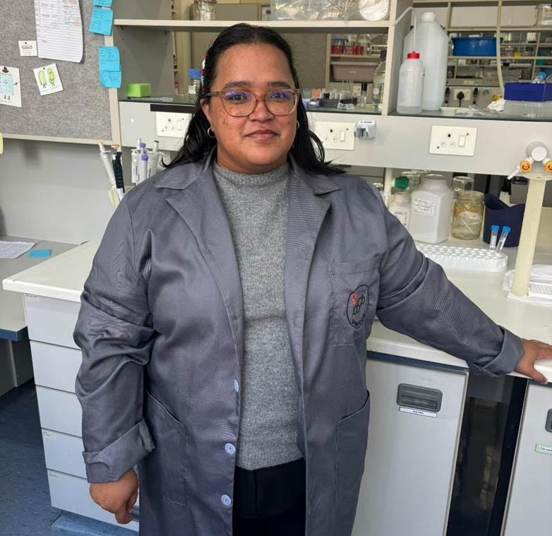
Rooibos, South Africa’s beloved herbal tisane, with proven benefits for the heart, diabetes management and brain health, is now revealing even more potential in preliminary breast cancer research led by Dr Nicky Verhoog, senior lecturer of biochemistry at Stellenbosch University.

Caption: Dr Nicky Verhoog, senior lecturer of biochemistry at Stellenbosch University.

Oestrogen’s role in breast cancer

While the research is still in its early stages, initial findings suggest that Rooibos extracts may counteract the activity of oestrogen in breast cancer cells – a discovery that has prompted the South African Rooibos Council (SARC) and the Department of Science, Technology and Innovation (DSTI) Sector Innovation Fund Programme to continue to support the work of Dr Verhoog with further studies in animal models scheduled to begin next year.

Oestrogen is often described as a kind of driver of breast cancer growth, because many breast cancer tumours carry receptors that respond to it. Although oestrogen doesn’t always start the cancer, it can speed up its growth once the disease has developed. Some newer studies also show that it may sometimes play a more direct role in triggering changes in cells that lead to cancer. Because so many breast cancers are sensitive to oestrogen, the possibility that Rooibos might help block its effect makes these findings especially significant.

Breast cancer in South Africa

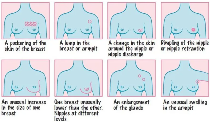
Breast cancer remains a major health concern in the country. According to the latest statistics, between 11 000[1] and 15 000[2] women are diagnosed with breast cancer annually and the disease accounts for approximately 24% of all cancer cases in South African women. Early detection is critical – when identified at an early stage, breast cancer survival rates can exceed 90%, highlighting the importance of regular screening and self-examination.

Caption: Warning signs of breast cancer. Source: CANSA (cansa.org.za)

Rooibos under the microscope

Against this backdrop, Dr Verhoog’s team conducted an in vitro (cell) study to examine whether Rooibos could influence oestrogen receptor-positive breast cancer cells. In the past, there has been some uncertainty about whether Rooibos might interact with oestrogen in ways that could affect women with hormone-sensitive breast cancer. This study set out to clarify its role – whether Rooibos could potentially act like estrogen or conversely, help block its activity – and to assess whether it influences cancer cell behaviour.

“To better understand how Rooibos interacts with natural hormones like estrogen, our team wanted to gather clearer scientific evidence,”

Dr Verhoog explains. “We also looked at whether Rooibos could influence the growth and spread of cancer cells, which is especially important for women with hormone-sensitive breast cancer.”

In the laboratory, the researchers worked with well-known breast cancer cell models that carry two types of estrogen receptors: one that can encourage tumour growth (ERα) and another that helps to suppress it (ERβ). They then added Rooibos extracts to see how the cells would respond – in terms of growth, movement and spread.”

The results were encouraging. The extracts acted against oestrogen, in a way similar to fulvestrant, a treatment commonly used to block oestrogen activity in breast cancer treatment. Rooibos slowed down oestrogen-induced cell growth and prevented the cells from spreading. Interestingly, the extracts showed a preference for activating ERβ, the receptor associated with tumour suppression.

“Rooibos appears to act in a protective way against oestrogen-driven breast cancer cells in the lab. It doesn’t mimic oestrogen, but rather inhibits its action. Importantly, we also found that it didn’t interfere with the effect of tamoxifen, a standard breast cancer therapy, which suggests Rooibos could be safely consumed by women undergoing treatment. Of course, this needs confirmation through in vivo (animal) studies.”

Next steps: pre-clinical trials in 2026

While the findings are promising, Dr Verhoog stresses the limitations of in vitro research.

“Lab studies involve single cell types and don’t replicate the complexity of the human body,” she says. “We need to see how Rooibos behaves when metabolised in the body, which is why our next step is pre-clinical trials planned for 2026.”

The next phase of research will move beyond cell studies to look at how Rooibos behaves in a pre-clinical model system. This step is important to confirm its safety for women with oestrogen-sensitive breast cancer and to better understand its effects in the body. The aim is not to test Rooibos as a treatment, but to ensure that it can be safely enjoyed alongside conventional therapies.

Rooibos’s broader health benefits

Rooibos is already celebrated for its wide range of health benefits. Rich in antioxidants and free from caffeine and added sugars, it is a popular alternative to sugary drinks, contributing to overall hydration and general wellness. Previous studies have also highlighted its potential protective effects against other cancer types, including prostate, liver and colon cancers, thanks to its potent antioxidant properties.

“While we are still in the early stages, our research adds to the growing evidence of Rooibos’ multifaceted biological actions. It’s a step toward understanding how this uniquely South African tisane may support health in a variety of ways,” emphasises Dr Verhoog.

Experts stress that these findings should not be interpreted as evidence that Rooibos can prevent or treat breast cancer. Rather, they reinforce its safety for consumption and provide a foundation for future research.

With October recognised as Breast Cancer Awareness Month, the study also underscores the urgent need for continued scientific investigation into the disease, while reminding women that regular screening, awareness of family history and early detection remain the most effective strategies for reducing breast cancer risk and improving outcomes.


[1] https://www.nicd.ac.za/wp-content/uploads/2025/04/NCR_ASR_tables_2023.pdf According to the National Cancer Registry (NCR), 10 980 cases of breast cancer were recorded in 2023. However, CANSA has highlighted significant underreporting, particularly in rural and under-serviced communities, suggesting that the actual number of cases is likely much higher. The available figures are based on pathological diagnoses.

[2] https://pmc.ncbi.nlm.nih.gov/articles/PMC10881925/

The statistics provided by the International Agency for Research on Cancer (IACR) differ from those reported by the South African NCR, with the number of new breast cancer cases in 2020 being much higher in the IACR data than in the NCR data (15,491 versus 9259). These differences are due to the different strategies used to tally the statistics. The NCR is a collection of pathology-based cancer surveillance figures based on the curation of cancer diagnosis data from histology, cytology and bone marrow aspirate and trephine samples; these samples have been collected, analysed and then reported on annually [4]. The IACR data are based on the South African NCR and Eastern Cape Province Cancer Registries.

Missing First Mammogram Raises Breast Cancer Death Risk

Photo by National Cancer Institute on Unsplash

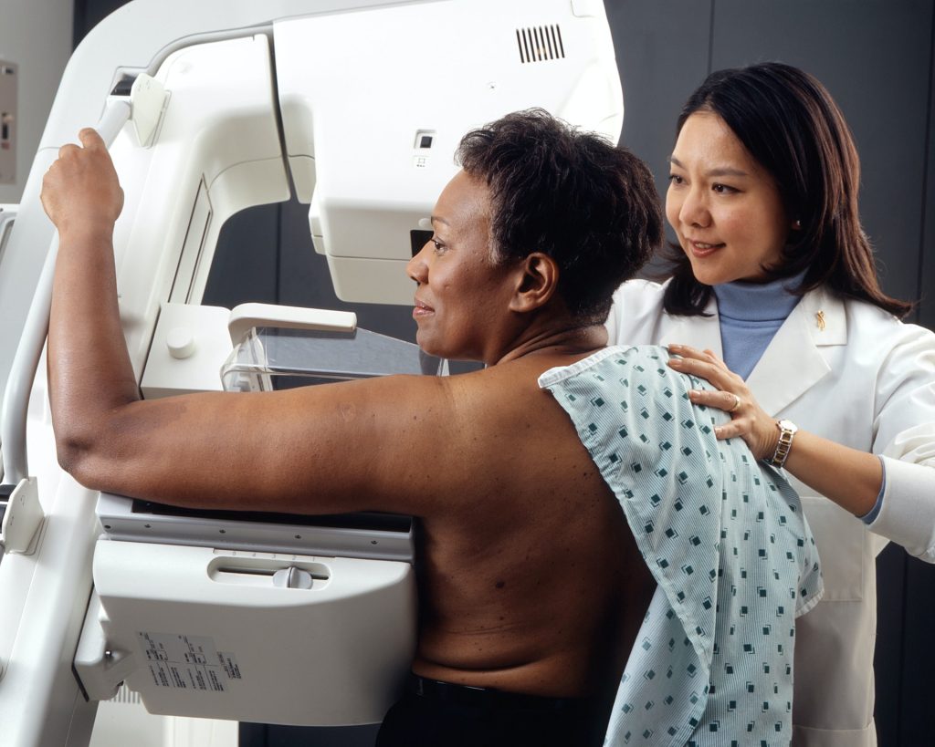
Women who miss their first mammogram run a higher risk of being diagnosed with advanced breast cancer and dying from the disease. This is shown in a new study from Karolinska Institutet published in The BMJ.

Since the early 1990s, women in Sweden have been offered regular mammograms, which has contributed to a decrease in breast cancer mortality. Despite this, a significant proportion choose not to attend their first examination. The researchers behind the new study wanted to investigate the long-term consequences of this. 

The study is based on data from the Swedish mammography screening program and national health registries, and covers almost 433 000 women in Stockholm between 1991 and 2020, with follow-up for up to 25 years. 

The results show that 32% of all women who were invited to their first screening declined. These women were also less likely to participate in future examinations, which often led to a later diagnosis and poorer prognosis.

“Skipping the first mammogram is a strong indicator of who is at risk of late detection and higher mortality. Our results show that missing the first mammogram is not just a one-time choice, but often marks the beginning of a long-term pattern of not attending check-ups,” says the study’s first author, Ziyan Ma, a doctoral student at the Department of Medical Epidemiology and Biostatistics, Karolinska Institutet.

Were detected at a more advanced stage

When women who skipped their first screening were later diagnosed with breast cancer, the disease was more often detected at a more advanced stage. The risk of developing stage III cancer was approximately 1.5 times higher, and for stage IV, the risk was as much as 3.6 times higher compared to those who participated in the first mammogram. Over a 25-year follow-up period, almost 1 percent of those who did not participate had died of breast cancer, compared with 0.7 percent among the participants – a difference that corresponds to a 40 percent higher risk of dying from the disease. 

However, the total proportion of women who developed breast cancer was almost the same in both groups, approximately 7.7%. According to the researchers, this shows that the increased mortality is mainly due to delayed detection rather than more cases of the disease.

“Family history is a well-known, unchangeable risk factor for breast cancer. Our study shows that missing the very first screening examination carries a similar mortality risk – but unlike family history, this is a behaviour that we can change. Since over 30 percent of women skip their first screening, increased participation could save many lives. Since this group can be identified early, decades before deaths occur, healthcare providers have a chance to intervene with reminders or support to encourage participation, says the study’s last author, Kamila Czene, professor at the Department of Medical Epidemiology and Biostatistics, Karolinska Institutet

Source: Karolinska Institutet

At-home Melanoma Testing with Skin Patch Test

A microneedle patch captures cancer biomarkers in the top-most layer of skin to detect melanoma in animal tissue samples

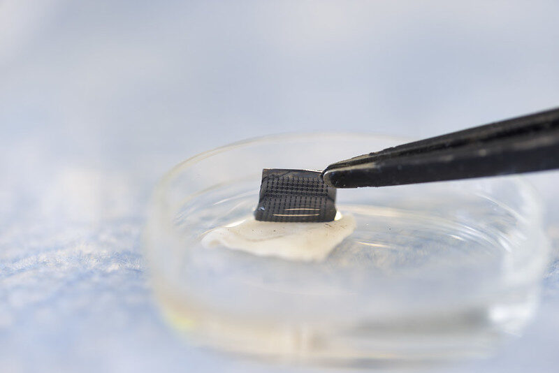
The newly designed ExoPatch being removed from a sample of mouse skin successfully distinguished melanoma from healthy skin in mice. A gel coating the microneedles picks up cancer indicators from the top-most layer of the skin. Dissolving the gel releases exosomes into a solution, which is then used on a two-lined test strip, similar to an at-home COVID-19 test. Image credit: Jeremy Little, Michigan Engineering.

Melanoma testing could one day be done at home with a skin patch and test strip with two lines, similar to COVID-19 home tests, according to University of Michigan researchers. Developed with funding from the National Institutes of Health, the new silicone patch with star-shaped microneedles, called the ExoPatch, distinguished melanoma from healthy skin in mice.

The patch and test move toward rapid at-home melanoma testing, helping patients catch the most aggressive form of skin cancer early without a biopsy or blood draw.

“The star-shaped needles make puncture easier and less painful, but they are so small that they only go through the top-most layer of the skin, the epidermis, and do not draw blood,” said Sunitha Nagrath, the Dwight F. Benton Professor of Chemical Engineering at U-M and co-corresponding author of the study published in Biosensors and Bioelectronics.

The ExoPatch microneedles, at just 0.6mm long with a width of less than 100 nm (0.0001 mm) at the tip, are coated with a gel that picks up exosomes, tiny packages released by cells, from the interstitial fluid that fills the spaces between cells in the epidermis.

Once thought to be trash ejected from cells for cleanup, exosomes actually contain DNA and RNA fragments that cells use to communicate with each other. Cancer cell exosomes can help tumours spread by preparing tissues to accept tumour cells before arrival, and detecting them can catch cancer earlier than past methods.

The gel that coats the ExoPatch contains a protein called Annexin V that attracts and sticks exosomes to the microneedles’ surface. Once removed from the skin, placing the patch in an acid dissolves the gel, which releases the exosomes into a solution. After dipping a test strip into the solution, two lines form if the sample contains melanoma exosomes, and one line forms for a negative test – the same way an at-home COVID-19 test strip works.

“A fair-skinned person with moles must go to the doctor about every six months to send off a biopsy to see if they’re malignant or benign. With this test, they could instead test at home, get the results right away and follow up with a dermatologist for a positive result,” Nagrath said.

As a first step in the proof-of-concept study, the researchers tested the ExoPatch on a tissue sample of pig skin, which closely resembles human skin in thickness and composition. Using a microscope, they found the microneedles penetrated about 350 to 600nm into the skin. For scale, the epidermis on the human forearm is about 18 300nm thick.

To test whether the ExoPatch could capture melanoma exosomes from skin tissue, the research team tested tissue samples of mouse skin, half from healthy mice and half from mice injected with a fragment of a human melanoma tumour. After a 15-minute application, the ExoPatch was placed under a powerful microscope.

“When looking at microscopy images, I was happy to see how nicely the exosomes adhered to the microneedles and were within the 30 to 150 nanometre size range we expect,” said Scott Smith, U-M doctoral student of chemical engineering and co-lead author of the study.

After confirming the exosomes stuck to the ExoPatch, the researchers dissolved the gel and ran the sample through the test strips. The test successfully distinguished between melanoma and healthy tissues with a 3.5-fold darker line in melanoma samples.

The ExoPatch isolated 11.5 times more exosomal protein from melanoma tissue samples compared to healthy tissue, showing it can specifically target cancerous exosomes.

A pilot study in humans followed by a series of clinical trials will be the next steps to move the technology toward use. Beyond melanoma, the ExoPatch gel coating could be modified to detect exosomes released by other cancers with a solid tumour, including lung, breast, colon, prostate and brain cancer.

“This is the first patch designed to capture disease-specific exosomes from fluid under the skin. The potential applications are huge,” said Nagrath.

Source: University of Michigan

Combination of Diet and Medication Reprograms Paediatric Neuroblastoma

Credit: National Cancer Institute

Researchers at Children’s Hospital of Philadelphia (CHOP) found that combining a specialised diet with an approved medication interrupts the growth of high-risk neuroblastoma, a deadly paediatric cancer, by reprogramming tumour behaviour. The findings were published in the journal Nature.

Neuroblastoma originates from primitive cells meant to form nerve tissues but that remain “undifferentiated,” indicating cancer cells that haven’t specialized, often suggesting a more aggressive and unfavourable prognosis. These tumours rely on a steady supply of chemicals called polyamines that are essential for rapid cell growth and tumour progression. A medicine called difluoromethylornithine (DFMO) was approved by the Food and Drug Administration (FDA) to treat children with high-risk neuroblastoma, as DFMO blocks polyamine production. However, researchers sought to improve the effectiveness of the drug by using it at high doses and combining it with a diet that is depleted of the nutrients used by the body to make polyamines (arginine). This two-step approach was anticipated to lower polyamines substantially more than low dose DFMO alone.

“Our findings show that this treatment reduced polyamines in tumours to roughly 10% of their usual levels. This reduction greatly slowed tumour growth, and in many cases, completely eliminated the tumours,” said Michael D. Hogarty, MD, a lead author and an Attending Physician in the Division of Oncology at Children’s Hospital of Philadelphia. “Notably, the treatment altered the way the tumour cells make proteins, making it harder for them to grow and easier for them to mature, or differentiate.”

Hogarty and his team used a preclinical model to mimic MYCN-driven neuroblastoma, directly addressing the strong association between extra MYCN gene copies and aggressive neuroblastoma with poor prognosis. Animal models with tumours were divided into groups: one fed a normal diet and the other lacking amino acids for polyamine production. Each group either received DFMO in their drinking water or did not. The special diet or DFMO alone partially lowered polyamines and extended survival, but the combination had the most significant impact on tumours due to the profound polyamine depletion it caused.

The researchers plan to conduct additional preclinical studies, followed hopefully by clinical trials in children to determine the safety and efficacy of targeting this specific metabolic dependency of neuroblastoma cells. By complementing existing treatments, they hope to substantially improve patient outcomes, and because the therapy targets polyamines it may be effective in many other types of cancer that have frequent MYC gene activation. 

Source: Children’s Hospital of Philadelphia

Study Explains How Lymphoma Rewires Human Genome

Source: Pixabay CC0

Translocations are chromosomal “cut and paste” errors that drive many lymphomas, a type of blood cancer and the sixth most common form of cancer overall. This includes mantle cell lymphoma, a rare but aggressive subtype diagnosed in about one in every 100 000 people each year.

Translocations are known to spark cancer by altering the activity of the genes near the breakpoints where chromosomes snap and rejoin. For example, a translocation can accidentally cut a gene in half, silencing its activity, or create new hybrid proteins that help promote cancer.

A study published today in Nucleic Acids Research shows a new way translocations promote cancer. The translocation most typically found in mantle cell lymphoma drags a powerful regulatory element into a new area of the human genome, where its new position allows it to boost the activity of not just one but 50 genes at once.

The discovery of this genome rewiring mechanism shows the traditional focus on the handful of genes at chromosomal breakpoints is too narrow. The study also greatly expands the list of potential drug targets for mantle cell lymphoma, for which there is no known cure.

“We did not expect to see a single translocation boosting the expression of almost 7% of all genes on a single chromosome. The ripples of disruption are much bigger than expected, and also identify new cancer driver genes, each of which represents a new potential therapeutic target,” says Dr. Renée Beekman, corresponding author of the study and researcher at the Centre for Genomic Regulation (CRG) in Barcelona.

In mantle cell lymphoma, a piece of chromosome 14 swaps places with a piece of chromosome 11. A gene regulatory element called the IGH enhancer, which normally boosts the activity of antibody production in healthy B cells, lands right beside CCND1, a gene which helps cells divide. The enhancer treats CCND1 as if it were a gene encoding for antibodies, boosting its activity and fuelling the disease.

Previous research has shown that boosting CCND1 expression alone is insufficient to kickstart the formation of mantle cell lymphoma. To understand why, the scientists first created translocations in cells in a dish. They used CRISPR to replicate the exact chromosome break seen in patients.

“We built a system to generate translocations in healthy B cells. Because these are engineered cells, we can carry out experiments that are technically or ethically unfeasible with patient tissues, making it a really useful early disease model,” explains Dr. Roser Zaurin, co-author of the study.

The experiments revealed that over fifty genes along the entire chromosome 11 were much more active after the translocation took place. The translocation affected gene activity across 50 million base pairs, a significantly larger space than previously thought.

How DNA folds inside the engineered cells revealed why the translocation affects so many genes at once. “DNA loops inside cells. It’s what brings two segments of DNA that are far away from each other in two-dimensional space closer together in three-dimensional space. The translocation drags the strong IGH enhancer into a preexisting loop, placing it in a privileged position of control, enabling it to have a widespread impact on dozens of genes at the same time,” explains Dr. Anna Oncins, first author of the study.

Intriguingly, most of the genes affected by the enhancer were not silent to begin with. The IGH enhancer simply dials their activity up. This biological nuance may explain why the same translocation can have different consequences in different cell types or stages of development. Only genes which were already active are boosted.

The findings could lead to new strategies for the early-stage detection of mantle cell lymphomas. “Because the enhancer mainly supercharges genes that were already active in the very first B cell that acquires the swap, epigenetic profiling of at-risk cells could spot dangerous combinations before a mantle cell lymphoma appears,” explains Dr. Beekman.

The authors of the study next plan on studying exactly how the newly identified genes contribute to the initiation and progression of lymphoma. Understanding and eventually interrupting the effects of the chromosomal translocation could yield broader, more durable therapies for mantle-cell lymphoma and other types of cancers driven by chromosome swaps.

Source: Centre for Genomic Regulation

Sugary Drinks May Increase Risk of Metastasis in Advanced Colorectal Cancer

Photo by Breakingpic on Pexels

A new study from researchers at The University of Texas MD Anderson Cancer Center shows that the glucose-fructose mix found in sugary drinks directly fuels metastasis in preclinical models of advanced colorectal cancer. The study was published in Nature Metabolism.

A research team led by Jihye Yun, PhD, assistant professor of Genetics, studied how sugary drinks may affect late-stage colorectal cancer. Using laboratory cancer models, they compared the effects of the glucose-fructose mix found in most sugary drinks with those of glucose or fructose alone. Only the sugar mix made cancer cells more mobile, leading to faster spread to the liver – the most common site of colorectal cancer metastasis.

The sugar mix activated an enzyme called sorbitol dehydrogenase (SORD), which boosts glucose metabolism and triggers the cholesterol pathway, ultimately driving metastasis. This is the same pathway targeted by statins, common heart drugs that inhibit cholesterol production. Blocking SORD slowed metastasis, even with the sugar mix present. These findings suggest that targeting SORD could also offer an opportunity to block metastasis.

“Our findings highlight that daily diet matters not only for cancer risk but also for how the disease progresses once it has developed,” Yun said. “While these findings need further investigation, they suggest that reducing sugary drinks, targeting SORD or repurposing statins may benefit patients with colorectal cancer.”

The Yun Laboratory is interested in studying how diet affects the intestine and cancer development, and they have made important discoveries on the impacts of sugary drinks on colorectal cancer.

Sugar has long been indirectly linked to an increase in cancer risk through obesity. However, a previous study by Yun’s lab challenged that view, showing that even moderate intake of sugary drinks directly fuelled tumour growth in early-stage colorectal cancer, independent of obesity. The current study was done to determine how sugary drinks may impact later-stage disease.

While this study needs further clinical investigation, the results suggest that reducing sugary drinks and targeting the SORD enzyme may offer opportunities to reduce colorectal cancer metastasis. Additional studies are warranted to confirm these results outside of preclinical models.

Further, Yun explained it may be worthwhile to consider revisions to current dietary recommendations to reduce sugary drink consumption in this patient population. To meet nutritional needs, many patients with cancer are encouraged to have nutritional supplement drinks and concentrated juices that contain high glucose and fructose content.

Source: The University of Texas MD Anderson Cancer Center

Study Affirms Efficacy of Nicotinamide for Skin Cancer Prevention 

Overall, there was a 14% reduction in skin cancer risk. When nicotinamide was taken after a first skin cancer, the risk reduction rose to 54%.

3D structure of a melanoma cell derived by ion abrasion scanning electron microscopy. Credit: Sriram Subramaniam/ National Cancer Institute

The dietary supplement nicotinamide has been recommended by dermatologists for people with a history of skin cancer since 2015, when a clinical study with 386 participants showed that those who took the vitamin B3 derivative developed fewer new occurrences. 

But data to validate those findings in a larger study group has been lacking because nicotinamide can be purchased over the counter without being entered into patients’ medical records.

In a new study published in JAMA Dermatology, researchers found a way to get that data by analysing records from the Veterans Affairs Corporate Data Warehouse. Nicotinamide is on the VA’s official formulary, so the researchers checked the outcomes of 33 833 patients for their next skin cancer diagnosis following baseline treatment with 500 milligrams of nicotinamide twice daily for longer than 30 days. They looked for occurrences of basal cell carcinoma and cutaneous squamous cell carcinoma. 

The researchers compared 12 287 patients who received the treatment with 21 479 who did not. Overall, there was a 14% reduction in skin cancer risk. When nicotinamide was taken after a first skin cancer, the risk reduction rose to 54%, but the benefit declined with treatment initiation following subsequent skin cancers. The risk reduction was much larger for squamous cell carcinoma.  

“There are no guidelines for when to start treatment with nicotinamide for skin cancer prevention in the general population. These results would really shift our practice from starting it once patients have developed numerous skin cancers to starting it earlier. We still need to do a better job of identifying who will actually benefit, as roughly only half of patients will develop multiple skin cancers,” said the study’s corresponding author, Lee Wheless, MD, PhD, assistant professor of Dermatology and Medicine at Vanderbilt University Medical Center and a staff physician at VA Tennessee Valley Healthcare System. 

The researchers were also able to ascertain the outcomes of 1,334 patients who were immunocompromised due to having received solid organ transplants. Among solid organ transplant recipients, no overall significant risk reduction was observed, although early nicotinamide use was associated with reduced occurrences of cutaneous squamous cell carcinoma. 

Source: Vanderbilt University Medical Center