Author: ModernMedia

Getting Vital Creatine into the Brain is a Weighty Problem

Photo by Jonathan Borba on Unsplash

Creatine is popularly known as a muscle-building supplement, but its influence on human muscle function can be a matter of life or death. But getting it to one particular organ that needs it – the brain – is challenging.

“Creatine is very crucial for energy-consuming cells in skeletal muscle throughout the body, but also in the brain and in the heart,” said Chin-Yi Chen, a research scientist at Virginia Tech’s Fralin Biomedical Research Institute at VTC.

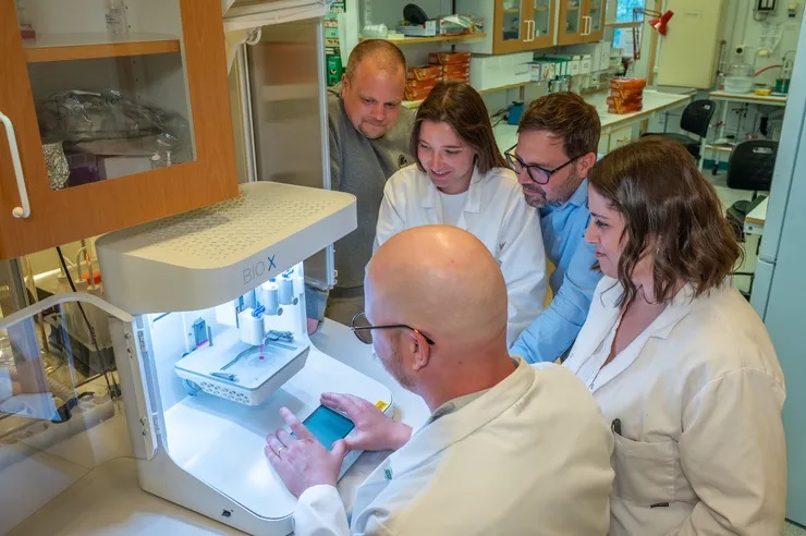
Chen is part of a research team working to develop a technique that uses focused ultrasound to deliver creatine directly to the brain. The work, being conducted in the lab of Fralin Biomedical Research Institute Assistant Professor Cheng-Chia “Fred” Wu, will be supported by a $30 000 grant from the Association for Creatine Deficiencies.

Creatine plays a vital role in the brain, where it interacts with phosphoric acid to help create the key energy molecule adenosine triphosphate (ATP). In addition to its role in energy production, creatine also influences neurotransmitter systems.

For example, creatine influences the brain’s major inhibitory pathways that use the neurotransmitter gamma-aminobutyric acid (GABA), which limits neuronal excitability in the central nervous system. It may play a role in a variety of functions, including seizure control, learning, memory, and brain development.

A growing body of research suggests that creatine may itself function as a neurotransmitter, as it is delivered to neurons from glial cells in the brain and can influence signalling processes between other neurons. While creatine deficiency disorders can weaken the skeletal muscle and the heart, they can also severely affect the brain. Many patients see increased muscle mass and body weight with creatine supplements, but they often continue to face neurodevelopmental challenges that can hinder their ability to speak, read, or write.

This is largely caused by the brain’s protective blood-brain barrier preventing creatine entry.

Wu studies therapeutic focused ultrasound, which precisely directs sound waves to temporarily accessed areas of the brain. The process allows drugs to reach diseased tissue without harming surrounding healthy cells. While Wu is investigating this method as a potential treatment for paediatric brain cancer, he also sees potential in applying it to creatine deficiency.

“Through the partnership between Virginia Tech and Children’s National Hospital, I was able to present our work in focused ultrasound at the Children’s National Research & Innovation Campus,” Wu said. “There, I met Dr Seth Berger, a medical geneticist, who introduced me to creatine transporter deficiency. Together, we saw the promise that focused ultrasound had to offer.”

The Focused Ultrasound Foundation has recognised Virginia Tech and Children’s National as Centers of Excellence. Wu said the two organisations bring together clinical specialists, trial experts, and research scientists who can design experiments that could inform future clinical trials.

“It was a moment that made me really excited – that I had found a lab where I could move from basic research to something that could help patients,” Chen said. “When Fred asked me, ‘Are you interested in this project?’ I said, ‘Yes, of course.’”

Because creatine deficiencies can impair brain development, the early stages of Chen’s project will concentrate on using focused ultrasound to deliver creatine across the blood-brain barrier. Chen hopes the technique will restore normal brain mass in models of creatine deficiency.

Source: Virginia Tech

Chicken and Egg: Which Comes First, Epstein–Barr Virus or Crohn’s Disease?

Irritable bowel syndrome. Credit: Scientific Animations CC4.0

Crohn’s disease, a debilitating inflammatory bowel disease, has many known contributing factors, including bacterial changes in the microbiome that foster an inflammatory environment. Now, for the first time, Crohn’s disease has been tied to a virus – specifically, Epstein-Barr virus (EBV), best known for causing infectious mononucleosis (mono).

Researchers had already observed increased levels of EBV in the intestine of patients with Crohn’s disease and had also found associations between EBV and autoimmune diseases other than Crohn’s, including lupusmultiple sclerosis, and rheumatoid arthritis.

But which came first? EBV or Crohn’s disease?

‘It was a classic chicken and egg problem,” says Anubhab Nandy, PhD, a research fellow in the Division of Gastroenterology, Hepatology, and Nutrition at Boston Children’s Hospital.

A longitudinal study just published in Gastroenterology systematically looked at the role of viruses in Crohn’s and found strong evidence that EBV infection predisposes people to develop Crohn’s.

Tracking EBV and Crohn’s

Nandy and colleagues analyzed data from a cohort of initially healthy military recruits, aged 20 to 24, who provided periodic serum samples throughout their service. They tested the samples with VirScan, a high-throughput assay developed by study coauthor Stephen Elledge, PhD, at Harvard Medical School. VirScan enables detection of antibodies against a wide range of viruses, providing insights into viral exposures.

Compared with healthy controls, military personnel whose samples were positive for anti-EBV antibodies were three times more likely to eventually develop Crohn’s disease. Interestingly, evidence of EBV exposure preceded their Crohn’s diagnosis by five to seven years.

“We went into this study not looking for EBV; we were looking for any virus that might elicit inflammatory bowel disease,” says Scott Snapper, MD, PhD, the study’s senior investigator and director of the Inflammatory Bowel Disease Center at Boston Children’s. “Then, when EBV was a hit, we looked very specifically at immune responses to EBV with more detailed tests.”

These tests bore out and strengthened the VirScan findings.

The team next looked for anti-EBV antibodies in a second cohort – more than 5000 children (median age; 11 years) who were first-degree relatives of people with Crohn’s disease. In this cohort, EBV was not a statistically significant predictor of a subsequent Crohn’s disease diagnosis. Snapper speculates that having first-degree relatives with Crohn’s could already have put them at increased risk due to shared genetic or environmental factors, muddying the association with EBV.

EBV and the immune system

Another possibility is that EBV affects children’s immune systems differently, perhaps because children are less likely to develop infectious mono when exposed. “Responding to certain organisms early in life may boost the immune system in a way that prevents immune-mediated disease,” Snapper says.

Nandy and Snapper now want to figure out what EBV does to people at a molecular level to make them more susceptible to Crohn’s disease.

One possibility is that the virus has certain genes or molecules that interact with human genes involved in susceptibility to autoimmune conditions. Another may relate to an anti-inflammatory protein produced by EBV, remarkably similar to mammalian IL-10. People exposed to that protein may make antibodies against it, preventing their own IL-10 from working and leaving them susceptible to inflammatory disease.

“Mechanistically, we need to understand exactly how EBV alters the immune system leading to Crohn’s disease,” Snapper says. “If you could figure out the mechanisms, you could come up with new therapies.”

Source: Boston Children’s Hospital

Decoding How Immune Cells Communicate in Autoimmune Disease and Cancer

Squamous cancer cell being attacked by cytotoxic T cells. Image by National Cancer Institute on Unsplash

By measuring interactions between cells, the method offers insights into how the human body fights viral infections, how malfunctions can lead to autoimmune diseases and why immunotherapies work for some people but not others.    

A healthy immune system is trained to detect and destroy infections and cancer cells. This defence is based on a complex communication system at cellular level, in which different immune cells each perform a specialised task: recognising infectious agents, alerting other immune cells, and eliminating harmful cells or pathogens. Problems arise when communication between different cell types is disrupted, potentially leading to a variety of diseases.  

For example, cancer cells often develop strategies to specifically disrupt or circumvent the exchange of information in the immune system – this allows them to evade immune surveillance and grow unhindered. “Modern immunotherapies have fundamentally changed the treatment of certain types of cancer by restoring or specifically strengthening communication between immune cells,” explains Prof Simon Haas, one of the leaders of the study.  

Dr Daniel Hübschmann, also head of the study, adds: “However, not all patients respond equally well to these therapies and reliable methods for predicting which patients will benefit most are still lacking.”   

Decoding immune cell communication for personalised cancer therapies    

Scientists have now developed a technology that overcomes many of these hurdles through a better understanding of immune cell communication. With this method, made possible by interdisciplinary collaboration, millions of cell-cell interactions can be measured quickly and cost-effectively, both in research laboratories and in the clinic.   

The scientists are using the newly developed technology to investigate the behaviour and kinetics of immunotherapies and to gain insights into how these therapies work at the level of cell-cell interactions. They were able to show that the approach enables the prediction of individual therapy responses and can thus create a central basis for personalised immunotherapies and targeted therapy decisions.   

In addition, the researchers were able to use their new technology to visualise, in high resolution, how cells of the immune system interact with each other during viral infections and autoimmune diseases. The results allow them to develop dynamic maps of immune cell networks, illustrating for the first time how the immune defence is coordinated in different tissues.   

Together with clinical partners, the team is now working on translating these findings from research into practice, for example to better predict treatment success and utilise immunotherapies in a more personalised manner.   

The study was published in Nature Methods.  

Source: Queen Mary University London

Sanofi Steps up Investment in SA Healthcare with New Leadership and Stronger Local Partnerships

Photo by Sora Shimazaki

Sanofi has reaffirmed its commitment to South African healthcare with the appointment of two senior leaders and a renewed focus on supporting healthcare professionals, expanding local partnerships, and improving access to essential treatments in key areas such as vaccines, immunology, and rare diseases.

“This is a new chapter for Sanofi in South Africa,” says Jean-Baptiste Bregeon, Sanofi South Africa’s Country Lead and Head of Vaccines. “We are focused on making a real impact through practical support for healthcare professionals, expanding access to important medicines and vaccines, and strengthening our ties with partners across the system.”

Focused leadership

Bregeon brings over 20 years of international experience, with senior roles in commercial and clinical operations across the Middle East, North Africa, and Eurasia. As former General Manager for North Africa at Sanofi Vaccines, he led efforts to improve vaccine access and build local capacity. He has also advised Sanofi’s global leadership on operational strategy and commercial planning.

Bregeon will oversee Sanofi’s overall strategy in the country and lead its vaccine division, which remains a core part of the company’s healthcare offering.

Lenisha Maharaj, newly appointed Head of Pharma for South Africa, Namibia and Botswana, is a seasoned Pharmaceutical Executive with over 20 years’ experience in the pharmaceutical and healthcare sectors. At Sanofi, she will oversee growth across the general medicines and specialty care portfolios, supporting both primary and specialist care.

“Sanofi understands the realities that healthcare professionals face,” says Maharaj. “Our goal is to support them with the right tools, training, and access to innovative treatments so they can keep delivering high-quality care.”

Strengthening partnerships and access

A key part of Sanofi’s long-term strategy is to build strong, lasting partnerships. Its collaboration with Biovac, a local vaccine manufacturer, is a core example. This will help to boost domestic vaccine production capacity and reduce reliance on global supply chains. Such public-private partnerships support healthcare resilience as well as local economic and scientific development.

Beyond vaccines, Sanofi is investing in other areas that have a direct impact on healthcare professionals and the patients they serve:

  • Clinical trials in South Africa: Expanding its local clinical research footprint to increase representation of African patients in global trials and ensure that treatment decisions are informed by local data.
  • Access to innovative medicines: Working to bring advanced treatments in rare diseases and immunology to South Africa, along with patient support programmes and diagnostic assistance, to help doctors and patients navigate access challenges.
  • Medical education and digital engagement: Offering CPD-accredited education, clinical webinars, and digital tools to help healthcare professionals stay updated on the latest treatment protocols and deliver more streamlined care.
  • Policy engagement and system support: Actively involved in discussions with policymakers and funders to help shape health policies that improve medicine access and promote fair, sustainable healthcare.

Supporting healthcare professionals

Sanofi says it recognises the ongoing pressures facing South African healthcare workers, from dealing with supply shortages and reimbursement hurdles to managing the growing burden of chronic and complex diseases.

“We see our role as being part of the solution,” says Bregeon. “We are here to support healthcare professionals, not only by ensuring a reliable supply of medicines, but by providing the training, tools, and partnerships they need to do their jobs effectively.”

The company is also prioritising engagement with healthcare professionals through regular updates, field team support, and opportunities for joint problem-solving around system-level challenges.

Sanofi says it is committed to staying in close conversation with the healthcare community and building a more resilient, inclusive, and patient-focused system.

“We want to work with doctors, nurses, pharmacists, policymakers and funders to build something long-term,” Bregeon adds. “We believe that by working together, we can improve outcomes for patients and strengthen the foundation of healthcare in this country.”

Pembrolizumab–Axitinib Duo Extends Survival in Advanced Kidney Cancer

Photo by Robina Weermeijer on Unsplash

A two-drug combination for treating advanced kidney cancer had sustained and durable clinical benefit in more than five years of follow-up, according to a study published August 1 in Nature Medicine

The study reports final clinical data and biomarker analyses from the Phase 3 KEYNOTE-426 trial, which compared the drug combination pembrolizumab plus axitinib versus the single drug sunitinib for patients with previously untreated advanced clear cell renal cell carcinoma, the most common type of kidney cancer.

“KEYNOTE-426 was the first trial to combine a PD-1 inhibitor immunotherapy (pembrolizumab) with a VEGF receptor inhibitor antiangiogenic drug (axitinib) in the first-line setting for advanced renal cell carcinoma. It therefore has the longest follow-up duration among the various trials comparing these types of drug combinations,” said Brian Rini, MD, a medical oncologist at Vanderbilt-Ingram Cancer Center, Professor of Medicine and the study’s lead and corresponding author. 

Immunotherapy drugs like pembrolizumab stimulate the immune system to kill tumour cells. VEGF receptor inhibitors like axitinib and sunitinib block angiogenesis — the development of blood vessels that tumours need to grow and spread. Pembrolizumab plus axitinib and other immunotherapy-antiangiogenic drug combinations are now standard first-line treatments for advanced kidney cancer. 

“Before the development of antiangiogenic drugs and immunotherapies, advanced renal cell carcinoma had a very poor prognosis. These drug combinations have dramatically improved treatment options and outcomes for patients,” said Rini. 

The first interim analysis of outcomes from KEYNOTE-426, published Feb. 16, 2019, in the New England Journal of Medicine, demonstrated that trial participants treated with pembrolizumab plus axitinib had longer overall and progression-free survival, and higher objective response rates compared to those taking sunitinib. The median follow-up was 12.8 months. 

Now, with a median follow-up of 67.2 months, the current analysis confirms and extends the interim analysis and provides valuable information about biomarkers that could help guide treatment decisions. 

The study in Nature Medicine reports that pembrolizumab plus axitinib had longer overall survival (47.2 months versus 40.8 months for sunitinib) and longer progression-free survival (15.7 months versus 11.1 months for sunitinib). The objective response rate was 60.6% for pembrolizumab plus axitinib and 39.6% for sunitinib. 

The researchers reported a variety of associations between the expression of biomarkers and outcomes (overall survival, progression-free survival, objective response rate). The biomarkers they evaluated included an 18-gene T-cell-inflamed expression profile, angiogenesis signature, and PD-1 ligand expression. 

“There is an unmet need for biomarkers that are predictive of patient outcomes following treatment with available first-line therapies for advanced renal cell carcinoma,” Rini said. “Although our analysis showed potential clinical utility of some RNA signatures in identifying patients who are likely to benefit the most from each treatment, further prospective clinical studies are needed.” 

Pembrolizumab plus axitinib is a first-line treatment option for patients with advanced renal cell carcinoma regardless of biomarker subtypes, he noted. 

Source: Vanderbilt University Medical Center

Bubonic Plague Treatment Proven Highly Effective and Safe in Global First

Scanning electron micrograph of Yersinia pestis, which causes bubonic plague, on proventricular spines of a Xenopsylla cheopis flea. Credit: NIAID

Researchers from the UK and Madagascar, in collaboration with Madagascar’s health services and national plague programme, have conducted the world’s first rigorous clinical trial of treatments for bubonic plague.

The IMASOY trial provides the first robust evidence of the efficacy and safety of two treatment regimens. They also found that a ten-day course of an oral antibiotic (ciprofloxacin) is a highly effective and safe alternative to existing treatment with injectable antibiotic requiring hospitalisation.

Plague has scourged humanity for millennia. Though cases have been declining, it remains a high-threat pathogen of pandemic potential, due to its widespread animal reservoir and potential for being weaponised via deliberate release. Most continents see sporadic cases of bubonic plague every year, with Madagascar reporting approximately 80% of global cases. Bubonic plague is a life-threatening disease with mortality ranging from 15-25%.

The results from the trial, published in the New England Journal of Medicine, are the outcome of a hugely ambitious clinical trial conducted over the last five years in rural Madagascar.

With many cases occurring in remote villages and with outbreaks being unpredictable, the team deployed dozens of research assistants, and trained over 230 local doctors and nurses and 1300 village health workers. The trial was embedded within Madagascar’s national health service and was conducted with the support of the Ministry of Public Health.

Several treatment options are included in the current international and national guidelines, but none of them have been rigorously tested in humans. Regulatory approval has been based solely on data from animal studies and human safety data.

Researchers designed the IMASOY trial to test two treatments for plague. Patients were randomly allocated to receive either a ten-day oral regimen of ciprofloxacin (an antibiotic tablet which can be taken at home), or a regimen requiring three days’ injectable gentamicin followed by seven days’ ciprofloxacin, (with the intravenous (IV) therapy requiring patients to be hospitalised).

450 patients with clinically suspected bubonic plague were enrolled between 2020-2024 at 47 sites in 11 districts in Madagascar and treated with either regimen. Of these, 222 plague infections were confirmed by laboratory testing.

Key findings:
•    Both regimens were found to be highly effective and safe, with overall treatment success rates of about 90% and an overall mortality rate of about 4%.
•    A ten-day ciprofloxacin regimen has many advantages over a regimen requiring three days of injectable antibiotics. It alleviates bed space (particularly in health centres with minimal bed availability for the surrounding villages), reduces healthcare worker workload, and is much cheaper – costing about one-tenth of the current treatment regimen, depending on the route of administration.

With funding from Wellcome and the UK Foreign, Commonwealth, and Development Office, researchers from the University of Oxford, Institut Pasteur de Madagascar, Centre Hospitalier Universitaire Joseph Raseta Befelatanana, Centre d’Infectiologie Charles Mérieux, London School of Hygiene and Tropical Medicine, along with Madagascar’s health services and national plague programme, collaborated through the International Severe Acute Respiratory and emerging Infection Consortium (ISARIC) to fill this gap in knowledge.

Piero Olliaro, Professor at the Pandemic Science Institute, University of Oxford and senior author of the study said: ‘Despite its deadly history, we’ve had little clinical evidence on treating bubonic plague – until now. Thanks to the patients and healthcare workers in the trial, we now have real-world proof of effective, safe treatment. Ongoing data analysis will deepen our understanding of the disease, including risk factors, symptoms, and diagnostics.’

Mihaja Raberahona, physician at CHU Joseph Raseta Befelatanana and researcher at the Centre d’Infectiologie Charles Mérieux Madagascar said: ‘In Madagascar, where plague cases occur in remote rural locations with limited healthcare infrastructure, taking a straightforward oral antibiotic is vastly preferable to a treatment requiring injections. It alleviates healthcare worker workload and is also much cheaper – costing about one-tenth of the current treatment regimen.’

Rindra Vatosoa Randremanana, Medical epidemiologist at Institut Pasteur de Madagascar said: ‘The trial was a massive operational undertaking. Many patients were in remote villages and the unpredictable nature of the outbreaks made the research very challenging. Recruiting the 450 patients, of whom about half were confirmed with bubonic plague, took five plague transmission seasons and an army of research assistants, doctors and community health workers. We are hugely grateful to everyone who took part.’

The impact of this study is far-reaching. As a result of the trial, the researchers will be working with national and international bodies like the World Health Organization (WHO) to provide the evidence that may be required to update clinical guidelines, thereby translating the results into practice and saving lives.

The full paper, ‘Ciprofloxacin versus Aminoglycoside-Ciprofloxacin for Bubonic Plague‘, is published in the New England Journal of Medicine.

Source: University of Oxford

When Should Preventive Mastectomy be Offered in High Breast Cancer Risk?

Photo by National Cancer Institute on Unsplash

More women at higher risk of breast cancer should be offered a mastectomy, according to researchers at Queen Mary and London School of Hygiene and Tropical Medicine.

A new analysis, published in JAMA Oncology, has found that the surgical technique was a cost-effective way of reducing the likelihood of developing breast cancer compared to breast screening and medication. Current guidelines on who is offered mastectomy may need to be revised to reflect these new findings. 

Clinicians currently use personalised risk prediction models which combine genetic and other data to identify those women who are at a higher risk of developing breast cancer (BC). Subsequent treatment options – including mammograms, MRI screening, surgery, and medication – are then offered dependent on each woman’s level of risk. 

Risk reducing mastectomy (RRM) is recommended for women at high risk, but in practice this surgery is only clinically offered to those carrying faults (called pathogenic variants) in genes that are known to increase the likelihood they will develop the disease (BRCA1/ BRCA2/ PALB2 PV). 

Professor Ranjit Manchanda from Queen Mary University of London, Dr Rosa Legood from London School of Hygiene and Tropical Medicine, along with colleagues from Manchester University and Peking University created a new economic evaluation model to accurately predict the level of risk that would make RRM a more cost-effective treatment. 

For their model, researchers used guidelines from the National Institute for Health and Care Excellence (NICE) to determine whether a treatment is considered cost-effective. Their model showed that mastectomy was a cost-effective treatment for women aged 30 or above who have a lifetime breast cancer risk greater than or equal to 35%. Offering RRM to women in this cohort could potentially prevent 6,500 of the 58,500 cases of breast cancer that are diagnosed every year in the UK. 

Professor Manchanda, Professor of Gynaecological Oncology at Queen Mary and  Consultant Gynaecological Oncologist, said: “We for the first time define the risk at which we should offer RRM. Our results could have significant clinical implications to expand access to mastectomy beyond those patients with known genetic susceptibility in high penetrance genes- BRCA1/ BRCA2/ PALB2 – who are traditionally offered this. This could potentially prevent can potentially prevent ~6500 breast cancer cases annually in UK women. We recommend that more research is carried out to evaluate the acceptability, uptake, and long-term outcomes of RRM among this group”.. 

Dr Legood, Associate Professor in health economics at the London School of Hygiene & Tropical Medicine, said: “Undergoing RRM is cost-effective for women 30-55 years with a lifetime breast cancer risk of 35% or more. These results can support additional management options for personalized breast cancer risk prediction enabling more women at increased risk to access prevention.”  

Dr Vineeth Rajkumar, Head of Research at Rosetrees, said: “Rosetrees is delighted to fund this truly groundbreaking research that could have a positive impact on women worldwide.” 

The researchers used data from women aged between 30 and 60 with varying lifetime breast cancer risks between 17% and 50%, and who were either undergoing RRM or receiving screening with medical prevention according to currently used predictive models. 

NICE deems a treatment cost-effective if it typically brings one additional year of health for no more than £20 000–£30 000 per patient (known as the ‘willingness to pay’ threshold, or WTP). The researchers’ model used a threshold of £30 000/Quality Adjusted Life Year. 

Source: Queen Mary University London

RSV Infection Significantly Affects Cardiac Risks of Older Adults

Photo by Anna Shvets

Respiratory syncytial virus (RSV) is a well-known cause of infections in children, but it’s understudied in older individuals. In a retrospective study published in the Journal of the American Geriatrics Society, adults aged 65 and older hospitalised for RSV in Ontario, Canada experienced significantly higher rates of adverse outcomes such as longer length of hospital stay, transfer to intensive care, and 30-day mortality, compared with patients hospitalised with influenza, urinary tract infection, or fracture.

Interestingly, RSV hospitalisation was also associated with higher rates of heart failure and atrial fibrillation up to 1-year post-discharge, regardless of prior cardiovascular conditions.

“Unlike other respiratory viruses, immunity against RSV tends to decline relatively quickly. This means that a previous infection will not afford the same long-term protection as it might for influenza or COVID-19,” said corresponding author Chris Verschoor, PhD, HSN, Foundation Research Chair in Healthy Aging at Health Sciences North Research Institute. “Our findings reinforce the importance of RSV vaccination in older adults and suggest that monitoring for signs of heart disease following an RSV illness may be pragmatic.”

Source: Wiley

Does Metformin Possibly Help Prevent Dementia?

Created with Gencraft. CC4.0

New research in Diabetes, Obesity and Metabolism reveals that metformin, a medication traditionally prescribed to treat diabetes, is linked to lower risks of dementia and early death.

In the study by investigators at Taipei Medical University that included 452,777 adults with varying degrees of overweight and obesity, 35,784 cases of dementia and 76,048 deaths occurred over 10 years. Metformin users exhibited significantly lower risks of both dementia and all-cause death than nonusers.

The benefits of metformin were seen across all categories of overweight, obesity, and severe obesity, with 8–12% lower risks of dementia and 26–28% lower risks of death.

“Although our study results are promising for metformin’s effects on dementia and mortality, further research is required to explore the mechanisms involved,” said co-corresponding author Chiehfeng Chen, MD, PhD, MPH.

Source: Wiley

“Skin in a Syringe” a Step Towards a New Way to Heal Burns

Researchers in fields such as regenerative medicine and materials science have collaborated to develop a gel containing living cells that can be 3D-printed into a transplant. Photographer: Magnus Johansson

Finding a way to replicate the skin’s complicated dermis layer has long been a goal of healing burn wounds, as it would greatly reduce scarring and restore functionality. Researchers at Linköping University have developed a gel containing living cells that can be 3D-printed onto a transplant, which then sticks to the wound and creates a scaffold for the dermis to grow.

Large burns are often treated by transplanting a thin layer of the top part of the skin, the epidermis, which is basically composed of a single cell type. Transplanting only this part of the skin leads to severe scarring.

“Skin in a syringe”

Beneath the epidermis is the dermis, which has the blood vessels, nerves, hair follicles and other structures necessary for skin function and elasticity. However, transplanting also the dermis is rarely an option, as the procedure leaves a wound as large as the wound to be healed. The trick is to create new skin that does not become scar tissue but a functioning dermis.

“The dermis is so complicated that we can’t grow it in a lab. We don’t even know what all its components are. That’s why we, and many others, think that we could possibly transplant the building blocks and then let the body make the dermis itself,” says Johan Junker, researcher at the Swedish Center for Disaster Medicine and Traumatology and docent in plastic surgery at Linköping University, who led the study published in Advanced Healthcare Materials.

The most common cell type in the dermis, the connective tissue cell or fibroblast, is easy to remove from the body and grow in a lab. The connective tissue cell also has the advantage of being able to develop into more specialised cell types depending on what is needed. The researchers behind the study provide a scaffold by having the cells grow on tiny, porous beads of gelatine, a substance similar to skin collagen. But a liquid containing these beads poured on a wound will not stay there.

The researchers’ solution to the problem is mixing the gelatine beads with a gel consisting of another body-specific substance, hyaluronic acid. When the beads and gel are mixed, they are connected using what is known as click chemistry. The result is a gel that, somewhat simplified, can be called skin in a syringe.

“The gel has a special feature that means that it becomes liquid when exposed to light pressure. You can use a syringe to apply it to a wound, for example, and once applied it becomes gel-like again. This also makes it possible to 3D print the gel with the cells in it,” says Daniel Aili, professor of molecular physics at Linköping University, who led the study together with Johan Junker.

3D-printed transplant

In the current study, the researchers 3D-printed small pucks that were placed under the skin of mice. The results point to the potential of this technology to be used to grow the patient’s own cells from a minimal skin biopsy, which are then 3D-printed into a graft and applied to the wound.

“We see that the cells survive and it’s clear that they produce different substances that are needed to create new dermis. In addition, blood vessels are formed in the grafts, which is important for the tissue to survive in the body. We find this material very promising,” says Johan Junker.

Blood vessels are key to a variety of applications for engineered tissue-like materials. Scientists can grow cells in three-dimensional materials that can be used to build organoids. But there is a bottleneck as concerns these tissue models; they lack blood vessels to transport oxygen and nutrients to the cells. This means that there is a limit to how large the structures can get before the cells at the centre die from oxygen and nutrient deficiency.

Step towards labgrown blood vessels

The LiU researchers may be one step closer to solving the problem of blood vessel supply. In another article, also published in Advanced Healthcare Materials, the researchers describe a method for making threads from materials consisting of 98 per cent water, known as hydrogels.

“The hydrogel threads become quite elastic, so we can tie knots on them. We also show that they can be formed into mini-tubes, which we can pump fluid through or have blood vessel cells grow in,” says Daniel Aili.

The mini-tubes, or the perfusable channels as the researchers also call them, open up new possibilities for the development of blood vessels for eg, organoids.

Source: Linköping University