As many as half of nursing home residents are cognitively impaired and may be unable to communicate symptoms such as pain or anxiety to the staff and clinicians caring for them. Therefore, information needed for the evaluation of symptoms and subsequent treatment decisions typically does not reliably exist in nursing home electronic health records (EHRs).
A new paper published in the International Journal of Geriatric Psychiatry reports on the novel adaptation of a commonly used symptom assessment instrument to more comprehensively acquire this difficult-to-obtain data with the ultimate goal of enabling knowledge-based expansion of palliative care services in nursing homes to address residents’ symptoms.
In the paper, part of the large, multi-state, multi-facility Utilizing Palliative Leaders in Facilities to Transform care for people with Alzheimer’s Disease (UPLIFT-AD) study researchers, including Regenstrief Institute, the Indiana University School of Medicine and the University of Maryland School of Social Work faculty, describe how they revamped and subsequently validated a symptom assessment tool used worldwide. The UPLIFT-AD researchers modified the instrument, originally designed for reporting by family members of individuals with dementia following their death, to enable reporting on the symptoms of current residents living with moderate to severe dementia by nursing home staff as well as family.
Led by Kathleen T. Unroe, MD, MHA, and John G. Cagle, PhD, the UPLIFT-AD team reports in the peer-reviewed paper that the tool they enhanced reliably addressed physical and emotional distress as well as well-being and symptoms that are precursors to end of life. This validation was critical as the researchers develop guidance for expansion of symptom recognition and management in any nursing home. Employing instruments used in other studies helps researchers to directly compare findings.
Dr. Unroe, Dr. Cagle and colleagues, including Wanzhu Tu, PhD, of the Regenstrief Institute and the IU School of Medicine, are in the late stages of the UPLIFT-AD clinical trial to enhance quality of care individuals with dementia by building capacity for palliative care within nursing homes.
“People receive care in nursing homes because they have significant needs – support for activities of daily living – as well as for complex, serious and multiple chronic conditions. But measuring symptoms of residents, especially those who are cognitively impaired, to address these needs is challenging,” said paper senior author Dr. Unroe, a Regenstrief Institute research scientist and an IU School of Medicine professor of medicine. “In my two decades of working as a clinician in nursing homes as well as a researcher, I have seen that often the information on symptoms that we want isn’t available consistently in the data that’s already collected or it isn’t collected at the frequency that we need to measure the impact of programs and approaches. And the gold standard for knowing if someone has a symptom, for example, if someone has pain or anxiety, to ask that person directly to assess the symptom, isn’t always possible for cognitively impaired residents. That’s why we took steps to validate a commonly used instrument in a wider population – individuals currently living with cognitive impairment – and added additional needed data points.
“While hospice care is typically available, there is widespread recognition that broader palliative care is needed in nursing homes. But there is no roadmap for how to provide it well. We hope that when we have our final results in 2026, UPLIFT-AD will prove to be a replicable model for implementing this much needed type of care.”
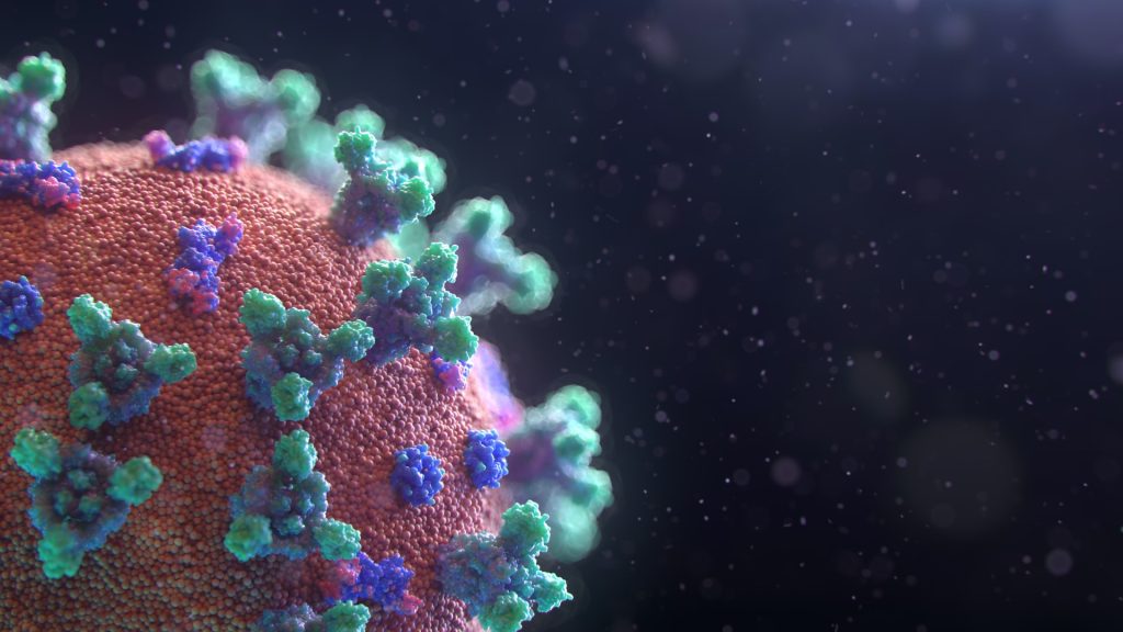
SARS-COV-2 has been very good at mutating to keep infecting people – so good that most antibody treatments developed during the pandemic are no longer effective. Now a team led by Stanford University researchers may have found a way to pin down the constantly evolving virus and develop longer-lasting treatments.
The researchers discovered a method to use two antibodies, one to serve as a type of anchor by attaching to an area of the virus that does not change very much and another to inhibit the virus’s ability to infect cells. This pairing of antibodies was shown to be effective against the initial SARS-CoV-2 virus that caused the pandemic and all its variants through omicron in laboratory testing. The findings are detailed in the journal Science Translational Medicine.
“In the face of an ever-changing virus, we engineered a new generation of therapeutics that have the ability to be resistant to viral evolution, which could be useful many years down the road for the treatment of people infected with SARS-CoV-2,” said Christopher O. Barnes, the study’s senior author, an assistant professor of biology.
An overlooked option
The team led by Barnes and first author Adonis Rubio, a doctoral candidate in the Stanford School of Medicine, conducted this investigation using donated antibodies from patients who had recovered from COVID-19. Analysing how these antibodies interacted with the virus, they found one that attaches to a region of the virus that does not mutate often.
This area, within the Spike N-terminal domain, or NTD, had been overlooked because it was not directly useful for treatment. However, when a specific antibody attaches to this area, it remains stuck to the virus. This is useful when designing new therapies that enable another type of antibody to get a foothold and attach to the receptor-binding domain, or RBD, of the virus, essentially blocking the virus from binding to receptors in human cells.
An illustration of the bispecific antibodies the Stanford-led research team developed to neutralise the virus that causes COVID-19. Named “CoV2-biRN,” these two antibodies work together by attaching to different areas of the virus.The bispecific antibodies target two areas of the virus: One attaches to the “NTD,” or Spike N-terminal domain, an area on the virus that does not change very much. This allows the second antibody to attach to the “RBD,” or receptor-binding domain, essentially preventing the virus from infecting human cells. | Christopher O. Barnes and Adonis Rubio using Biorender stock images
The researchers designed a series of these dual or “bispecific” antibodies, called CoV2-biRN, and in laboratory tests they showed high neutralisation of all the variants of SARS-CoV-2 known to cause illness in humans. The antibodies also significantly reduced the viral load in the lungs of mice exposed to one version of the omicron variant.
More research, including clinical trials, would have to be done before this discovery could be used as a treatment in human patients, but the approach is promising – and not just for the virus that causes COVID-19.
Next, the researchers will work to design bispecific antibodies that would be effective against all coronaviruses, the virus family including the ones that cause the common cold, MERS, and COVID-19. This approach could potentially also be effective against influenza and HIV, the authors said.
“Viruses constantly evolve to maintain the ability to infect the population,” Barnes said. “To counter this, the antibodies we develop must continuously evolve as well to remain effective.”
In a decades-long study following twins, researchers from the University of Jyväskylä, Finland, investigated the links between long-term leisure-time physical activity and mortality. They also sought to determine whether physical activity can mitigate the increased risk of mortality due to genetic predisposition to diseases. Moreover, they examined the relationship between physical activity and later biological aging.
The study included 22 750 Finnish twins born before 1958 whose leisure-time physical activity was assessed in 1975, 1981 and 1990. Mortality follow-up continued until the end of 2020.
Moderate activity yields maximum longevity benefits
Four distinct sub-groups were identified from the data, which was based on leisure-time physical activity over the 15-year follow-up: sedentary, moderately active, active and highly active groups. When the differences in mortality between the groups were examined at the 30-year follow-up, it was found that the greatest benefit – a 7% lower risk of mortality – was achieved between the sedentary and moderately active groups. A higher level of physical activity brought no additional benefit.
When mortality was examined separately in the short and long term, a clear association was found in the short-term: the higher the level of physical activity, the lower the mortality risk. In the long term, however, those who were highly active did not differ from those who were sedentary in terms of mortality.
“An underlying pre-disease state can limit physical activity and ultimately lead to death, not the lack of exercise itself.”
“This can bias the association between physical activity and mortality in the short term”, says Associate Professor Elina Sillanpää from the Faculty of Sports and Health Sciences.
Meeting physical activity guidelines does not guarantee a lower mortality risk
The researchers also investigated whether following the World Health Organization’s physical activity guidelines affects mortality and genetic disease risk. The guidelines suggest 150 to 300 minutes of moderate or 75 to 150 minutes of vigorous activity weekly. The study found that meeting these guidelines did not lower mortality risk or alter genetic disease risk. Even for twins who met the recommended levels of PA over a 15-year period, no statistically significant difference in mortality rates was found compared to their less active twin pair.
“The widely observed favorable association between physical activity and mortality are based on observational studies that are prone to bias from different sources.”
“In our studies, we aimed to account for various sources of biases, and combined with the long follow-up period, we could not confirm that adhering to physical activity guidelines mitigates genetic cardiovascular disease risk or causally reduces mortality”, says postdoctoral researcher Laura Joensuu from the Faculty of Sports and Health Sciences.
Link between physical activity and biological aging is U-shaped
For the subsample of twins, biological aging was determined from blood samples using epigenetic clocks. Epigenetic clocks allow a person’s biological aging rate to be estimated based on methyl groups that regulate gene expression and are linked to aging process.
“We found that the association between leisure-time physical activity and biological aging was U-shaped: Biological aging was accelerated in those who exercised the least and the most,” says Sillanpää.
Other lifestyles, such as smoking and alcohol consumption, largely explained the favourable associations of physical activity with biological aging.
Genetic data were available for 4897 twins. The genetic susceptibility of twins to coronary artery disease, as well as systolic and diastolic blood pressure was assessed using new polygenic risk scores, which sum the genome-wide susceptibility to morbidity. In addition, all-cause and cardiovascular mortality was followed in 180 identical twin pairs. The biological aging rate of 1153 twins was assessed from a blood sample.
Photo by Miguel Á. Padriñán: https://www.pexels.com/photo/syringe-and-pills-on-blue-background-3936368/
By Ufrieda Ho
Amid major disruptions caused by aid cuts from the United States government, the health department aims to enrol a record number – an additional 1.1 million – of people living with HIV on life-saving antiretroviral medicine this year. Experts tell Spotlight it can’t be business as usual if this ambitious programme is to have a chance of succeeding.
Government’s new “Close the Gap” campaign launched at the end of February has set a bold target of putting an additional 1.1 million people living with HIV on antiretroviral treatment by the end of 2025.
Around 7.8 million people are living with HIV in the country and of these, 5.9 million are on treatment, according to the National Department of Health. The target is therefore to have a total of seven million people on treatment by the end of the year. Specific targets have also been set for each of the nine provinces.
The initiative is aimed at meeting the UNAIDS 95–95–95 HIV testing, treatment and viral suppression targets that have been endorsed in South Africa’s National Strategic Plan for HIV, TB, and STIs 2023 – 2028. The targets are that by 2030, 95% of people living with HIV should know their HIV status, 95% of people who know their status should be on treatment, and 95% of people on treatment should be virally suppressed (meaning there is so little HIV in their bodily fluids that they are non-infectious).
Currently, South Africa stands at 96–79–94 against these targets, according to the South African National Aids Council (SANAC). This indicates that the biggest gap in the country’s HIV response lies with those who have tested positive but are not on treatment – the second 95 target.
But adding 1.1 million people to South Africa’s HIV treatment programme in just ten months would be unprecedented. The highest number of people who started antiretroviral treatment in a year was the roughly 730 000 in 2011. In each of the last five years, the number has been under 300 000, according to figures from Thembisa, the leading mathematical model of HIV in South Africa. According to our calculations, if South Africa successfully adds 1.1 million people to the HIV treatment programme by the end of 2025, the score on the second target would rise to just above 90%.
The record for the most people starting antiretroviral treatment in a single year was approximately 730 000 in 2011. (Graph by Spotlight, based on Tembisa data.)
The ambitious new campaign launches at a moment of crisis in South Africa’s HIV response. Abrupt funding cuts from the United States government – the PEPFAR funding – has meant that the work of several service-delivery NGOs have ground to a halt in recent weeks.
These NGOs played an important role in getting people tested and in helping find people and supporting them to start and restart treatment. The focus of many of these NGOs was on people in marginalised but high-risk groups, including sex workers, people who use drugs and those in the LGBTQI community. As yet, government has not presented a clear plan for how these specialised services might continue.
“We will need bridging finance for many of these NGOs to contain and preserve the essential work that they were doing till we can confer these roles and responsibilities to others,” says Professor Francois Venter, of the Ezintsha Research Centre at the University of the Witwatersrand.
He says good investment in targeted funding for NGOs is a necessary buffer to minimise “risks to the entire South African HIV programme” and the looming consequences of rising numbers of new HIV cases, more hospitalisations, and inevitably deaths.
Disengaging from care
South Africa’s underperformance on the second 95 target is partly due to people stopping their treatment. The reasons for such disengagement from HIV care can be complex. Research has shown it is linked to factors like frequent relocations, which means people have to restart treatment at different clinics over and over. They also have to navigate an inflexible healthcare system. A systematic review identified factors including mental health challenges, lack of family or social support, long waiting times at clinics, work commitments, and transportation costs.
Venter adds that while people are disengaged from care, they are likely transmitting the virus. The addition of new infections for an already pressured HIV response contributes to South Africa’s sluggish creep forward in meeting the UNAIDS targets.
The health department has not been strong on locating people who have been “lost” to care, says Venter. This role was largely carried out by PEPFAR-supported NGOs that are now unable to continue their work due to the withdrawal of crucial US foreign aid.
Inexpensive interventions
Other experts working in the HIV sector, say the success of the Close the Gap campaign will come down to scrapping programmes and approaches that have not yielded success, using resources more efficiently, strategic investment, and introducing creative interventions to meet the service delivery demands of HIV patients.
Key among these interventions, is to improve levels of professionalism in clinics so patients can trust the clinics enough to restart treatment.
Professor Graeme Meintjes of the Department of Medicine at the University of Cape Town says issues like improving staff attitudes and updating public messaging and communications are inexpensive interventions that can boost “welcome back” programmes.
“The Close the Gap campaign must utilise media platforms and social media platforms to send out a clear message, so people know the risks of disengagement and the importance of returning to care. The longer someone interrupts their treatment and the more times this happens, the more they are at risk of opportunistic infections, severe complications, getting very sick and needing costly hospitalisations,” he says.
Clinics need to provide friendly, professional services that encourage people to return to and stay on treatment, Meintjes says, and services need to be flexible. These could include more external medicine pick-up points, scripts filled for longer periods, later clinic operating hours, and mobile clinic services.
“We need to make services as flexible as possible. People can’t be scolded for missing an appointment – life happens. Putting these interventions in place are not particularly costly, in fact it is good clinical practice and make sense in terms of health economics by avoiding hospitalisations that result from prolonged treatment interruptions,” he says.
The Close the Gap campaign, Meintjes adds, should reassure people that HIV treatment has advanced substantially over the decades. The drugs work well and now have far fewer side effects, with less risk of developing resistance. More patients are stable on the treatment for longer and most adults manage their single tablet once-a-day regime easily.
Insights from our experiences
Professor Linda-Gail Bekker, Chief Executive Officer at the Desmond Tutu Health Foundation, says to get closer to the target of 1.1 million people on treatment by year-end will mean using resources better.
“Additional funding is always welcome, so are new campaigns that catalyse and energise. But we also need to stop doing the things we know don’t have good returns. For instance, testing populations of people who have been tested multiple times and aren’t showing evidence of new infections occurring in those populations,” she says.
There is also a need for better data collection and more strategic use of data, Bekker says. Additionally, she suggests a status-neutral approach, meaning that if someone tests positive, they are referred for treatment, while those who test negative are directed to effective prevention programmes, including access to pre-exposure prophylaxis (PrEP) for people at high risk of exposure through sex or injection drug use.
But Bekker adds: “We need to be absolutely clear; these people aren’t going to come to us in our health facilities, or we would have found them already. We have to do the work that many of the PEPFAR-funded NGOs were doing and that is going to the last mile to find the last patient and to bring them to care.”
She says the impact of the PEPFAR funding cuts can therefore not be downplayed. “The job is going to get harder with fewer resources that were specifically directed at solving this problem.”
Venter names another approach that has not worked. This, he says, is the persistence of treating HIV within an integrated health system. Overburdened clinics have simply not coped, he adds, with being able to fulfil the ideal of a “one-stop-shop” model of healthcare.
Citing an example, he says: “Someone might come into a clinic with a stomach ache and be vomiting, they might be treated for that but there’s no investigation or follow-up to find out if it might be HIV-related, for instance. And once that person is out of the door, they’re gone.”
Campaign specifics still lacking
The Department of Health did not answer Spotlight’s questions about funding for the Close the Gap campaign; what specific projects in the campaign will look like; or how clinics and clinic staff will be equipped or supported in order to find the 1.1 million people. There is also scant details of the specifics of the campaign online.
Speaking to the public broadcaster after the 25 February campaign launch, Health Minister Dr Aaron Motsoaledi said South Africa is still seeing 150 000 new infections every year. He said they will reach their 1.1 million target through a province-by-province approach. He used the Eastern Cape as an example.
“When you look at the 1.1 million, it can be scary – it’s quite big. But if you go to the provinces – the Eastern Cape needs to look for 140 000 people. Then you come to their seven districts, that number becomes much less. So, one clinic could be looking for just three people,” he said.
Nelson Dlamini, SANAC’s communications manager, says the focus will be to bring into care 650 000 men, as men are known to have poor health-seeking habits. Added to this will be a focus on adolescents and children who are living with HIV.
He says funding for the Close the Gap campaign will not be shouldered by the health department alone.
“This is a multisectoral campaign. Other departments have a role to play, these include social development, basic education, higher education and training, etc, and civil society themselves,” Dlamini says.
The province-by-province approach to reach the target of finding 1.1 million additional people is guided by new data sources.
“Last year, SANAC launched the SANAC Situation Room, a data hub which pulls data from multiple sources in order for us to have the most accurate picture on the status of the epidemic,” says Dlamini.
These include the Thembisa and Naomi model outputs and data from the District Health Information System and Human Sciences Research Council, he says adding that SANAC is working to secure data sharing agreements with other sectors too.
Dlamini however says the health department, rather than SANAC, will provide progress reports on the 10-month project.
A brain rhythm working in tandem with the body’s natural sleep-wake cycle may explain why bipolar patients alternate between mania and depression, according to new research.
The McGill University-led study published in Science Advances marks a breakthrough in understanding what drives shifts between the two states, something that, according to lead author Kai-Florian Storch, is considered the “holy grail” of bipolar-disorder research.
“Our model offers the first universal mechanism for mood switching or cycling, which operates analogously to the sun and the moon driving spring tides at specific, recurring times,” said Storch, an Associate Professor in McGill’s Department of Psychiatry and a researcher at the Douglas Research Centre.
The findings suggest that regularly occurring mood switches in bipolar disorder patients are controlled by two “clocks”: the biological 24-hour clock, and a second clock that is driven by dopamine-producing neurons that typically influence alertness. A manic or depressed state may arise depending on how these two clocks, which run at different speeds, align at a given time.
Notably, the authors say this second dopamine-based clock probably stays dormant in healthy people.
To carry out their study, the scientists activated the second clock in mice to create behavioral rhythms similar to the mood swinging in bipolar disorder. When they disrupted dopamine-producing neurons in the brain’s reward centre, these rhythms ceased, highlighting dopamine as a key factor in the mood swings of bipolar disorder.
Hope for new treatments: Silencing the clock
Current treatments for bipolar disorder focus on stabilizing moods but often don’t address the root causes of mood swings, the researchers said.
“Our discovery of a dopamine-based arousal rhythm generator provides a novel and distinct target for treatment, which should aim at correcting or silencing this clock to reduce the frequency and intensity of mood episodes,” said Storch.
What remains unknown is the exact molecular workings of the dopamine clock, as well as the genetic and environmental factors that may activate it in humans. The research team’s next step will be to focus on these molecular “gears” and investigate these potential triggers.
A study led by University of Oxford researchers has developed an advanced physics-based AI-driven tool to aid traumatic brain injury (TBI) investigations in forensics and law enforcement. The findings have been published in Communications Engineering.
TBI is a critical public health issue, with severe and long-term neurological consequences. In forensic investigations, determining whether an impact could have caused a reported injury is crucial for legal proceedings, yet there is currently no standardised, quantifiable approach to do this. The new study demonstrates how machine learning tools informed by mechanistic simulations could provide evidence-based injury predictions. This would help police and forensic teams accurately predict TBI outcomes based on documented assault scenarios.
The study’s AI framework, trained on real, anonymised police reports and forensic data, achieved remarkable prediction accuracy for TBI-related injuries:
94% accuracy for skull fractures
79% accuracy for loss of consciousness
79% accuracy for intracranial haemorrhage
In each case, the model showed high specificity and high sensitivity (a low rate of false positive and false negative results).
This research represents a significant step forward in forensic biomechanics. By leveraging AI and physics-based simulations, we can provide law enforcement with an unprecedented tool to assess TBIs objectively.
The framework uses a general computational mechanistic model of the head and neck, designed to simulate how different types of impacts—such as punches, slaps, or strikes against a flat surface—affect various regions. This provides a basic prediction of whether an impact is likely to cause tissue deformation or stress. However, it does not predict on its own any risk of injury. This is done by an upper AI layer which incorporates this information with any additional relevant metadata, such as the victim’s age and height, before providing a prediction for a given injury.
Lead researcher Antoine Jérusalem, Professor of Mechanical Engineering in the Department of Engineering Science, University of Oxford
The researchers trained the overall framework on 53 anonymised real police reports of assault cases. Each report included information about a range of factors which could affect the blow’s severity (e.g., age, sex, body build of the victim/offender). This resulted in a model capable of integrating mechanical biophysical data with forensic details to predict the likelihood of different injuries occurring.
When the researchers assessed which factors had the most influence on the predictive value for each type of injury, the results were remarkably consistent with medical findings. For instance, when predicting the likelihood of skull fracture, the most important factor was the highest amount of stress experienced by the scalp and skull during an impact. Similarly, the strongest predictor of loss of consciousness was the stress metrics for the brainstem.
Understanding brain injuries using innovative technology to support a police investigation, previously reliant on limited information, will greatly enhance the interpretation required from a medical perspective to support prosecutions.
Ms Sonya Baylis, Senior Manager at the National Crime Agency
The research team insists that the model is not intended to replace the involvement of human forensic and clinical experts in investigating assault cases. Rather, the intention is to provide an objective estimate of the probability that a documented assault was the true cause of a reported injury. The model could also be used as a tool to identify high-risk situations, improve risk assessments, and develop preventive strategies to reduce the occurrence and severity of head injuries.
Lead researcher Antoine Jérusalem, Professor of Mechanical Engineering in the Department of Engineering Science at the University of Oxford said: ‘Our framework will never be able to identify without doubt the culprit who caused an injury. All it can do is tell you whether the information provided to it is correlated with a certain outcome. Since the quality of the output depends on the quality of the information fed into the model, having detailed witness statements is still crucial.’
Dr Michael Jones, Researcher at Cardiff University, and Forensics Consultant, said: ‘An “Achilles heel” of forensic medicine is the assessment of whether a witnessed or inferred mechanism of injury, often the force, matches the observed injuries. With the application of machine learning, each additional case contributes to the overall understanding of the association between the mechanism of cause, primary injury, pathophysiology and outcome.’
The study ‘A mechanics-informed machine learning framework for traumatic brain injury prediction in police and forensic investigations’ has been published in Communications Engineering. It was conducted by an interdisciplinary team of engineers, forensic specialists, and medical professionals from the University of Oxford, Thames Valley Police, the National Crime Agency, Cardiff University, Lurtis Ltd., the John Radcliffe Hospital and other partner institutions.
Good news for tea lovers: That daily brew might be purifying the water, too. In a new study, Northwestern University researchers demonstrated that brewing tea naturally adsorbs heavy metals like lead and cadmium, effectively filtering dangerous contaminants out of drinks. Heavy metal ions stick to, or adsorb to, the surface of the tea leaves, where they stay trapped.
“We’re not suggesting that everyone starts using tea leaves as a water filter,” said Northwestern’s Vinayak P. Dravid, the study’s senior author. “In fact, we often utilise model experiments and tweak diverse parameters to probe and understand the scientific principles and phenomena involved in capture/release cycles of contaminants. For this study, our goal was to measure tea’s ability to adsorb heavy metals. By quantifying this effect, our work highlights the unrecognised potential for tea consumption to passively contribute to reduced heavy metal exposure in populations worldwide.”
“I’m not sure that there’s anything uniquely remarkable about tea leaves as a material,” said Benjamin Shindel, the study’s first author. “They have a high active surface area, which is a useful property for an adsorbent material and what makes tea leaves good at releasing flavor chemicals rapidly into your water. But what is special is that tea happens to be the most consumed beverage in the world. You could crush up all kinds of materials to get a similar metal-remediating effect, but that wouldn’t necessarily be practical. With tea, people don’t need to do anything extra. Just put the leaves in your water and steep them, and they naturally remove metals.”
Exploring different variables
To conduct the study, the Northwestern team explored how different types of tea, tea bags and brewing methods affect heavy metal adsorption. The various varieties tested included “true” teas such as black, green, oolong and white, as well as chamomile and rooibos teas. They also examined the differences between loose-leaf and commercially bagged tea.
The researchers created water solutions with known amounts of lead and other metals (chromium, copper, zinc and cadmium), and then heated the solutions to just below boiling temperature. Next, they added the tea leaves, which steeped for various time intervals, from mere seconds to 24 hours.
After steeping, the team measured how much of the metal content remained in the water. By comparing metal levels before and after adding the tea leaves, they were able to calculate how much was effectively removed.
Cellulose bags work best — and don’t release microplastics
After multiple experiments, Dravid, Shindel and their team identified several trends. Perhaps somewhat unsurprising: The bag matters. After testing different types of bags without tea inside, the researchers found cotton and nylon bags only absorbed trivial amounts of the contaminants. The cellulose bags, however, worked incredibly well.
The key to a successful sorbent material is high surface area. Similar to how a magnet attaches to a refrigerator door, metal ions cling to the surface of a material. So, the more area for the particles to stick to, the better. Shindel posits that cellulose, which is a biodegradable natural material made from wood pulp, has higher surface area – and therefore more binding sites – than sleeker synthetic materials.
“The cotton and nylon bags remove practically no heavy metals from water,” Shindel said. “Nylon tea bags are already problematic because they release microplastics, but the majority of tea bags used today are made from natural materials, such as cellulose. These may release micro-particles of cellulose, but that’s just fiber which our body can handle.”
Longer steeping time, fewer metals
When comparing different varieties of tea, the researchers discovered tea type and grind played minor roles in adsorbing contaminants. Finely ground tea leaves, particularly black tea leaves, absorbed slightly more metal ions than whole leaves. Again, the researchers attributed this to surface area.
“When tea leaves are processed into black tea, they wrinkle and their pores open,” Shindel explained. “Those wrinkles and pores add more surface area. Grinding up the leaves also increases surface area, providing even more capacity for binding.”
Out of all the experiments, one factor stood out most. Steeping time played the most significant role in tea leaves’ ability to adsorb metal ions. The longer the steeping time, the more contaminants were adsorbed.
“Any tea that steeps for longer or has higher surface area will effectively remediate more heavy metals,” Shindel said. “Some people brew their tea for a matter of seconds, and they are not going to get a lot of remediation. But brewing tea for longer periods or even overnight – like iced tea – will recover most of the metal or maybe even close to all of the metal in the water.”
Future opportunities
Although results depend on several factors – steeping time and water-to-tea ratio, for example – tea preparation removes an amount of lead from water that should be significant from a public health perspective.
From their experiments, the researchers estimate that tea preparation can remediate about 15% of lead from drinking water, even up to lead concentrations as high as 10 parts per million. That estimate applies only to a “typical” cup of tea, which includes one mug of water and one bag of tea, brewed for three to five minutes. Changing the parameters remediates different levels of lead. Steeping for longer than five minutes, for example, adsorbs more lead compared to the average steeping time.
“Ten parts lead per million is obviously incredibly toxic,” Shindel said. “But with lower concentrations of lead, tea leaves should remove a similar fraction of the metal content in the water. The primary limiting factor is how long you brew your tea for.”
In high-resource areas of the world, it’s unlikely that concentrations will reach such high levels. And if there is a water crisis, brewing tea will not solve the problem. But Shindel said the study’s results provide useful new information that could be applied to public health research.
“Across a population, if people drink an extra cup of tea per day, maybe over time we’d see declines in illnesses that are closely correlated with exposure to heavy metals,” he said. “Or it could help explain why populations that drink more tea may have lower incidence rates of heart disease and stroke than populations that have lower tea consumption.”
Epidemiologists in the School of Public Health conducted a meta-analysis to assess whether red wine protects against cancer, comparing the cancer risks of red wine vs. white wine. It is published in the journal Nutrients.
Alcohol – specifically, the ethanol in alcoholic beverages – metabolises into compounds that damage DNA and proteins, contributing to cancer risk. In 2020, excessive alcohol consumption was linked to more than 740 000 cancer cases worldwide, accounting for 4.1% of all cases.
Despite the classification of alcoholic beverages as Group 1 carcinogens, meaning they are carcinogenic to humans, a common perception is that not all alcoholic beverages are alike. Red wine, in particular, is often considered a healthier choice, and its consumption is on the rise. The popularity of red wine may stem from the widespread belief that its high resveratrol content, an antioxidant with anti-inflammatory properties, offers protective effects against cancer.
Researchers from the Brown University School of Public Health have conducted a study that scours “the vast and often contradictory literature on the carcinogenicity of red and white wine” to assess whether this assumption holds up, and to compare the cancer risks associated with wine type.
“In an effort to better understand the potential impact of wine consumption on cancer risk, we conducted a comprehensive meta-analysis to assess whether red wine is truly a healthier choice than white wine,” said Eunyoung Cho, co-lead author of the study and associate professor of epidemiology and of dermatology at Brown. “Our analysis included as many published epidemiological studies as possible that separately explored the relationship between red and white wine consumption and cancer risk.”
Analyzing 42 observational studies (20 cohort and 22 case-control) involving nearly 96,000 participants, Cho and her team found no overall increased cancer risk from wine consumption, regardless of type. However, they also found no clear evidence that red wine mitigates cancer risk.
Paradoxically, when focusing on cohort studies that follow participants over a long period of time, researchers found that white wine is associated with a 22% increased risk of skin cancer compared to red wine intake.
“The results of our meta-analysis revealed no significant difference in cancer risk between red and white wine overall,” Cho said. “However, we did observe a distinction when it came to skin cancer risk. Specifically, the consumption of white wine, but not red wine, was associated with an increased risk of skin cancer.”
The reasons for this are indeterminate. Researchers suggest that heavy consumption of wine may correlate to high-risk behaviors, such as indoor tanning and inadequate sunscreen use. However, it is unclear why white wine, in particular, is the culprit.
In an additional twist, the study also found a stronger association between white wine intake and increased overall cancer risk among women. This finding warrants further investigations into potential underlying mechanisms.
The meta-analysis, the first study of its kind, challenges the belief that red wine is healthier than white. It also points to the need for further study into the association between white wine consumption and cancer risk, particularly in women.
Credit: Darryl Leja National Human Genome Research Institute National Institutes Of Health
Prostate cancer statistics can look scary: 34 250 U.S. deaths in 2024. 1.4 million new cases worldwide in 2022. Dr Bruce Montgomery, an oncologist at University of Washington, hopes that patients won’t see these numbers and just throw up their hands in fear or resignation.
“Being diagnosed with prostate cancer is not a death knell,” said Montgomery, senior author of a literature and trial review that appeared in JAMA.Montgomery is the clinical director of genitourinary oncology at Fred Hutch Cancer Center and University of Washington Medical Center, and a professor of medicine and urology at the UW School of Medicine.
He encourages patients to ask their primary-care doctor specific questions about this cancer too. Montgomery also encourages his fellow doctors to bring up the question of prostate cancer screening with their patients.
“Knowing whether there is prostate cancer and how risky it is can be the first step. Not every cancer needs to be treated,” he said. “Sometimes it’s safe to just watch and use active surveillance.”
A 2024 study coauthored by UW Medicine urologist Dr Daniel Linshowed that active surveillance can be extremely safe: 0.1% of men who opted for surveillance died of prostate cancer after 10 years.
“We need to realise that prostate cancer is not one disease,” Montgomery said. “As a provider, you need to personalise your approach to the patient you’re seeing and to the disease that they personally are dealing with.”
For example, if a 50-year-old man develops prostate cancer that is only in the prostate, then more aggressive measures may need to be considered. However, if the disease, which can be slow-moving, develops in an 80-year-old patient, the discussion may be quite different.
“I’ve seen men that age (80s) develop prostate cancer and they’ve opted for no therapy,” he said. “They know that treatment, such as radiation, might make them feel terrible … so they just say ‘no.’
You, as their physician, he noted, must respect that.
“But if you’re 50 and have 25 to 30 years in which prostate cancer can become a bigger issue, even with the downsides, most patients should get therapy,” he said.
For more advanced prostate cancer, the number of effective treatments developed has markedly increased, as has the survival rate of men with whose prostate cancer has spread to other parts of their bodies.
“Metastatic prostate cancer needs therapy and research over the past 10 to 20 years has improved and continues to improve survival substantially,” he said. “Knowing who needs treatment, which treatment to use and when is both an art and a science.”
The article covered facts that men and their doctors should know, including:
Approximately 1.5 million new cases of prostate cancer are diagnosed annually worldwide. Approximately 75% of cases are first detected when the cancer is still localised to the prostate. This early detection was associated with a five-year survival rate of nearly 100%.
Management includes active surveillance, prostatectomy surgical removal of the prostate, or radiation therapy, depending on risk of progression.
Approximately 10% of cases are diagnosed after the cancer has spread. This stage of prostate cancer has a five-year survival rate of 37%.
The most common prostate cancer is adenocarcinoma, a type that starts in gland cells, and the median age at diagnosis is 67 years.
More than 50% of prostate cancer risk is attributable to genetic factors and older age.
Prostate cancer came to public attention, both nationally and internationally last year, when famed local travel writer, Rick Steves, announced he had developed prostate cancer. He proclaimed last month via his X account, formerly Twitter, that after radiation and surgery at UW Medicine and Fred Hutch, he was cancer free.
Researchers at the Francis Crick Institute have identified genetic changes in blood stem cells from frequent blood donors that support the production of new, non-cancerous cells.
Understanding the differences in the mutations that accumulate in our blood stem cells as we age is important to understand how and why blood cancers develop and hopefully how to intervene before the onset of clinical symptoms.
As we age, stem cells in the bone marrow naturally accumulate mutations and with this, we see the emergence of clones, which are groups of blood cells that have a slightly different genetic makeup. Sometimes, specific clones can lead to blood cancers like leukaemia.
When people donate blood, stem cells in the bone marrow make new blood cells to replace the lost blood and this stress drives the selection of certain clones.
Blood donation impacts makeup of cell populations
In research published in Blood, the team at the Crick, in collaboration with scientists from the DKFZ in Heidelberg and the German Red Cross Blood Donation Centre, analysed blood samples taken from over 200 frequent donors – (three donations a year over 40 years, more than 120 times in total) – and sporadic control donors who had donated blood less than five times in total.
Samples from both groups showed a similar level of clonal diversity, but the makeup of the blood cell populations was different.
For example, both sample groups contained clones with changes to a gene called DNMT3A, which is known to be mutated in people who develop leukaemia. Interestingly, the changes to this gene observed in frequent donors were not in the areas known to be preleukaemic.
A balancing act
To understand this better, the Crick researchers edited DNMT3A in human stem cells in the lab. They induced the genetic changes associated with leukaemia and also the non-preleukaemic changes observed in the frequent donor group.
They grew these cells in two environments: one containing erythropoietin (EPO), a hormone that stimulates red blood cell production which is increased after each blood donation, and another containing inflammatory chemicals to replicate an infection.
The cells with the mutations commonly seen in frequent donors responded and grew in the environment containing EPO and failed to grow in the inflammatory environment. The opposite was seen in the cells with mutations known to be preleukaemic.
This suggests that the DNMT3A mutations observed in the frequent donors are mainly responding to the physiological blood loss associated with blood donation.
Finally, the team transplanted the human stem cells carrying the two types of mutations into mice. Some of these mice had blood removed and then were given EPO injections to mimic the stress associated with blood donation.
The cells with the frequent donor mutations grew normally in control conditions and promoted red blood cell production under stress, without cells becoming cancerous. In sharp contrast, the preleukaemic mutations drove a pronounced increase in white blood cells in both control or stress conditions.
The researchers believe that regular blood donation is one type of activity that selects for mutations that allow cells to respond well to blood loss, but does not select the preleukaemic mutations associated with blood cancer.
Interactions of genes and the environment
Dominique Bonnet, Group Leader of the Haematopoietic Stem Cell Laboratory at the Crick, and senior author, said: “Our work is a fascinating example of how our genes interact with the environment and as we age. Activities that put low levels of stress on blood cell production allow our blood stem cells to renew and we think this favours mutations that further promote stem cell growth rather than disease.
“Our sample size is quite modest, so we can’t say that blood donation definitely decreases the incidence of pre-leukaemic mutations and we will need to look at these results in much larger numbers of people. It might be that people who donate blood are more likely to be healthy if they’re eligible, and this is also reflected in their blood cell clones. But the insight it has given us into different populations of mutations and their effects is fascinating.”
Hector Huerga Encabo, postdoctoral fellow in the Haematopoietic Stem Cell Laboratory at the Crick, and first joint author with Darja Karpova from the DKFZ in Heidelberg, said: “We know more about preleukaemic mutations because we can see them when people are diagnosed with blood cancer.
“We had to look at a very specific group of people to spot subtle genetic differences which might actually be beneficial in the long-term. We’re now aiming to work out how these different types of mutations play a role in developing leukaemia or not, and whether they can be targeted therapeutically.”