Gut Microbiome. Credit Darryl Leja National Human Genome Research Institute National Institutes Of Health
Nitrogen metabolism of gut bacteria can provide health benefits. Specifically, gut microbes metabolise dietary nitrates and nitrites and prevent the formation of cancer-causing compounds called nitrosamines. New research published in The FEBS Journal sheds light on these processes and pinpoints which types of bacteria are most important.
Investigators found that Escherichia coli – and to a lesser extent, species of the genera Lactobacillus, Bacteroides and Phocaeicola – can efficiently metabolise different forms of nitrogen, thus preventing carcinogenic nitrosamine formation. They also demonstrated that this bacterial processing is critical to enable microorganisms to survive and colonise the intestinal tract, likely preventing harmful changes in the composition of the gut microbiota.
The findings highlight the importance of the gut microbiota in preventing the formation of harmful nitrogen metabolites, potentially decreasing the risk of certain cancers. The study also illustrates how the microbiota facilitates crosstalk between our diet and the gut, thus having important implications for both health and disease.
“The discovery that specific gut bacteria rapidly metabolise nitrite suggests a protective mechanism through which the microbiota contributes to the maintenance of intestinal and systemic health,” said corresponding author Prof Uwe Deppenmeier, of the University of Bonn, in Germany.
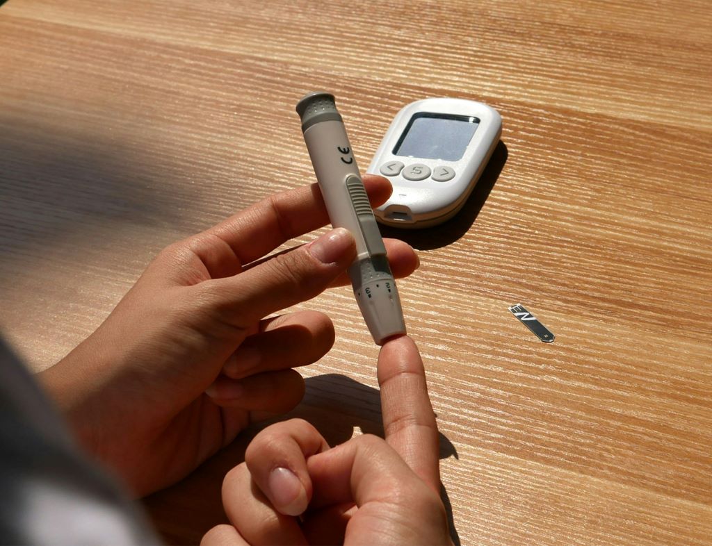
Getting to grips with rising diabetes rates is arguably one of the most urgent tasks for South Africa’s public healthcare system, but the setbacks keep coming. While some communities are facing shortages of blood sugar meters and insulin pens, a smaller wave of insulin vial shortages is now on the horizon.
In August, activist Eksoda Mazibuko was sure that years of community organising had finally yielded tangible results for people with diabetes in Hluvukani, a town in Mpumalanga.
The 35-year-old had just received R50 000 from Good Morning Angels, Jacaranda FM’s community upliftment project. It was more than enough for him to buy blood sugar meters and test strips for the fifty-person support group he runs at Tintswalo Hospital in Acornhoek, where stock had run out.
When the body can’t make or use insulin – the hormone that keeps blood sugar in check – people have to watch their levels, so they know how to eat and medicate themselves. It’s a process held together by medicines and an ecosystem of tools such as meters, strips, pens, lancets, needles, syringes, which unravels when one part is missing. Over time, poorly controlled blood sugar causes cumulative damage to one’s body that can result in severe complications such as amputation, blindness, kidney damage, and stroke.
Most people who take pills to treat diabetes need monitoring from time to time, but for the majority of those who are on insulin treatment, it is essential. People with diabetes who are taking insulin must check their blood sugar levels multiple times a day. To do this, they need glucometers – devices that measure the sugar levels in a drop of blood. But access to glucometers is a challenge. Spotlight previously reported that not everyone who needs these home testing devices is given one and those who do receive them rarely get enough test strips and lances to enable proper monitoring of their blood sugar levels.
Without tests and test strips, people in Hluvukani had no way of knowing how to adjust their insulin. Injecting the wrong amount could in extreme cases result in someone going into a coma or dying.
Mazibuko himself, who was diagnosed in 2003 and has always needed insulin, knows how terrifying it can be when monitoring tools are out of reach.
When the devices and test strips finally arrived, he shared a celebratory photo on social media. Excited messages streamed in on WhatsApp, but among them was an upsetting note from a government pharmacist: “You should have asked me before you ordered.”
Unbeknownst to the hospital staff that helped Mazibuko choose the device, the national government’s supplier would be changing, as it does every three years or so when a new tender is awarded. That means state pharmacies would soon stock a different kind of test strip.
Glucometers generally can’t interpret test strips from a different brand or model, so the glucometers that he’d already started to hand out would soon be useless.
“They were already open so I couldn’t send them back. After I worked so hard to get those machines for my community members,” said Mazibuko. “It was heartbreaking.”
According to a report from the Clinton Health Access Initiative, in poorer countries companies make most of their profit on the test strips rather than the glucometers used to read the strips. Spotlight understands that some companies go as far as giving away the devices to lock people into using their specific test strips. According to Cathy Haldane, who leads the non-communicable diseases team at FIND (a global diagnostics alliance), there have been some efforts toward encouraging universal interoperability of test strips, but these efforts haven’t gathered much steam.
Why diabetes is still a national guessing game
South Africa is one of the few countries that buys blood glucose meters and test strips en masse, but there are still lots of people who are treated with insulin who don’t have access to them.
One reason for this is that the national health department buys machines and strips for the public sector but it’s up to provinces to manage stock at pharmacies and clinics, explains Haldane.
A lack of good quality diabetes data could be making harder for health department staff to predict how much they’ll need, she says. Unlike the country’s digital HIV & TB tracking system, there’s no centralised database for diabetes and other chronic diseases such as high blood pressure and cancer. As Spotlight previously reported in-depth, there is a serious lack of reliable diabetes data for South Africa. Haldane says, “that’s how people on insulin treatment who should get a machine and monthly test strips end up going without”.
Not having reliable data leaves national planners, doctors and nurses in the dark about how many people need blood sugar monitors, where the system is failing and how the country is faring against targets outlined in the health department’s action plan for chronic diseases, which lapses in 2027. The plan states that by 2027, the health department wants at least 50% of people receiving care for diabetes to have their blood sugar under control. The available data though, all from pockets of academic research, suggests that we are falling far short of this target.
The diabetes data that is available paints a harrowing picture.
According to a StatsSA report on non-communicable diseases, diabetes was the leading underlying cause of death for women and second biggest underlying cause of death for men in 2018. While other reports suggest that diabetes is lower on the list of top killers, it clearly does claim many lives in the country. The International Diabetes Federation estimates that about half of people with diabetes in South Africa haven’t been diagnosed.
If trends continue, 2018 research suggests the treatment, management and complications of type two diabetes could cost the government as much as R35-billion by 2030.
In rural KZN, insulin pen stockouts persist
Meanwhile, more than 700 kilometers from Hluvukani, in KwaZulu-Natal’s rural King Cetshwayo district, some healthcare staff are using their own money to help keep diabetes services going.
Indira Govender, a doctor affiliated with the Rural Doctors Association of South Africa (Rudasa) who works in the area, says clinic managers are often the ones buying new batteries for blood sugar meters used in the facility and by patients.
The devices use the coin-like batteries also used in some watches, which aren’t easy to find in far flung areas.
Govender worries about the patients on insulin who still have to use a glass vial and syringe to inject themselves. “Not everybody has a fridge to store the insulin in. People struggle to draw up the right amount of insulin, sometimes because they can’t see well,” says Govender.
South Africa ran out of pens in 2024 when the health department’s longtime supplier, Novo Nordisk, stopped manufacturing pens prefilled with the cheapest form of insulin. The news came as global demand surged for one of Novo Nordisk’s long-acting diabetes medicines, semaglutide, because it was shown to also be effective for weight loss. Semaglutide is also provided in pens rather than vials.
In a 2024 letter to Novo Nordisk’s chief executive officer, MSF demanded that the pharma giant either ensure continued supply of the cheapest insulin pens in South Africa or that it offer a newer kind of pen at $1 each. That’s the amount that MSF’s research found would cover production costs, a fair profit margin and an allowance for tax.
The newer pens are filled with a form of insulin that takes effect faster and lasts for longer than previous versions. Novo Nordisk signed a deal in May in which it commits to providing these pens to South Africa until 2027. The department was charged just under $4 (around R75) per pen.
At the government clinic where Govender works in KwaZulu-Natal, however, insulin pens have reportedly not returned to pharmacy shelves.
“We haven’t had pens here since at least 2024,” says Govender.
The KwaZulu-Natal health department did not respond to Spotlight’s queries about the delivery delays.
Local consequences of global disruptions
While some communities are still waiting for insulin pens, a smaller wave of vial shortages is on its way for South Africa, according to an October circular.
Novo Nordisk told the health department to expect six to eight week delays in the delivery of short-acting, medium-acting and longer-acting insulin sold in 10ml vials. The department did not respond to Spotlight’s queries, but the circular listed four alternative prefilled pens that are available and expects stock to stabilise by January 2026.
One of the listed alternatives, Novo Nordisk’s NovoMix30, is also on a list of insulin pens and vials that will be discontinued in 2026, according to a directive issued by the health ministry in New Zealand.
No such directive has been issued by South Africa’s health department. Candice Sehoma, advocacy advisor for MSF Access in Southern Africa, says she would be surprised if the country avoids it.
It’s part of a concerning pattern of shortages of essential medicines worldwide, she says.
“We’re seeing more and more companies deprioritising insulin and discontinuing affordable medicines,” says Sehoma.
When there’s insulin but no food
While his stock of test strips lasts, Mazibuko takes them along when he visits members of his support group in Hluvukani.
They could technically find matching strips in the private sector, but they’re likely to be too expensive. A 2024 study found that for someone earning South Africa’s minimum wage, a single blood-sugar test in the private sector costs more than an hour of work, and a month of basic diabetes supplies can swallow three full days’ wages.
Many of the people on Mazibuko’s route are facing far more serious problems than the loss of glucometers. Those who aren’t working are often not taking their medication well either, Mazibuko says. “They don’t have food so they skip breakfast and also skip their insulin because they’re scared.”
Injecting insulin on an empty stomach can cause a sudden blood sugar crash that could lead to dizziness, confusion or a seizure.
Mazibuko is working on a skills programme to help these people make a living that might also protect them from lapses in basic supplies at government health facilities, which he claims happens often.
“Sometimes you go to the clinic, they tell you that they’ve run out of insulin, or they tell you to buy your own needles and syringes. You will have to do that with borrowed money,” says Mazibuko.
The Mpumalanga health department also did not respond to Spotlight’s requests for comment.
Republished from Spotlight under a Creative Commons licence.
Caffeine may have been unfairly portrayed as the villain in some heart rhythm disorders, according to a new study published in the Journal of the American Medical Association.
Longstanding medical advice has held that patients with atrial fibrillation (AF) should cut back on their caffeine intake – or eliminate it entirely – to improve their condition. Wong et al. conducted an investigation into the relationship between regular caffeinated coffee consumption and the recurrence of atrial fibrillation (AF) or atrial flutter.
The DECAF randomised clinical trial, conducted across five international centres, enrolled 200 patients with persistent AF who were successfully cardioverted and then randomised to either consume caffeinated coffee (averaging one cup daily) or abstain from coffee and caffeine for six months. But contrary to expectations, the caffeine group actually saw an improvement in symptoms.
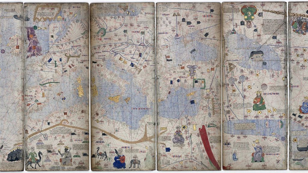
Catalan Atlas, 1375. Credit: Bibliothèque Nationale de France, via Wikimedia Commons
Clues contained in tree rings have identified mid-14th-century volcanic activity as the first domino to fall in a sequence that led to the devastation of the Black Death in Europe.
Researchers from the University of Cambridge and the Leibniz Institute for the History and Culture of Eastern Europe (GWZO) in Leipzig have used a combination of climate data and documentary evidence to paint the most complete picture to date of the ‘perfect storm’ that led to the deaths of tens of millions of people, as well as profound demographic, economic, political, cultural and religious change.
Their evidence suggests that a volcanic eruption – or cluster of eruptions – around 1345 caused annual temperatures to drop for consecutive years due to the haze from volcanic ash and gases, which in turn caused crops to fail across the Mediterranean region. To avoid riots or starvation, Italian city-states used their connections to trade with grain producers around the Black Sea.
This climate-driven change in long-distance trade routes helped avoid famine, but in addition to life-saving food, the ships were carrying the deadly bacterium that ultimately caused the Black Death, enabling the first and deadliest wave of the second plague pandemic to gain a foothold in Europe.
This is the first time that it has been possible to obtain high-quality natural and historical proxy data to draw a direct line between climate, agriculture, trade and the origins of the Black Death. The results are reported in the journal Communications Earth & Environment.
The Black Death was one of the largest disasters in human history. Between 1347 and 1353, it killed millions of people across Europe. In some parts of the continent, the mortality rate was close to 60%.
While it is accepted that the disease was caused by the bacterium Yersinia pestis, which originated from wild rodent populations in central Asia and reached Europe via the Black Sea region, it’s still unclear why the Black Death started precisely when it did, where it did, why it was so deadly, and how it spread so quickly.
“This is something I’ve wanted to understand for a long time,” said Professor Ulf Büntgen from Cambridge’s Department of Geography. “What were the drivers of the onset and transmission of the Black Death, and how unusual were they? Why did it happen at this exact time and place in European history? It’s such an interesting question, but it’s one no one can answer alone.”
Büntgen, whose research group uses information stored in tree rings to reconstruct past climate variability, worked with Dr Martin Bauch, a historian of medieval climate and epidemiology from the Leibniz Institute for the History and Culture of Eastern Europe, on the study.
“We looked into the period before the Black Death with regard to food security systems and recurring famines, which was important to put the situation after 1345 in context,” said Bauch. “We wanted to look at the climate, environmental and economic factors together, so we could more fully understand what triggered the onset of the second plague pandemic in Europe.”
Together, they combined high-resolution climate data and written documentary evidence with conceptual reinterpretations of the connections between humans and climate to show that a volcanic eruption – or series of eruptions – around 1345 was likely the first step in a sequence that ultimately led to the Black Death.
The researchers were able to approximate this eruption through information contained in tree rings from the Spanish Pyrenees, where consecutive ‘Blue Rings’ point to unusually cold and wet summers in 1345, 1346 and 1347 across much of southern Europe. While a single cold year is not uncommon, consecutive cold summers are highly unusual. Documentary evidence from the same period notes unusual cloudiness and dark lunar eclipses, which also suggest volcanic activity.
This volcanically forced climatic downturn led to poor harvests, crop failure and famine. However, the Italian maritime republics of Venice, Genoa and Pisa were able to import grain from the Mongols of the Golden Horde around the Sea of Azov in 1347.
“For more than a century, these powerful Italian city-states had established long-distance trade routes across the Mediterranean and the Black Sea, allowing them to activate a highly efficient system to prevent starvation,” said Bauch. “But ultimately, these would inadvertently lead to a far bigger catastrophe.”
The ships that carried grain from the Black Sea most likely also carried fleas infected with Yersinia pestis, as previous research has already pointed out. But why grain was so urgently needed by the Italians has now become much clearer. It is still unknown exactly where this deadly bacterium originated, but ancient DNA has suggested there may have been a natural reservoir in wild gerbils somewhere in central Asia.
Once the plague-infected fleas arrived in 14th-century Mediterranean ports on grain ships, they became a vector for disease transmission, enabling the bacterium to jump from mammalian hosts – mostly rodents, but potentially including domesticated animals – to humans. It rapidly spread across Europe, devastating the population.
“In so many European towns and cities, you can find some evidence of the Black Death, almost 800 years later,” said Büntgen. “Here in Cambridge, for instance, Corpus Christi College was founded by townspeople after the plague devastated the local community. There are similar examples across much of the continent.”
“And yet, we could also demonstrate that many Italian cities, even large ones like Milan and Rome, were most probably not affected by the Black Death, apparently because they did not need to import grain after 1345,” said Bauch. “The climate-famine-grain connection has potential for explaining other plague waves.”
The researchers say the ‘perfect storm’ of climate, agricultural, societal and economic factors after 1345 that led to the Black Death can also be considered an early example of the consequences of globalisation.
“Although the coincidence of factors that contributed to the Black Death seems rare, the probability of zoonotic diseases emerging under climate change and translating into pandemics is likely to increase in a globalised world,” said Büntgen. “This is especially relevant given our recent experiences with Covid-19.”
The researchers say that resilience to future pandemics requires a holistic approach to address a wide spectrum of health threats. Modern risk assessments should incorporate knowledge from historical examples of the interactions between climate, disease and society.
The research was supported in part by the European Research Council, the Czech Science Foundation and the Volkswagen Foundation.
A speedy new scan could improve how millions of people with hypertension are treated, suggests a new study
Credit: Pixabay CC0
About a quarter of people with high blood pressure have been estimated to have primary aldosteronism, a problem with their adrenal glands producing too much of the hormone aldosterone, which regulates levels of salt in the body.
This problem is often missed, as the path to diagnosis is complex, involving multiple tests and, to guide treatment, an invasive procedure that is not always reliable.
The new 10-minute scan, developed at University College London and described in a research letter in the New England Journal of Medicine (NEJM), reveals overactivity in adrenal glands that was invisible with conventional tests, showing exactly where too much aldosterone is being made.
This, the researchers say, will make it easier to decide on the best treatment approach – either removal of an adrenal gland that is producing too much aldosterone, or the use of new medications that block aldosterone production, targeting the cause of high blood pressure in many patients.
Professor Bryan Williams, Chair of Medicine at UCL and clinical lead for the study, said: “We have been waiting for a test like this for many decades. This British innovation is going to transform the diagnosis of aldosterone excess as an important and previously hidden cause of hypertension in many of our patients. It offers huge potential to completely change the way we make this diagnosis and enable us to provide better targeted treatment for our patients.”
The over-production of aldosterone, which raises high blood pressure by causing the body to retain too much salt, can result in a condition called primary aldosteronism, which increases the risk of heart disease, stroke and kidney disease. However, many people who do not meet the threshold for this condition are thought to have excess aldosterone raising their blood pressure.
Currently the condition is screened with a blood test and confirmed with a second test*. To decide on treatment, two catheters are inserted in veins on either side of the groin to measure levels of aldosterone on each side of the body. This helps clinicians determine if the problem is only located in one adrenal gland or both – but the test is not always accurate and not often offered as few hospitals have the expertise to perform this complex procedure.
To better detect the condition, researchers at UCL used a PET-CT scan, which creates detailed 3D images of parts of the inside of the body and maps the accumulation of a tiny amount of radioactive tracer injected into a person’s vein.
They built a new tracer compound designed to bind to the aldosterone-producing enzyme, aldosterone synthase. The tracer was highly selectively taken up by the parts of the adrenal gland that were over-producing aldosterone, lighting up these areas on the scan.
In their NEJM research letter, the researchers described how 17 patients were scanned in the world’s first use of this technique at UCLH. The team found the source of over-production of aldosterone in every patient and did not see any side effects.
Professor Williams added: “This is the first time we have been able to visualise this disease. We can see it light up on the scan. The intensity of the signal reflects the level of aldosterone over-production. This might allow us, in future, to more precisely target these over-producing areas.”
The achievement builds on more than a decade’s work by Professor Erik Arstad (UCL Division of Medicine and UCL Chemistry) and colleagues, who pioneered and patented a new method to make radioactive tracers.
Using this method, they were able to repurpose a drug-like molecule that bound to the aldosterone-producing enzyme for use as a tracer, replacing a single atom with a radioactive version of that atom – meaning this molecule would light up on a PET-CT scan.
Professor Arstad said: “It is very rewarding to be able to bring laboratory innovation into the clinic for the benefit of patients with hard-to-treat hypertension.”
The study was conducted at UCL and UCLH and was funded by the MRC and the NIHR University College London Hospitals Biomedical Research Centre.
The team is now embarking on a phase 2 clinical trial to gather sufficient data for the test to be approved for routine clinical use in the NHS.
In the UK, more than 14 million people are estimated to have high blood pressure (about one in three adults).
*For instance, a salt loading test, where a person increases their intake of salt (sodium), which would be expected to suppress aldosterone levels. If aldosterone levels are still high despite this increase, that confirms a primary hyperaldosteronism diagnosis.
Cedars-Sinai scientists have developed an experimental drug that repairs DNA and serves as a prototype for a new class of medications that fix tissue damage caused by heart attack, inflammatory disease or other conditions.
“By probing the mechanisms of stem cell therapy, we discovered a way to heal the body without using stem cells,” said Eduardo Marbán, MD, PhD, executive director of the Smidt Heart Institute at Cedars-Sinai and the study’s senior author. “TY1 is the first exomer – a new class of drugs that address tissue damage in unexpected ways.”
TY1 is a laboratory-made version of an RNA molecule that naturally exists in the body. The research team was able to show that TY1 enhances the action of a gene called TREX1, which helps immune cells clear damaged DNA. In so doing, TY1 repairs damaged tissue.
The development of TY1 has been more than two decades in the making. It started when Marbán’s previous laboratory at Johns Hopkins University developed a technique to isolate progenitor cells from the human heart. Like stem cells, progenitor cells can turn into new healthy tissue, but in a more focused manner than stem cells. Heart progenitor cells promote the regeneration of the heart, for example.
Later, at Marbán’s lab at Cedars-Sinai, Ahmed Ibrahim, PhD, MPH, discovered that these heart progenitor cells send out tiny molecule-filled sacs called exosomes. These sacs are loaded with RNA molecules that help repair and regenerate injured tissue.
Ahmed Ibrahim, PhD, MPH“Exosomes are like envelopes with important information,” said Ibrahim, who is associate professor in the Department of Cardiology in the Smidt Heart Institute and first author of the paper. “We wanted to take apart these coded messages and figure out which molecules were, themselves, therapeutic.”
Scientists genetically sequenced the RNA material inside the exosomes. They found that one RNA molecule was more abundant than the others, hinting it might be involved in tissue healing. The investigators found the natural RNA molecule to be effective in promoting healing after heart attacks in laboratory animals. TY1 is the synthetic, engineered version of that RNA molecule, designed to mimic the structure of approved RNA drugs already in the clinic. TY1 works by increasing the production of immune cells that reverse DNA damage, a process that minimises the formation of scar tissue after a heart attack.
“By enhancing DNA repair, we can heal tissue damage that occurs during a heart attack,” Ibrahim said. “We are particularly excited because TY1 also works in other conditions, including autoimmune diseases that cause the body to mistakenly attack healthy tissue. This is an entirely new mechanism for tissue healing, opening up new options for a variety of disorders.”
The investigators next plan to study TY1 in clinical trials.
A young boy born with a devastating, rare genetic condition has been given a new lease of life thanks to a team of UCL scientists who manufactured a pioneering gene therapy for him.
Ollie Chu was born with Hunter syndrome – or MPSII – an inherited, life-limiting genetic condition which causes progressive damage to the body and brain.
In the most severe cases, patients with the disease usually die before the age of 20. The effects are sometimes described as a type of childhood dementia.
Due to a faulty gene, before the treatment Ollie was unable to produce an enzyme crucial for keeping cells healthy.
But in a world first, scientists at UCL have altered Ollie’s cells using gene therapy in a bid to halt the disease.
His parents are thrilled at Ollie’s progress which they say they has been “exponential” since his transplant. He is being treated in hospital in Manchester.
Dr Karen Buckland (UCL Great Ormond Street Institute of Child Heath) said: “It’s really heart-warming to hear how well Ollie is doing – his progress is amazing.
“It’s also really, really rewarding for the team here. A lot of hard work has gone into manufacturing this new gene therapy. As well as the core team of five, around 50 staff were involved in total including all the clinical trial co-ordinators, project managers and quality assurance staff.
“It took two years just to check that the manufacturing process worked correctly. We then had to get regulatory approval before we could even begin treating Ollie’s cells.”
Dr Buckland and three other of her UCL Institute of Child Heath colleagues were directly involved with manufacturing his cells: Dr Winston Vetharoy, Edward Morgan and Raymond Nguyen. The fifth member of the team was Agrim Mahajan, from Great Ormond Street Hospital.
Dr Buckland said the project came about because she and colleagues had worked successfully on a previous project with the same Manchester team on a treatment for a similar condition called Mucopolysaccharidosis type III (MPS III) or San Filippo Syndrome.
In 2019 the Manchester team, then led by Professor Brian Bigger, approached Dr Buckland and her team again and asked them to manufacture the treatment for Hunter’s Syndrome.
In 2021 the UCL team began the process of checking the manufacturing process worked correctly.
That process, which is the first outside-of-the-body gene therapy to treat the condition, involves purifying a patient’s white blood cells to retrieve their stem cells, which are then added to a specialist growth medium.
A working copy of the gene is then added to a viral vector, which is used to transport the healthy copy of the gene into the patient’s own stem cells.
The cells are then washed and frozen and stored at -130 degrees C, in a process known as cryopreservation.
The cells are checked using several quality control tests to make sure they’re safe to be given back to the patient.
Having confirmed, in 2022, that the process worked correctly the University of Manchester applied successfully to the UK medicines regulator, the Medicines and Healthcare products Regulatory Agency (MHRA), for approval. They could then use the process on a patient.
Ollie, from California, is the first of five boys around the world to receive the treatment.
The team treating him at Royal Manchester Children’s Hospital (RMCH) sent Dr Buckland and her colleagues some of his white blood cells.
Dr Vetharoy, who led the manufacture of the gene therapy product for Ollie, said: “Every stage of the process required meticulous attention to detail. We needed to maintain absolute control over contamination risks and ensure that all reagents and materials met the highest-quality standards.
“Seeing how well Ollie is thriving after such a short time is incredibly fulfilling for the entire team, and we wish him continued progress and good health in the years to come.”
In November 2024, the UCL manufacturing team made the altered cells for him, using the validated manufacturing process. In February 2025, those altered cells were transplanted into Ollie’s body.
Nine months on, his father, Ricky, says the improvement in his son’s condition is remarkable.
He said: “I don’t want to jinx it, but I feel like it’s gone very, very well. His life is no longer dominated by needles and hospital visits. His speech, agility and cognitive development have all got dramatically better.
“It’s not just a slow, gradual curve as he gets older, it has shot up exponentially since the transplant.”
Currently the only licensed treatment that can help to improve life for children with Hunter syndrome is Elaprase – a weekly enzyme replacement therapy that takes approximately three hours to administer, that children must take for their whole life.
Dr Buckland and her colleagues manufactured the new gene therapy treatment at the specialist Cell and Gene Therapy Manufacturing Facility in The Zayed Centre for Research (ZCR) into Rare Diseases in Children, a facility jointly run by UCL and GOSH.
The clinical trial at RMCH is being done in collaboration with University of Manchester and the Manchester Centre for Genomic Medicine at Saint Mary’s Hospital.
First study to use worn face masks as a passive tool to monitor indoor air
Photo by Daniel Eledut on Unsplash
According to a new Northwestern University study, the ambient air on aeroplanes and in hospitals mostly contains harmless microbes typically associated with human skin.
In the first study of its kind, scientists used an unexpected sampling tool – used face masks and an aircraft air filter – to uncover the invisible world of microbes floating in our shared air. Their results revealed that the same types of harmless, human-associated bacteria dominate both aeroplane and hospital air.
Across all samples, the team detected 407 distinct microbial species, including common skin bacteria and environmental microbes. While a few potentially pathogenic microbes did appear, they were in extremely low abundance and without signs of active infection.
Not only does the study help illuminate what microbes exist in shared air, but it also demonstrates that face masks and air filters can be repurposed as non-invasive, cost-effective tools to monitor confined, high-traffic environments.
“We realized that we could use face masks as a cheap, easy air-sampling device for personal exposures and general exposures,” said Northwestern’s Erica M. Hartmann, who led the study. “We extracted DNA from those masks and examined the types of bacteria found there. Somewhat unsurprisingly, the bacteria were the types that we would typically associate with indoor air. Indoor air looks like indoor air, which also looks like human skin.”
An expert on indoor microbiomes, Hartmann is an associate professor of civil and environmental engineering at Northwestern’s McCormick School of Engineering.
A second life for used face masks
Hartmann and her team conceived the project in January 2022, amid the COVID-19 pandemic. At the time, travellers became increasingly concerned with how well aircraft cabins filtered and circulated air. Hartmann received a grant to collect aircraft cabin filters to look for evidence of pathogens.
Although Hartmann did procure an aircraft’s high-efficiency particulate air (HEPA) filter, which had been used for more than 8000 flight hours, she quickly realised the project might be impractical.
“At the time, there was a serious concern about Covid transmission on planes,” Hartmann said. “HEPA filters on planes filter the air with incredibly high efficiency, so we thought it would be a great way to capture everything in the air. But these filters are not like the filters in our cars or homes. They cost thousands of dollars and, in order to remove them, workers have to pull the airplane out of service for maintenance. This obviously costs an incredible amount of money, and that was eye opening.”
Searching for another method that passively traps microbes, Hartmann and her team pivoted to a much cheaper and much less disruptive tool: face masks. For the study, volunteers wore face masks on both domestic and international flights. After landing, they put the masks into sterile bags and sent them to Hartmann’s lab. For comparison, Hartmann also collected face masks that volunteers took on flights but never wore.
To understand how indoor environments differ, Hartmann and her team wanted to examine another high-traffic, enclosed environment with heavily filtered air. The team selected hospitals as the second testbed. After wearing a face mask during a shift, hospital workers submitted their masks to Hartmann’s lab.
“As a comparison group, we thought about another population of people who were likely wearing masks anyway,” Hartmann said. “We landed on health care providers.”
The sky is clear
After receiving masks from travellers and health care workers, Hartmann’s team collected DNA from the outsides of masks. They found the air in hospitals and in aircraft contains a diverse but mostly harmless mix of microbes, with only minimal traces of potentially pathogenic species.
In both environments, common human-associated bacteria – especially those found on skin and in indoor air – dominated the samples. Although the abundance of each microbe present was slightly different, the microbial communities from hospitals and aeroplanes were highly similar. The overlap suggests that people themselves – rather than the specific environment – are the main source of airborne microbes in both settings. And those microbes floating around indoor air come from people’s skin, not from illness.
Hartmann’s team also identified a handful of antibiotic resistance genes, linked to major classes of antibiotics. While these genes do not indicate the presence of dangerous microbes in the air, they highlight how widespread antibiotic resistance has become.
Although indoor air might not be as harmful as some people may have feared, Hartmann emphasises that airborne spread is just one way infections can travel. For many common illnesses, other routes – such as direct contact with an infected individual or interacting with high-touch surfaces – are far more important.
“For this study, we solely looked at what’s in the air,” Hartmann said. “Hand hygiene remains an effective way to prevent diseases transmission from surfaces. We were interested in what people are exposed to via air, even if they are washing their hands.”
The study, published in the journal Microbiome, was supported by the Walder Foundation.
When it comes to protecting babies from the sun, many parents wonder if sunscreen is safe and necessary. The truth is, experts advise against using sunscreen on infants under six months old as their skin is thinner and more sensitive, leading to greater absorption of chemicals and a higher risk of irritation and rashes.
Babies under six months have a higher surface-area-to-body-weight ratio, which increases their exposure to sunscreen chemicals. Some chemical ingredients, like oxybenzone, may cause allergic reactions or disrupt hormones. Sunscreen can also impede a baby’s ability to sweat and regulate their body temperature.
Instead, the best protection for young babies is to keep them out of direct sunlight, dress them in lightweight, long-sleeved clothing, and use hats and shade as natural barriers.
For babies over six months, a gentle, broad-spectrum baby sunscreen with at least SPF 30 can be safely applied. However, using sunscreen should complement, not replace, other sun safety measures, which are vital – especially in our sunny South African climate!
Karen Van Rensburg, spokesperson for Sanosan, explains, “Parents often struggle with knowing how much sunscreen to use on their babies. It’s important to understand that while sunscreen is a helpful tool, relying solely on it, especially for very young infants, can be risky. Using physical barriers like shade and protective clothing alongside sunscreen provides the safest approach to sun care for babies.”
To keep babies safe, parents should:
Avoid sun exposure during peak hours (10 a.m. to 4 p.m.)
Use shade and protective clothing as the first defence.
For babies over six months, reapply a suitable sunscreen on a regular basis to maintain protection, especially after going in the water, after drying off or after sweating.
Your baby should not stay in the sun too long even with sunscreen because every sunburn damages the skin and is a serious risk to their health.
This balanced approach highlights that cautious sunscreen use combined with physical protection methods is key to keeping baby skin healthy and safe from sun damage.
Sanosan Baby Sun Cream SPF 50+ is a top-tier sunscreen designed specifically for delicate baby skin including broad range of UVA+UVB protection SPF 50+. With its pleasant texture, this cream absorbs quickly for easy application and delivers 24 hours of nourishing care, making it suitable for babies, children, and adults alike. With its gentle formula, this sun cream helps maintain skin hydration while protecting against sun damage, allowing for worry-free outdoor playtime. Plus, its microplastic-free, and safe for our oceans!
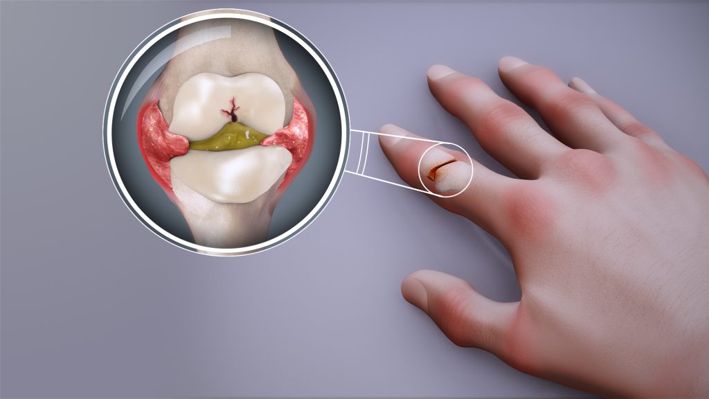
Cedars-Sinai investigators may have figured out why certain immunosuppressive treatments don’t work well in rheumatoid arthritis. In a study published inScience Immunology, scientists traced the problem to specific changes that occur in immune cells within the joints as the disease progresses.
The findings could lead to more effective therapies for the incurable autoimmune disease.
“Our discoveries point to the importance of the tissue environment in worsening rheumatoid arthritis and driving resistance to antirheumatic medications,” said Nunzio Bottini, MD, PhD, director of the Kao Autoimmunity Institute at Cedars-Sinai, professor of Medicine and corresponding author of the study.
Rheumatoid arthritis causes chronic inflammation in the joints. In other forms of autoimmune arthritis, inflammation can be relieved by targeting interleukin-17, one of several proteins that can contribute to joint inflammation.
In experiments involving human rheumatoid arthritis tissues and laboratory mice, investigators showed that, over time, the immune cells that produce interleukin-17 gradually stop making it. This finding helps explain why IL-17-targeted treatments do not work well against established rheumatoid arthritis.
“These immune cells can also change in ways that make them more aggressive and able to sustain inflammation even without interleukin-17,” Bottini said.
Changes to the immune cells appear to be driven by synoviocytes – nonimmune cells that produce the lubricating synovial fluid in the joints, according to the study.
Bottini said that the Department of Computational Biomedicine at Cedars-Sinai, particularly the laboratory of Kyoung Jae Won, PhD, played a key role in the study by contributing critical work in spatial biology, an emerging field that studies how cells function within their tissue environments.
The findings carry significant implications for treating rheumatoid arthritis, according to Joyce So, MD, PhD, chief genomics officer at Cedars-Sinai and medical director of the newly established Center for Genomic Medicine at Cedars-Sinai Guerin Children’s.
“This important new insight contributes to shifting the paradigm of how we understand rheumatoid arthritis progression and why IL-17 treatments haven’t worked as well as expected,” So said. “Only with a precise understanding of the biological mechanisms of disease can effective, precision therapies be developed. In the meantime, clinicians can help patients in early or presymptomatic stages make the most of treatments that may lose effectiveness over time.”