Month: May 2022

Sarin Gas Likely the Cause of Mysterious Gulf War Illness

Photo by Pablo Stanic on Unsplash

Since the 1990s, scientists have debated the underlying cause of Gulf War illness (GWI), a constellation of unexplained and chronic symptoms affecting veterans of the Persian Gulf War. Now researchers have solved the mystery, showing through a detailed genetic study that the nerve gas sarin was largely responsible for the syndrome. The findings were published in Environmental Health Perspectives.

Dr Haley’s research group not only discovered that veterans with exposure to sarin were more likely to develop GWI, but also found that the risk was modulated by a gene that helps break down the nerve gas. Sarin-exposed Gulf War veterans with a weak variant of the gene were more likely to develop symptoms of GWI than other exposed veterans with the strong form of the gene.

“Quite simply, our findings prove that Gulf War illness was caused by sarin, which was released when we bombed Iraqi chemical weapons storage and production facilities,” said Robert Haley, MD, at UT Southwestern, a medical epidemiologist who had led that study and has been investigating GWI for 28 years. “There are still more than 100 000 Gulf War veterans who are not getting help for this illness and our hope is that these findings will accelerate the search for better treatment.”

Multiple causes of Gulf War illness suggested

After the Gulf War, more than a quarter of the US and coalition veterans began reporting a range of chronic symptoms, including fatigue, fever, night sweats, memory and concentration problems, difficulty finding words, diarrhoea, sexual dysfunction, and chronic body pain. Since then, military and academic researchers have studied a list of possible causes of GWI, ranging from stress, vaccinations, and burning oil wells to exposure to pesticides, nerve gas, anti-nerve gas medication, and depleted uranium used in weapons.

“What makes this new study a game-changer is that it links GWI with a very strong gene-environment interaction that cannot be explained away by errors in recalling the environmental exposure or other biases in the data.”

Study leader Robert Haley, MD, medical epidemiologist

Over the years, these studies have identified statistical associations with several of these, but no cause has been widely accepted. Most recently, Dr Haley and a colleague reported a large study testing veterans’ urine for depleted uranium that would still be present if it had caused GWI and found none.

Studies have shown statistical associations with several of these causes, though none received wide acceptance. Dr Haley and a colleague recently reported a large study that found no depleted uranium in veterans’ urine, which would have still been present if it had caused GWI.

“As far back as 1995, when we first defined Gulf War illness, the evidence was pointing toward nerve agent exposure, but it has taken many years to build an irrefutable case,” said Dr Haley.

Sarin’s effects

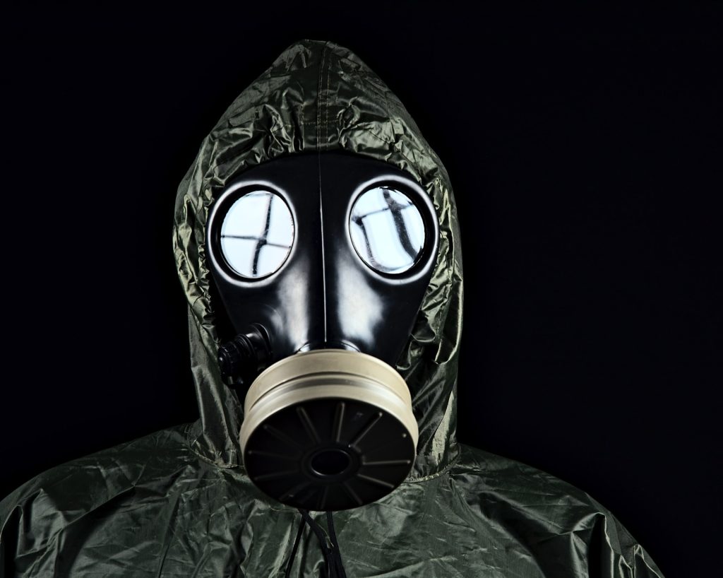
Sarin is a toxic nerve agent, production of which was banned in 1997. When people are exposed to either the liquid or gas form, sarin enters the body through the skin or breathing and attacks the nervous system. High-level sarin often results in death, but studies on survivors have revealed that lower-level sarin exposure can lead to long-term impairment of brain function. A large release of this gas occurred when a chemical weapons storage plant was bombed, causing thousands of nerve gas alarms to sound.

Previous studies have found an association between Gulf War veterans who self-reported exposure to sarin and GWI symptoms. However, this has raised criticisms of recall bias. “What makes this new study a game-changer is that it links GWI with a very strong gene-environment interaction that cannot be explained away by errors in recalling the environmental exposure or other biases in the data,” Dr Haley said.

In the new paper, Dr Haley and his colleagues studied 508 deployed veterans with GWI and 508 deployed veterans who did not develop any GWI symptoms. They asked whether the veterans had heard chemical nerve gas alarms, indicating sarin exposure, and also collected blood and DNA samples.

The role of PON1

The researchers tested the samples for variants of a gene called PON1, which has two variants. The Q variant generates a blood enzyme that efficiently breaks down sarin while the R variant helps the body break down other chemicals but is not efficient at destroying sarin. Everyone has either a QQ, RR or QR genotype.

For Gulf War veterans with the QQ genotype, hearing nerve agent alarms — a proxy for chemical exposure — raised their chance of developing GWI by 3.75 times, those with the QR genotype had an a 4.43 fold risk increase. And for those with RR genotype, the chance of GWI increased by 8.91 times. Those soldiers with both the RR genotype and low-level sarin exposure were over seven times more likely to get GWI due to the interaction per se, over and above the increase in risk from both risk factors acting alone. For genetic epidemiologists, this number leads to a high degree of confidence that sarin is a causative agent of GWI.

“Your risk is going up step by step depending on your genotype, because those genes are mediating how well your body inactivates sarin,” said Dr Haley. “It doesn’t mean you can’t get Gulf War illness if you have the QQ genotype, because even the highest-level genetic protection can be overwhelmed by higher intensity exposure.”

This kind of strong gene-environment interaction is considered a gold standard for showing that an illness like GWI was caused by a particular environmental toxic exposure, he added. The research doesn’t rule out that other chemical exposures could be responsible for a small number of cases of Gulf War illness. However, Dr. Haley and his team carried out additional genetic analyses on the new data, testing other factors that could be related, and found no other contributing causes.

“There’s no other risk factor coming anywhere close to having this level of causal evidence for Gulf War illness,” said Dr Haley.

The team is continuing research on GWI’s impacts on the body, particularly the immune system, whether any of its effects are reversible, and whether there are biomarkers to detect prior sarin exposure or GWI.

Source: UT Southwestern Medical Center

No Added Seizure Risk from Antidepressant Use in Pregnancy

Pregnant with ultrasound image
Source: Pixabay

A large Swedish study in the journal Neurology found that pregnant women taking selective serotonin reuptake inhibitors (SSRIs) or selective norepinephrine reuptake inhibitors (SNRIs) during the first trimester of was not linked to an increased risk for neonatal seizures and epilepsy in childhood.

Any increase in seizures or epilepsy is likely due to other factors, the researchers said.

“It’s not likely the medications themselves that are causing the seizures and epilepsy in children, but rather the reasons why these women are taking the medication,” according to Kelsey Kathleen Wiggs, a PhD candidate at Indiana University in Bloomington. There are also the other background factors that differ between women who do and do not use SSRI/SNRIs.

“When it rains, it pours,” Wiggs said. “Women who are taking antidepressants in pregnancy are doing that for lots of different reasons, and they might be at risk for different things than women who aren’t taking those medications in pregnancy.”

The study found an elevated risk for neonatal seizures (risk ratio [RR] 1.41) and epilepsy in early childhood (HR 1.21) among offspring of mothers who used antidepressants in pregnancy.

Adjustment for maternal indications for SSRI/SNRI use and background factors like smoking during pregnancy revealed that they were drivers for both associations: neonatal seizures (RR 1.10); epilepsy diagnosis at 5 years (HR 0.96). Parental history of epilepsy was not found to affect the association.

The findings provide a “conclusive answer” to these concerns with using SSRI/SNRIs during pregnancy, according to Anne Berg, PhD, and Torin Glass, BM, Bch, BAO.

“[SSRI/SNRIs] have been demonstrated to have serotonergic central nervous system effects and are associated with an observable withdrawal syndrome which may be seen in the neonate following in utero exposure,” noted Drs Berg and Glass, in an accompanying editorial.

“The authors understood that with a population-based data registry and huge sample size, they had more than sufficient statistical power to detect even a modest increase in risk,” the editorialists wrote. “They tested this hypothesis and were able to reject it, definitively!”

In order to determine whether antidepressants had a causal association with infant seizures and childhood epilepsy, the researchers analysed data from national Swedish healthcare registries on a total of 1 721 274 children in Sweden born between 1996 and 2011.

Participants were divided into two groups: one group of mothers who reported use of an SSRI (fluoxetine, citalopram, paroxetine, sertraline, fluvoxamine, escitalopram) or SNRI (venlafaxine, duloxetine) during the first trimester of pregnancy (n = 24 308), and another group with no reported antidepressant use (n = 1 696 966).

Source: MedPage Today

How Kaposi Sarcoma-associated Herpesvirus Evades the Immune System

Kaposi sarcoma on the skin of an AIDS patient. Credit: National Cancer Institute

A study published in Cell Reports has identified a protein in the cancer cell’s nucleus as a critical agent keeping Kaposi sarcoma-associated herpesvirus (KSHV) dormant and hidden from the immune system. The virus, in the same family as Epstein-Barr virus, is linked to AIDS-related Castleman’s disease and cancers such as Kaposi sarcoma.

Up to 50% of the population in some parts of Africa are affected with KSHV, though not everyone with KSHV will develop Kaposi sarcoma. Those who do typically have a weakened immune system due to HIV infection, organ transplant, being older or other factors.

The introduction of antiretroviral therapy significantly reduced AIDS-related Kaposi sarcoma prevalence in Western countries; however, in sub-Saharan Africa, the disease continues to have a poor prognosis.

On entry into a human cell the virus causes a hidden infection in the nucleus: the virus simply latches onto parts of the cell’s chromosomes without replicating.

Researchers studied KSHV’s latent-lytic switch, a process in which the virus exits its dormancy state to replicate in the host cell. This replication phase, called the lytic cycle, ends with the disintegration of the cell and the release of the viruses, infecting neighbouring cells.

“The virus likes to stay silent as long as possible to avoid being detected by the body’s immune system,” said Professor Yoshihiro Izumiya, the study’s senior author.

The team sought to understand the mechanisms behind this latent-lytic switch and the role the host cell environment played in this process.

“Where the virus latches onto the host cell, how it manages to stay dormant, and what triggers its activation were very exciting and important puzzles to solve,” Prof Izumiya said.

The study identified where the virus genome could be found on the host genome.

Izumiya and his team profiled and analysed chromosomal interactions on three cancer cell lines naturally infected with KSHV, locating the virus’s preferred chromosome docking sites. The binding patterns, similar among the three cancer cell lines, showed a nuclear ecosystem that can attract and help keep the virus in its silent form.

The team also found that CHD4 (chromodomain helicase DNA binding protein 4) binds to the virus’s genomic elements. CHD4, a protein in the host cell’s chromosomes, suppresses the work of the gene responsible for viral replication. The study showed that CHD4 is a key regulator of the KSHV latency-lytic switch.

“The location where the virus genome attaches to the host chromosome is not random,” said Ashish Kumar, a postdoctoral researcher in Izumiya Lab and the paper’s first author. “Without having enriched CHD4 protein, the virus starts to replicate, kicking in a cell destructive mode. For the virus to select CHD4 among many other host proteins, CHD4 must play a unique and important role in host cells.”

Virology can help identify cellular proteins essential for cell homeostasis. Over millions of years, the virus’s genome developed to encode or assemble a small number of very efficient proteins, which strategically connect to host cell proteins to keep viral chromatin dormant and impact the host cell’s tumour suppression function.

“We used virology as an entry point to shed light on the function of CHD4 in gene regulation in general. During virus-host co-evolution, KSHV cleverly learned to hijack host proteins that can help keep the gene responsible for viral replication dormant.”

The researchers found a viral protein which could serve as the basis for a replication inhibitor. Since CHD4 is critical for cancer cell growth in a variety of cancers, they hope this virus-host interaction could inform cancer treatment research.

Source: University of California – Davis Health

New Treatment Traps and Kills off Toxoplasma gondii in Host Cells

Toxoplasma gondii, an obligate intracellular human parasite, has a unique cytoskeletal apparatus that is probably used for invading host cells and for parasite replication. Shown here are images of T. gondii constructing daughter scaffolds within the mother cell. Green: YFP-α-Tubulin; bright yellow: mRFP-TgMORN1 (see Hu et al., Figure 6 A-C).
Credit: Image provided by Ke Hu and John M. Murray

A new method blocks the protein regulation of Toxoplasma gondii, causing the parasite to die off inside the host cell, a method which could be adapted to malaria. The approach is detailed in the journal Nature Microbiology.

Toxoplasmosis is one of the most widespread zoonoses worldwide. It is an infectious disease that can be transmitted from cats to humans, and from consumption of raw or undercooked meat. Infection is particularly dangerous for pregnant women, and immunosuppressed HIV/AIDS patients often manifest neurological symptoms.

The cause of the disease is the single-celled parasite T. gondii. Inside the host cell, it forms a parasitophorous vacuole, a tiny compartment facilitating nutrient exchange and synchronised cell division. Up to 64 daughter cells can form inside, connected with each other inside the vacuole via a network. As soon as the offspring are mature, a regulation mechanism prompts the dissolution of the vacuole and the structures that have formed inside it, releasing the daughter cells to invade new host cells.

Hope for the development of new drugs

Previously, it was not known which genes encode the proteins that control the exit from the host cell. To identify them, a team led by Professor Markus Meißner at LMU, collaborated with colleagues from the University of Glasgow in Scotland to develop a novel genetic screening technique based on Cas9 ‘genetic scissors’, and investigate a library of 320 parasite-specific genes. They discovered two genes without which cell egress is impossible.

The targeted destruction of these genes resulted in the exit being trapped and the next generation of parasites dying within the host cell. “This paves the way potentially for the development of active substances that could block the function of the corresponding proteins and so put a halt to propagation,” remarked Prof Markus Meißner.

T. gondii is closely related to the malaria pathogen Plasmodium falciparum. Therefore, the parasite serves as a model organism for the pathogen of the tropical disease, which kills hundreds of thousands of people worldwide every year. “We assume that similar processes control the propagation of the malaria pathogen,” explains LMU parasitologist Dr. Elena Jimenez-Ruiz. “Next, we will investigate what functions these proteins have in the malaria pathogen and whether there are possible starting points for the development of new drugs.”

Source: Ludwig-Maximilians-Universität München

CSF From Young Mice Improves Memory of Older Mice

Mouse
Photo by Kanasi on Unsplash

In a finding reminiscent of how vampires and zombies in fiction get sustenance from their victims, a team of researchers reported in the journal Nature that injecting cerebrospinal fluid (CSF) from young mice into old mice improves the memory and cognitive abilities of the older mice

Such an approach is nothing new, although the chief obstacle was safely harvesting such a tiny amount of CSF from the small animals. About two decades ago, studies had reported that transferring blood from younger mice to older ones notably improved the health of the older mice, giving them a ‘rejuvenating’ effect. It did not take long for people to take note of this discovery, with a startup company offering transfers of young people’s plasma for exorbitant amounts to wealthy older clients in the unproven hopes of reversing ageing. Fears of a dystopian future were averted when the US Food and Drug Administration released a statement stating such transfers had no clinical benefit, and the company folded. However, research continued.

Since ageing is too complex to measure in a clinical trial anyway, scientists have been focusing on tackling specific aspects of it, such as in neurodegenerative diseases like Alzheimer’s and research has continued in this direction. A few years ago, human umbilical cord plasma was shown to revitalise hippocampal function in aged mice, and previous work led by Tony Wryss-Coray, PhD had found that young mouse blood improved age-related impairments in cognition. Studies of fear conditioning had shown that proliferation of oligodendrocyte precursor cells (OPCs) was necessary for fear formation, which raised the question of whether CSF might affect this.

Infusing CSF taken from 10 week old mice over seven days, researchers trained 18 month old mice to associate a flashing light with an electric shock to the foot. The CSF infusion was shown to improve recall of the fear stimulus in the older mice and induce greater OPC proliferation.

“The broad message here is that the aging process is malleable, which of course is not new because of this paper,” senior author Dr Wyss-Coray said in an interview with MedPage Today. “But it adds to the idea that aging is a potential therapeutic target, a process we can start to understand better and start to manipulate.”

“The other message – one that’s more brain-specific – is that if you improve the environment in which neurons live, you can actually have a substantial improvement in function,” he added. “That may be as important, or even more important sometimes, than targeting neuronal processes themselves.”

The researchers isolated fibroblast growth factor 17 (Fgf17) infusion as being necessary for OPC proliferation, and blocking it in young mice impaired cognition.

“This suggests that Fgf17 is not only able to recapitulate some of the useful effects of CSF from young mice, but it also seems to be necessary to make a young brain function at its full capacity,” Dr Wyss-Coray said.

Reduce Blood Sugar ASAP After Diabetes Diagnosis, Study Suggests

Image source: Pixabay CC0

Findings from new research published in Diabetes, Obesity and Metablism suggest that people with type 2 diabetes may need to reduce their blood sugar levels sooner after diagnosis than previously thought, to prevent major cardiovascular events such as heart attacks.

The University of Surrey study Surrey suggests that achieving glycaemic control within the first year of diagnosis reduces the incidence of major cardiovascular events. Additionally, the team found that the greater the variation in blood levels 12-months after diagnosis, the more likely a patient was to experience dangerous cardiovascular events.

Dr Martin Whyte, co-author of the study and Reader in Metabolic Medicine at the University of Surrey, said: “The conventional wisdom has been to slowly and steadily treat type 2 diabetes with diet and medicine dose-escalation over years – the period over which it took people to reduce their sugar levels after diagnosis was thought less important for major vascular protection. However, our observational study suggests that getting blood levels under control quickly — within the first 12 months after diagnosis — will significantly help reduce cardiovascular events.”

Type 2 diabetes is a common condition that results in the level of sugar in the blood becoming too high. The condition is linked to obesity or a family history of type 2 diabetes and can increase a person’s risk of getting serious health conditions.

The University of Surrey’s study used Royal College of General Practitioners’ Research and Surveillance Centre database to perform a comprehensive examination of glycaemic control achieved within the first year of diagnosis and subsequent blood sugar level variability with cardiovascular disease incidents.

Source: University of Surrey

How Do You Do, Fellow Kids? Making Anti-vaping Messaging Work

Vaping with an e-cigarette
Photo by Toan Nguyen on Unsplash

Effective anti-vaping advertisements geared to teens have the greatest impact when they emphasise the adverse consequences and harms of vaping e-cigarettes, use negative imagery, and avoid memes, hashtags and other ‘teen-centric’ communication styles, according to a first-of-its-kind study by researchers in the journal Tobacco Control.

The researchers also found that certain messaging content currently being used, especially sweets and flavour-related imagery, increases the appeal of vaping and should be avoided when designing prevention messages.

“E-cigarettes and vaping have become a major public health concern, with nicotine addiction and other harmful outcomes looming large for youth,” said Seth M. Noar, PhD, the paper’s corresponding author and UNC Lineberger professor. “The percentage of teens vaping increased from about 5% in 2011 to over 25% in 2019,” Prof Noar said. “That is an alarming trend, making an understanding of effective vaping prevention messages especially urgent.”

Since the introduction of e-cigarettes, numerous US health departments have created their own anti-vaping messaging geared to teens, as have national health organisations such as the US Food and Drug Administration and Centers for Disease Control and Prevention.

The online study asked 1501 teens to rate seven randomly selected vaping prevention ads from a pool of more than 200 ads. Vaping prevention ads that clearly communicated the health harms of vaping, or compared vaping to cigarette smoking, were comparatively more effective. Neutral or less personally relevant content, such as referencing the environmental impact of vaping or the targeting of youth by the tobacco industry, was less impactful.

“Although we anticipated that vaping prevention ads with neutral or pleasant imagery would not be as effective, we were alarmed to find that flavour-related messages actually heightened the attractiveness of vaping,” said Marcella H. Boynton, PhD, first author

“In retrospect, it stands to reason that by reminding teens about pleasurable aspects of e-cigarettes, even within the context of a prevention ad, we run the risk of doing harm. Notably, we found that flavour-related prevention ad content was associated with vaping appeal among both users and non-users of e-cigarettes, which is a good reminder of how much candy and fruit flavours in e-cigarettes have driven the youth vaping epidemic.”

The researchers hope to next investigate the effects of other types of anti-vaping ads on a wide range of audiences. They also are developing a series of messages and a companion website to test the ability of a text message-driven intervention to reduce youth vaping. In that regard, Prof Noar noted that “We have been developing our own evidence-based messages based on the latest science about the harms of vaping. Our messaging approach has been greatly influenced by the insights generated by this study.”

The study used UNC’s Vaping Prevention Resource, a website designed to provide practitioners, researchers and communities with vaping prevention media content from around the world, as well as strategies and resources for youth vaping prevention. It is the largest repository of free, open-access vaping prevention materials, all available for download at https://vapingprevention.org/.

Source: UNC Lineberger Comprehensive Cancer Center

Steroids after Severe COVID Reduces One-year Mortality by 51%

Photo by Stephen Andrews on Unsplash

Researchers have shown that severe inflammation during hospitalisation for COVID increases post-recovery mortality risk by 61% – but this risk is reduced again by 51% if anti-inflammatory steroids are prescribed upon discharge. We need to think of COVID as a potentially chronic disease that requires long-term management, argue the authors, whose results are published in Frontiers in Medicine.

Evidence continues to gather that ‘long COVID’, that is, continued negative health impacts months after apparent recovery from severe COVID, is an important risk for some patients. For example, researchers showed last December that hospitalised patients who seemingly recovered from severe COVID run more than double the risk of dying within the next year, compared to those with only mild COVID or who never had COVID.

Now, the same research team shows that among patients hospitalised for COVID who seemingly recovered, severe systemic inflammation during their hospitalisation is a risk factor for death within one year.

“Here we show that the stronger the inflammation during the initial hospitalisation, the greater the probability that the patient will die within 12 months after seemingly ‘recovering’ from COVID.”

Professor Arch G Mainous III

“COVID is known to create inflammation, particularly during the first, acute episode. Our study is the first to examine the relationship between inflammation during hospitalisation for COVID and mortality after the patient has ‘recovered’,” said first author Professor Arch G Mainous III at the University of Florida Gainesville.

“Here we show that the stronger the inflammation during the initial hospitalisation, the greater the probability that the patient will die within 12 months after seemingly ‘recovering’ from COVID.”

Prof Mainous and colleagues analysed electronic health records of 1207 adults hospitalised with COVID in 2020 or 2021 within the University of Florida health system, with at least a one year follow-up after discharge. As a proxy for the severity of systemic inflammation during hospitalisation, they used a common and validated measure: C-reactive protein (CRP), secreted by the liver in response to a signal by active immune cells.

Widespread inflammation in the body

As expected, the blood concentration of CRP during hospitalisation was strongly correlated with the severity of COVID: 59.4mg/L for patients not needing supplemental oxygen, 126.9 mg/L for those who needed extra oxygen without mechanical ventilation, and 201.2 mg/L for the most severe cases, who required ventilation through a ventilator or through ECMO.

After correcting for risk factors, patients with the highest CRP concentration measured their during their hospital stay had a 61% greater risk of all-cause mortality within one year of discharge than patients with the lowest CRP concentration.

Prof Mainous said: “Many infectious diseases are accompanied by an increase in inflammation. Most times the inflammation is focused or specific to where the infection is. COVID is different because it creates inflammation in many places besides the airways, for example in the heart, brain, and kidneys. High degrees of inflammation can lead to tissue damage.”

Importantly, the authors showed that the increased all-cause mortality risk associated with severe inflammation was reduced again by 51% if the patient was prescribed anti-inflammatory steroids after their hospitalisation.

These results mean that the severity of inflammation during hospitalisation for COVID can predict the risk of subsequent serious health problems, including death, from ‘long COVID’. They also imply that current recommendations for best practice may need to be changed, to include more widespread prescription of orally taken steroids to COVID patients upon their discharge.

COVID as a chronic disease?

The authors propose that COVID should be seen as a potentially chronic disease.

“When someone has a cold or even pneumonia, we usually think of the illness being over once the patient recovers. This is different from a chronic disease, like congestive heart failure or diabetes, which continue to affect patients after an acute episode. We may similarly need to start thinking of COVID as having ongoing effects in many parts of the body after patients have recovered from the initial episode,” said Prof Mainous.

“Once we recognise the importance of ‘long Covid’ after seeming ‘recovery’, we need to focus on treatments to prevent later problems, such as strokes, brain dysfunction, and especially premature death.”

Source: Frontiers

A Blindness Gene That Also Increases Intelligence

DNA repair
Source: Pixabay/CC0

A new study published in Brain shows that a genetic mutation which causes blindness in humans also increases intelligence, possibly through an increase in synaptic activity between the very same neurons damaged by the mutation.

The present study came about when Professors Tobias Langenhan and Manfred Heckmann, came across a paper on a mutation that damages a synaptic protein. The mutation caused patients to go blind, but then doctors noticed that the patients were also of above-average intelligence, something which piqued the two neurobiologists’ interest. “It’s very rare for a mutation to lead to improvement rather than loss of function,” said Prof Langenhan.

The two neurobiologists have been using fruit flies to analyse synaptic functions for many years. “Our research project was designed to insert the patients’ mutation into the corresponding gene in the fly and use techniques such as electrophysiology to test what then happens to the synapses. It was our assumption that the mutation makes patients so clever because it improves communication between the neurons which involve the injured protein,” explained Prof Langenhan. “Of course, you can’t conduct these measurements on the synapses in the brains of human patients. You have to use animal models for that.”

“75 per cent of genes that cause diseases in humans also exist in fruit flies”

Professor Tobias Langenhan

First, in collaboration with Oxford researchers, the scientists showed that the fly protein called RIM looks molecularly identical to that of humans. This was essential in order to be able to study the changes in the human brain in the fly. In the next step, the neurobiologists inserted the genetic mutation into flies. They then took electrophysiological measurements of synaptic activity. “We actually observed that the animals with the mutation showed a much increased transmission of information at the synapses. This amazing effect on the fly synapses is probably found in the same or a similar way in human patients, and could explain their increased cognitive performance, but also their blindness,” concludes Professor Langenhan.

The scientists also found out how the increased transmission at the synapses occurs: the molecular components in the transmitting nerve cell that trigger the synaptic impulses move closer together as a result of the mutation effect and lead to increased release of neurotransmitters. A novel method, super-resolution microscopy, was one of the techniques used in the study. “This gives us a tool to look at and even count individual molecules and confirms that the molecules in the firing cell are closer together than they normally are,” said Prof Langenhan.

“The project beautifully demonstrates how an extraordinary model animal like the fruit fly can be used to gain a very deep understanding of human brain disease. The animals are genetically highly similar to humans. It is estimated that 75% of the genes involving disease in humans are also found in the fruit fly,” explained Professor Langenhan, pointing to further research on the topic: “We have started several joint projects with human geneticists, pathologists and the team of the Integrated Research and Treatment Center (IFB) Adiposity Diseases; based at Leipzig University Hospital, they are studying developmental brain disorders, the development of malignant tumours and obesity. Here, too, we will insert disease-causing mutations into the fruit fly to replicate and better understand human disease.”

Source: Universität Leipzig

Tumours Use Lactate to Bully Surrounding Cells into Helping Them

Cancer-associated fibroblasts surrounding a prostate tumour. Credit: Moscat/Diaz-Meco labs.

Some tumours can force neighbouring cells into supporting cancer growth by releasing lactate into their local environment, according to a study published in Cell Reports. The findings could lead to drugs that target that defence mechanism to help cancer patients ؘ– and also boost a current class of cancer drugs.

In the study, the researchers determined how developing tumours recruit nearby cells called fibroblasts to work as their enablers. Fibroblasts form part of the stroma, organs’ connective tissue, and normally have important repair and maintenance functions. But cancer-associated fibroblasts (CAFs) acquire properties that allow them to assist tumours in ways that make the tumours more malignant and harder to kill.

The researchers also discovered that PARP-1 inhibitors, a widely used class of cancer drugs, mimic one of the key steps in CAF recruitment, and can hamstring their own effectiveness by inadvertently switching local fibroblasts to this cancer-enabling mode.

“Future therapeutics that block this cancer-associated state of fibroblasts might be useful on their own or as a way to improve the effectiveness of PARP-1 inhibitors,” said study co-senior author Dr Maria Diaz-Meco.

Dr Diaz-Meco collaborated in the study with the laboratory of co-senior author Dr Jorge Moscat.

Developing tumours are known to often modify their local environments for their own survival and growth. Cancer-associated fibroblasts are a central component of the tumour microenvironment in prostate, lung, colon and other cancer types. Targeting these cells is therefore seen as a promising complementary approach to standard cancer treatment – and one that could work very broadly against cancers of different cellular and genetic origins.

“Cancer-associated fibroblasts support tumour growth by providing growth factors and essential metabolites to the tumour, by fending-off anti-tumour immune cells, and in many other ways,” Dr Moscat said. “The result is a tumour that is more malignant and treatment-resistant.”

Several years ago, the researchers discovered that a protein called p62, produced in fibroblasts, normally suppresses the CAF state, though many tumours find a way to restore this state by reducing fibroblast p62 production. In the new study, they showed that tumours achieve this by secreting high levels of an organic compound called lactate, also known as lactic acid.

Lactate is a normal by-product of certain energy-production processes in cells, which are often hyperactive in tumours. In experiments with prostate cancer cells, the researchers detailed the molecular chain of events by which tumour-secreted lactate disrupts the normal metabolism of fibroblasts, leading to reduced p62 gene activity and the activation of the tumour-enabling CAF state.

The finding is significant because it illuminates a major cancer-promoting pathway, which in principle can be targeted with future drugs as a standalone or add-on treatment strategy.

A second, surprising finding was that a key step leading from tumour lactate secretion to fibroblast p62 suppression turned out to be the inhibition of a DNA-repair enzyme called PARP-1, which has the same effect as PARP-1 inhibitors – suggesting that these drugs might be working partly against themselves by creating a more tumour-friendly microenvironment.

In vitro and animal testing confirmed that the PARP-1 inhibitor olaparib does reduce p62 in fibroblasts, and pushes them into the CAF state, in turn increase tumours’ resistance to the drug’s primary cancer-killing effect.

Thus, the researchers emphasised, future treatments that reprogram CAF cells to the non-cancer state or prevent their development might greatly enhance PARP-1 inhibitors’ anti-tumour effectiveness.

“We’re now studying several potential CAF-blocking therapeutics in our labs,” Dr Moscat said.

Source: Weill Cornell Medical College