Tag: stroke

Study Reveals How Shingles Elevates the Risk of Stroke
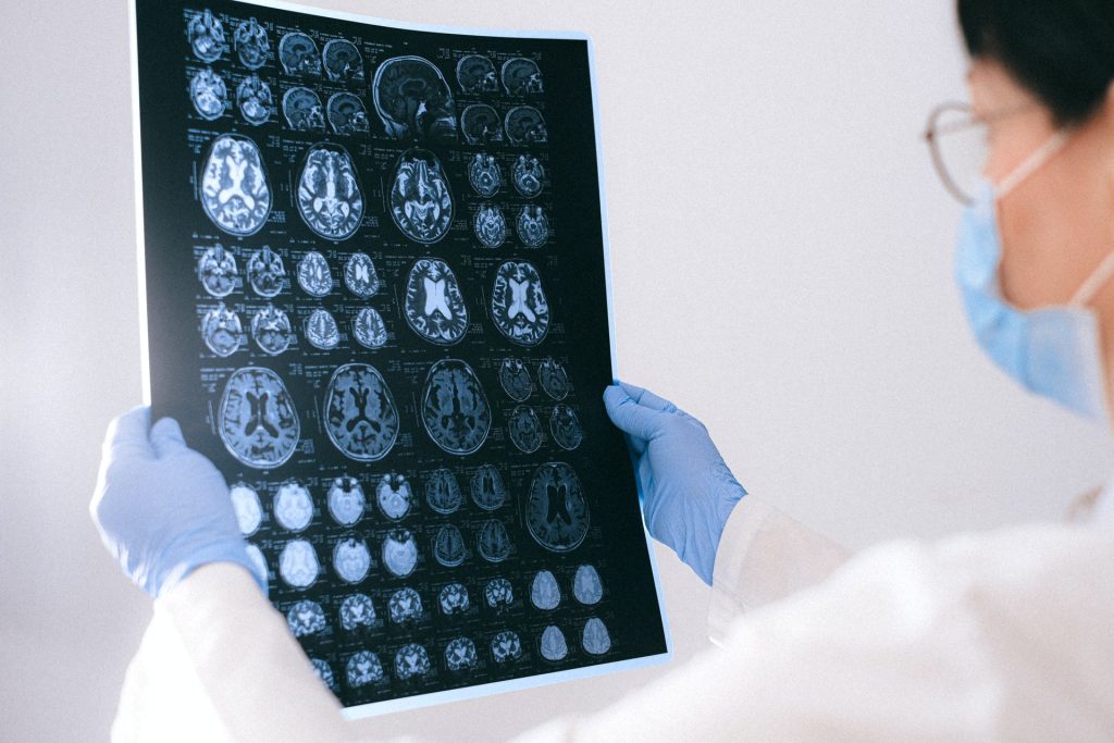
MRI images of the brain
Photo by Anna Shvets on Pexels

Scientists investigating why people who have had shingles have an increased stroke risk now believe the answer lies within, exosomes, lipid vesicles called that shuttle proteins and genetic information between cells. Their study, published The Journal of Infectious Diseases, details the mechanisms behind the link between shingles and strokes.

“Most people know about the painful rash associated with shingles, but they may not know that the risk of stroke is elevated for a year after infection,” said the study’s lead author Andrew Bubak, PhD, assistant research professor in the Department of Neurology at the University of Colorado School of Medicine. “Importantly, the rash is often completely healed and individuals feel normal but nonetheless are walking around with this significant elevation in stroke risk.”

Herpes zoster (HZ) or shingles is caused by the varicella zoster virus which causes chicken pox. The virus lingers in the ganglionic neurons and can reactivate, causing excruciating pain. But researchers have found that shingles can also increase the risk of stroke especially for those under age 40 where the shingles vaccine is not typically recommended.

The risk is greatest in people with the rashes on their faces, perhaps due to the proximity to the brain.

To better understand how this works, Bubak and his team began looking more closely at exosomes.

“Exosomes carry pathogenic cargo that can cause thrombosis and inflammation distant from site of actual infection,” Bubak said. “That could ultimately lead to a stroke in patients.”

Researchers collected plasma samples from 13 patients with shingles and 10 without. The samples were taken at time of infection and at 3-month follow-ups for a subset of patients and exosomes were extracted from the plasma.

The researchers found prothrombotic exosomes which could cause blood clots in those with the infection. They also discovered proinflammatory exosomes that also pose risks for stroke at the 3-month follow-up.

Bubak said the findings suggest that in a subset of people with shingles, the virus may not return to latency or the circulating exosomes that induce a prolonged prothrombotic state may persist even after therapy is done and the rash is gone. He said using antiviral agents longer with the addition of antiplatelet and anti-inflammatory agents could help.

“As well as initiatives to increase HZ vaccine uptake to decrease stroke risk, particularly in individuals with known preexisting stroke risk factors,” said Bubak. “If these findings are confirmed with a larger longitudinal study, then this could change clinical practice.”

Most physicians are unaware of the connection between shingles (which has an effective vaccine) and stroke.

“But it’s really important and so easily mitigated,” Bubak said. “Send them home with antiplatelet agents.”

Source: University of Colorado Anschutz Medical Campus

Smartphone Video of Carotid Arteries Predicts Stroke Risk

Credit: American Heart Association

Narrowed arteries in the neck – a major risk factor for stroke – may be detected by analysing smartphone video that picks up the motion of blood flowing just beneath the skin, a small study shows.

The research, published Wednesday in the Journal of the American Heart Association, may be useful in developing a non-invasive, early screening tool for detecting blockages in the carotid arteries that can lead to strokes.

“Between 2% and 5% of strokes each year occur in people with no symptoms, so better and earlier detection of stroke risk is needed,” study author Dr. Hsien-Li Kao said in a news release. He is an interventional cardiologist at National Taiwan University Hospital in Taipei.

“This was an exciting ‘eureka’ moment for us,” he said. “Existing diagnostic methods – ultrasound, CT and MRI – require screening with specialised medical imaging equipment and personnel. Analysis of video recorded on a smartphone is non-invasive and easy to perform, so it may provide an opportunity to increase screening.”

The carotid arteries, found in the neck, can become blocked by a buildup of fatty deposits known as plaque. That condition – carotid artery stenosis – restricts blood flow to the brain and may lead to an ischemic stroke. Nearly 87% of all strokes in the US are this type of stroke.

The carotid artery is just below the skin’s surface. When velocity and blood flow patterns change, those changes are reflected in the motion of the overlying skin, Kao said. However, those differences cannot be detected by the naked eye.

In the study, researchers used motion magnification and pixel analysis to detect subtle changes in pulse characteristics on the skin’s surface captured in 30-second smartphone video recordings. An older-generation smartphone was used to make video clips of the necks of 202 Taiwanese adults, who were an average 68 years old when the study took place between 2016 and 2019. While recordings were being made, participants lay on their backs with their heads tilted back in a custom-made box that restricted movement.

Among participants, 54% had previously been diagnosed with a blockage of 50% or more in the carotid artery. The phone videos were 87% accurate in predicting who had a blockage in the artery. Narrowing in the arteries was confirmed using a Doppler ultrasound test.

Kao said further research could determine whether it is possible to take recordings and perform the motion analysis remotely, in conjunction with a downloadable app.

“More research is needed to determine whether video recorded on smartphones is a promising approach to help expedite and increase stroke screening,” he said. “Carotid artery stenosis is silent until a stroke happens. With this method, clinicians may be able to record a video of the patient’s neck with a smartphone, upload the videos for analysis and receive a report within five minutes. The early detection of carotid artery stenosis may improve patient outcomes.”

Source: American Heart Association

Falling Victim to Fraud Has a Lasting Impact on Men’s Blood Pressure

A new study published in the Journal of the American Geriatrics Society suggests that experiencing financial exploitation, fraudulent schemes, and scams may raise a person’s blood pressure, especially in later life. A key difference in the findings was that fraud victimisation was linked with elevated blood pressure in men, but not in women.

Instead of focusing on subjective measures of health after fraud vicitimisation, this study included objective measures of physical health, specifically, systolic and diastolic blood pressure, pulse pressure, and mean arterial pressure. Chronic elevation of these measures are known to contribute to end organ damage including stroke, cardiovascular disease morbidity, and mortality. 

The study participants consisted of 1200 older adults from the Rush Memory and Aging Project. During up to 11 years of annual observations, participants were asked about fraud victimisation and underwent serial blood pressure measurements.

In men, blood pressure elevations were observed after they had been the victims of fraud. Those elevations, compounded over time, could indicate future poor health. The rise in blood pressure persisted for years after the fraud had taken place, especially in old age.

“These findings show that fraud victimisation has important public health consequences and underscore the need for efforts to prevent exploitation,” said lead author Melissa Lamar, PhD, of Rush University Medical Center.

Source: Wiley

Gout Flare-ups Linked to Increased MI and Stroke Risk

Photo by freestocks.org

The risk of myocardial infarction (MI) and strokes temporarily increases in the four months after a gout flare, suggests a study published in the journal JAMA.

The findings showed that gout patients who suffered from an MI or stroke were twice as likely to have had a gout flare in the 60 days prior to the event, and one and a half times more likely to have a gout flare in the 61-120 days prior.

Gout is a common form of arthritis that is caused by high levels of uric acid, a chemical produced by breakdown of body tissues and present in certain foods and drinks.

At high levels, uric acid is deposited in and around joints as needle shaped urate crystals. Once released from their deposits, these crystals cause severe inflammation that manifest as joint pain, swelling, redness, and tenderness that often lasts for 1–2 weeks. These episodes, called gout flares, often recur. Inflammation is also a risk factor for heart attack and stroke.

While gout patients tend to have more cardiovascular risk factors, there have been no previous studies about whether gout flares are linked with an increased risk of MI and stroke.

To address this, the team used data from 62 574 patients with gout treated in the NHS. Of these, 10 475 experienced heart attack or stroke after the diagnosis of gout, while matched controls did not experience such events. They evaluated the association between heart attacks or strokes and recent gout flares and adjusted these results for possible confounding factors. They found that gout patients who suffered an MI or stroke were twice as likely to have had a gout flare in the 60 days prior to the event, and one and a half times more likely to have a gout flare in the preceding 61–120 days.

They found a similar high rate of MI or stroke in the 0–60 and 61–120 days after gout flares compared with other time periods, when they used information from only patients who consulted for a gout flare and also experienced either MI or stroke. This further strengthened the finding that gout flares are associated with a transient increase in cardiovascular events following flares. The increased odds and rates persisted when people with pre-existing heart disease or stroke before their gout diagnosis were excluded, and when shorter exposure periods such as 0-15 and 16-30 days prior to MI or stroke, were considered.

Gout patients who died from a MI or stroke had over four times the odds of experiencing a gout flare in the preceding 0-60 days and over twice the odds of gout flare in the preceding 61-120 days.

The study’s lead author, Professor Abhishek at the University of Nottingham, said: “This is the first study of its kind to examine whether there is an association between recent gout flares and heart attacks and strokes.

“The results show that among patients with gout, patients who experienced a heart attack or stroke had significantly increased odds of a gout flare during the preceding 120-days compared with patients who did not experience such events. These findings suggest that gout flares are associated with a transient increase in cardiovascular events following flares.

“People with recurrent gout flares should be considered for long-term treatment with urate lowering treatments such as allopurinol. This is a reliable way of removing urate crystal deposits and providing freedom from gout flares. Patients should also be considered for concurrent treatment with anti-inflammatory medicines such as colchicine for the first few months because urate lowering treatments may trigger gout flares in the short term.

“People with gout should be encouraged to adopt a healthy lifestyle with appropriate treatment of conditions such as high blood pressure, high cholesterol, obesity and diabetes to minimise their background risk of heart attack and stroke.”

Source: University of Nottingham

Blood Vascular Network Retains the ‘Memory’ of a Stroke

Credit: American Heart Association

A study into the structure of blood vascular network structure found that it is dynamic and can adapt to external factors, resulting in a kind of memory of certain events such as an ischaemic stroke. In particular, the study researchers found that rarely used connections incrementally weaken until they disappear eventually.

Researchers from the Max Planck Institute for Dynamics and Self-Organization in Göttingen and the Technical University of Munich used computer simulations to model vascular networks and identified adaptation rules for their connections.

“We found that the strength of a connection within a network depends on the local flow,” explained Karen Alim, corresponding author of the study. “This means that links with a low flow below a certain threshold will decay more and more until they eventually vanish,” she continued. Since the limited amount of material available to build the vascular system needs to be efficiently used, this mechanism offers an elegant way to streamline the vascular system.

Persistent changes in the network

Once a connection has become very weak due to a low flow rate, recovering that connection is very difficult. For example, a blood vessel blockage of the type that could lead to an ischaemic stroke. During an ischaemic stroke, some blood vessels in the affected region are weakened by the blockage.

“We found that in such a case, adaptations in the network are permanent and are maintained after the obstacle is removed. One can say that the network prefers to reroute the flow through existing stronger connections instead of re-growing weaker connections – even if the flow would require the opposite,” explained Komal Bhattacharyya, principal author of the study.

The researchers have thus shown that blood flow permanently changes even after successful removal of the clot. This memory capability of networks can also be found in other living systems: for example, the slime mould Physarum polycephalum uses its adaptive network to navigate its environment based on imprints by food stimuli, as demonstrated previously.

The study was published in Physical Review Letters.

Source: Max Planck Institute for Dynamics and Self-Organization

New Study Launched to Examine How Sleeps Aids Stroke Recovery

Sleeping woman
Photo by Cottonbro on Pexels

Researchers at the University of East Anglia are launching a new study to investigate how sleep can aid in stroke recovery, by examining whether people’s sleep patterns influence recovery of neuromuscular function.

Lead researcher Prof Valerie Pomeroy, from UEA’s School of Health Sciences, said: “We want to better understand how the brain recovers after a stroke – so we will be investigating how stroke survivors regain movement, and how this is influenced by sleep and time.

“We hope to find out more about sleep patterns that are beneficial for movement recovery after stroke.”

The team are looking for people in the region who have had a stroke to take part in the study. Participants will undertake measures of daily activity, sleep and movement.

The project will involve measuring people’s movement using small sensors placed on the skin’s surface that record natural muscle activity whilst they carry out a daily task – picking up a telephone.

Participants will be asked to attend two visits at the university, during which participants will undertake the movement measures and complete questionnaires about how they sleep. 

In-between visits, participants will wear a motion watch on each wrist for seven days to measure their everyday activity at home.

Prof Valerie Pomeroy said: “There is strong evidence that physiotherapy improves the ability of people to move and be independent after a stroke.  But at six months after stroke many people remain unable to produce the movement needed for everyday activity such as answering a telephone. 

“We are undertaking this study to understand more about whether this situation could be improved by using interventions to change a patient’s sleep pattern and thus improve recovery of movement ability.”

Source: University of East Anglia

The American Heart Association’s New Intracerebral Haemorrhage Guideline

Credit: American Heart Association

A new guideline published in the journal Stroke reveal that home treatments or preventive therapies used to manage intracerebral haemorrhages (ICH) are not as effective as previously believed.

The guideline from the  American Heart Association/American Stroke Association includes recommendations on surgical techniques, individual activity levels after an ICH, and additional education and training for at-home caregivers. It reflects advances in the intracerebral haemorrhage field since the last guideline on ICH management was published in May 2015.

“Advances have been made in an array of fields related to ICH, including the organisation of regional health care systems, reversal of the negative effects of blood thinners, minimally invasive surgical procedures and the underlying disease in small blood vessels,” said Steven M. Greenberg, M.D., Ph.D., FAHA, chair of the guideline writing group.

Updates to Standard Care Practices

The new guideline suggests that many techniques widely considered “standard care” are unnecessary. For example, wearing compression socks or stockings to prevent deep vein thrombosis after ICH was not found to be effective. Instead, use of intermittent pneumatic compression may be helpful if started on the same day of an ICH diagnosis.

“This is an area where we still have a lot of exploration to do. It is unclear whether even specialised compression devices reduce the risks of deep vein thrombosis or improve the overall health of people with a brain bleed. Even more research is needed on how new blood clot prevention medications may help, especially within the first 24 to 48 hours of the first symptoms,” said Dr Greenberg.

Use of anti-seizure medicines or anti-depressants after ICH is also updated; neither of these classes of medications helps a person’s overall health unless a seizure or depression is already present, therefore, they are not advised for most people. Anti-seizure medication did not contribute to improvements in functionality or long-term seizure control, and the use of anti-depressants increased the chance of bone fractures.

The guideline writing group also addresses previously standard in-hospital therapies. They suggest administering steroids to prevent complications from a bleeding stroke is ineffective and highlight that platelet transfusions, unless used during an emergency surgery, may worsen the stroke survivor’s condition.

Surgical Intervention

Some research suggests procedures with a less invasive approach are less likely to damage brain tissue while removing the fluid build-up.

“The evidence is now reasonably strong that minimally invasive surgery may improve the likelihood that a patient will survive following a moderate or large ICH,” says Greenberg. “It is less clear, however, whether this or any other kind of surgical procedure improves the chances of survival and recovery from ICH, which are our ultimate goals.”

Recovery and Rehabilitation

Stroke rehabilitation includes several strategies to help restore the individual’s quality of life, and the guideline reinforces the importance of having a multi-disciplinary team to develop a plan for recovery. Research suggests a person with a mild or moderate ICH may begin activities like stretching, dressing, bathing and other normal daily tasks 24–48 hours after the stroke to improve survival rate and recovery time; however, moving too much or too intensely within 24 hours is linked to an increased risk of death within 14 days after an ICH.

Home Caregivers

The guideline also recommends education, practical support and training for family members so they may be involved and knowledgeable about what to expect during rehabilitation.

Other Highlights

The guideline suggests there may be an opportunity to prevent ICH in some people by using MRI which can image small blood vessel damage. In addition, major risk factors for small vessel damage are high blood pressure, Type 2 diabetes and older age. Blood thinners remain an important topic since the use of these medications may increase complications and death from a bleeding stroke. Updated guidance is provided for immediate reversal of the newer blood thinners like apixaban, rivaroxaban, edoxaban and dabigatran, as well as older medications like warfarin or heparin.

Renewed emphasis is placed on the complexities of a do-not-attempt-resuscitation (DNAR) status versus the decision to limit other medical and surgical interventions. The writing group highlights the need to educate medical professionals, stroke survivors and/or the individual’s caregiver about the differences. The guideline recommends the severity of a hemorrhage, as measured by the standard scales, not be used as the sole basis for determining life-saving treatments.

Source: American Heart Association

Intensive Hypertension Treatment may Prevent Strokes in Older Adults

Photo by Kindel Media on Pexels

More intensive hypertension treatment could help prevent or delay strokes in older adults, according to an analysis of results from randomised clinical trials published in the Journal of the American Geriatrics Society.

The researchers initially screened 22 trials for inclusion. Nine trials involving 38 779 adults with an average age ranging from 66 to 84 years were included in the analysis, with follow-up times ranging from 2.0 to 5.8 years.

On average, the researchers found that it took 1.7 years to prevent 1 stroke for 200 older persons treated with more intensive hypertension treatment.

For older adults with baseline systolic blood pressures below 150 mmHg, the time to benefit from more intensive hypertension treatment was longer than 1.7 years; for older adults with baseline systolic blood pressure above 190 mmHg, the time to benefit was shorter than 1.7 years.

In their discussion, the researchers noted the risks of aggressive hypertension treatment, including hypotension, syncope and falls. However, they noted that emerging evidence shows that the increase in fall risk is transient.

“While the 2017 American College of Cardiology/American Heart Association guidelines recommend individual risk discussions about hypertension treatment for primary prevention in older adults, there is a critical gap in data about how long a patient needs to receive blood pressure treatment before they will benefit – or the blood pressure treatment’s time to benefit,” said lead author Vanessa S. Ho, MS, of California Northstate University College of Medicine. “A treatment’s time to benefit is an especially important consideration for patients with a limited life expectancy who may experience immediate burdens or harms from any additional medication.”

Source: Wiley

American Heart Association’s In-hospital Stroke Evaluation and Treatment Recommendations

Image copyright American Heart Association

Despite the fact that hospitalised patients are in a monitored environment, stroke evaluation and treatment are often delayed compared to patients arriving with a stroke at the emergency department, contributing to higher rates of morbidity and mortality for in-hospital stroke. 

This is according to an American Heart Association scientific statement published in Stroke. This scientific statement was discussed at the Association’s International Stroke Conference in New Orleans. An American Heart Association scientific statement is an expert analysis of current research and may inform future clinical practice guidelines. This follows on from a previous 2019 update on recommendations systems of care to improve patient outcomes in stroke.

The statement outlines five elements for the development of hospital systems of care and targeted quality improvement to reduce delays and optimise treatment to improve outcomes for patients who experience an in-hospital stroke. In-hospital stroke is a stroke that occurs during a hospitalisation for another diagnosis and affects between 35 000 and 75 000 hospitalised patients annually in the United States.

The five core elements of the statement are:

  • training all hospital staff on stroke signs, symptoms and activation protocols for in-hospital stroke alerts;
  • creating rapid response teams with dedicated stroke training and immediate access to neurologic expertise;
  • standardising the evaluation of potential in-hospital stroke patients with physical assessment and imaging;
  • eliminating and addressing potential treatment barriers including interfacility transfer to advanced stroke treatment; and
  • establishing an in-hospital stroke quality oversight program delivering data-driven performance feedback and driving targeted quality improvement efforts.

The statement encourages institutions to develop a plan for in-patient stroke response teams that includes education, quality review and specified oversight.

The statement was developed by the writing committee on behalf of the American Heart Association’s Stroke Council; the Council on Arteriosclerosis, Thrombosis and Vascular Biology; the Council on Cardiovascular and Stroke Nursing; the Council on Clinical Cardiology; and the Council on Lifestyle and Cardiometabolic Health. The diverse committee included experts in nursing, neurology, internal medicine, neurocritical care, neurosurgery and neurointerventional radiology. The American Academy of Neurology affirms the value of this statement as an educational tool for neurologists, and the American Association of Neurological Surgeons/Congress of Neurological Surgeons Cerebrovascular Section affirms the educational benefit of this statement.

American Heart Association scientific statements promote greater awareness about cardiovascular diseases and stroke issues and help facilitate informed health care decisions. Scientific statements outline what is currently known about a topic, and what areas need additional research. While scientific statements inform the development of guidelines, they do not make treatment recommendations. American Heart Association guidelines provide the Association’s official clinical practice recommendations.

Source: American Heart Association

Selenium Could Help Reverse Cognitive Decline

Photo source: Pixabay

The trace metal selenium could help reverse the cognitive impact of stroke and boost learning and memory in ageing brains, according to a study published in Cell Metabolism.

Previous studies on the impact of exercise on the ageing brain found levels of a protein key to transporting selenium in the blood were elevated by physical activity.

Lead researcher Dr Tara Walker said: “We’ve known for the last 20 years that exercise can create new neurons in the brain, but we didn’t really understand how,” Dr Walker said.

The research team sought to find out whether dietary selenium supplements could replicate the effects of exercise.

“Our models showed that selenium supplementation could increase neuron generation and improve cognition in elderly mice,” Dr Walker said. “The levels of new neuron generation decrease rapidly in aged mice, as they do in humans. When selenium supplements were given to the mice, the production of neurons increased, reversing the cognitive deficits observed in ageing.”

Selenium is an essential trace metal which can play an important role in human health. It is absorbed from soil and water and is found in foods such as grains, meat and nuts, with the highest levels found in Brazil nuts. The researchers also investigated whether selenium would have an impact on post-stroke cognitive decline.

“Young mice are really good at the learning and memory tasks, but after a stroke, they could no longer perform these tasks,” Dr Walker said. “We found that learning and memory deficits of stroke affected mice returned to normal when they were given selenium supplements.”

Dr Walker said the results opened a new therapeutic avenue to boost cognitive function in people who were unable to exercise due to poor health or old age.

“However, selenium supplements shouldn’t be seen as a complete substitute for exercise, and too much can be bad for you,” she said. “A person who is getting a balanced diet of fruits, nuts, veggies and meat usually has good selenium levels. But in older people, particularly those with neurological conditions, selenium supplements could be beneficial.”

Source: University of Queensland