Tag: MRI

New Antenna Upgrade Boosts MRI Image Quality

Photo by Mart Production on Pexels

Magnetic resonance imaging (MRI) is one of medicine’s most powerful diagnostic tools. But certain tissues deep inside the body – including brain regions and delicate structures of the eye and orbit that are of particular relevance for ophthalmology – are difficult to image clearly. The problem is not the scanner itself, but the hardware that sends and receives radio signals. 

Now, researchers at the Max Delbrück Center have developed an advanced materials-based MRI antenna that overcomes these limitations – delivering enhanced images more quickly and that can be used in existing MRI machines. The research, led by Nandita Saha, a doctoral student in the Experimental Ultrahigh Field Magnetic Resonance lab of Professor Thoralf Niendorf, was published in Advanced Materials.

Niendorf and his team worked closely with researchers at Rostock University Medical Center, combining expertise in MRI physics with clinical ophthalmology and translational imaging. The Rostock team is also supporting clinical validation of the technology.

“By using concepts from metamaterials, we were able to guide radiofrequency fields more efficiently and demonstrate how advanced physics can directly improve medical imaging,” says Niendorf, senior author of the paper. “This work shows a pathway toward faster, clearer MRI scans that could benefit patients in many clinical areas.” 

Rethinking MRI hardware with metamaterials

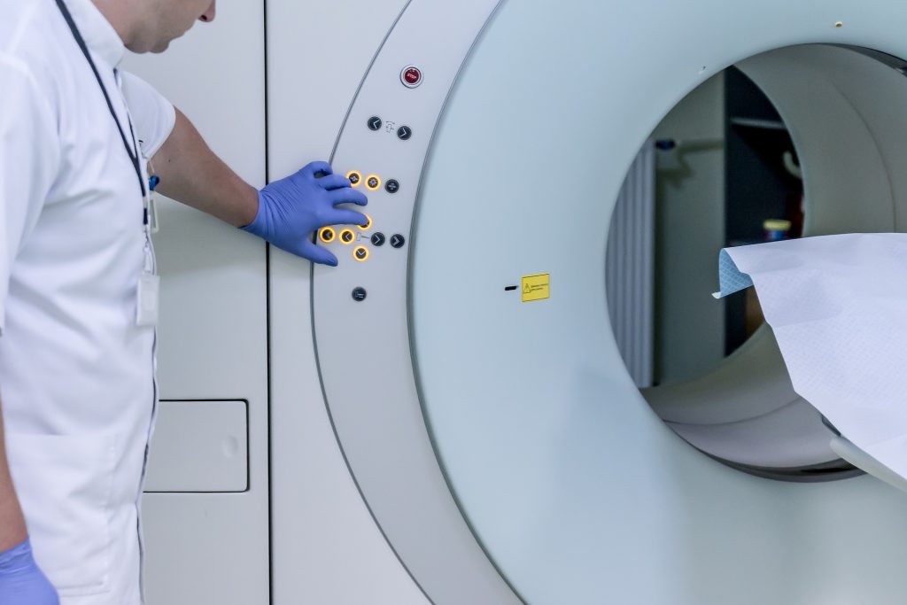
MRI works by sending radiofrequency (RF) signals into the body and detecting how tissues respond inside a strong magnetic field. The stronger the signal response, the better the image. Conventional MRI antennas – also called RF coils – often struggle to collect enough signal from deep or anatomically complex regions. This leads to images that lack detail and prolongs scan times.

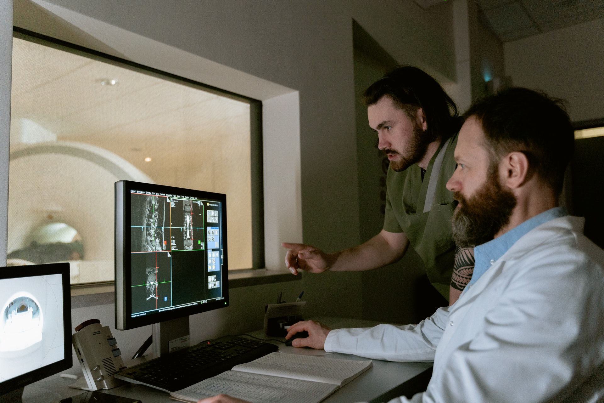
The research team addressed this bottleneck by integrating metamaterials directly into the MRI antenna. Metamaterials are engineered structures that interact with electromagnetic waves in ways not found in natural materials. The engineered RF antenna increases signal strength from targeted tissues, improves spatial resolution and image sharpness and enables faster data acquisition. Crucially, the antenna fits into existing MRI systems, avoiding the need for new infrastructure. The team validated the technology by imaging the eye and orbit region in a group of volunteers at 7.0 Tesla.

MRI image of an eye, the eye socket and the brain.
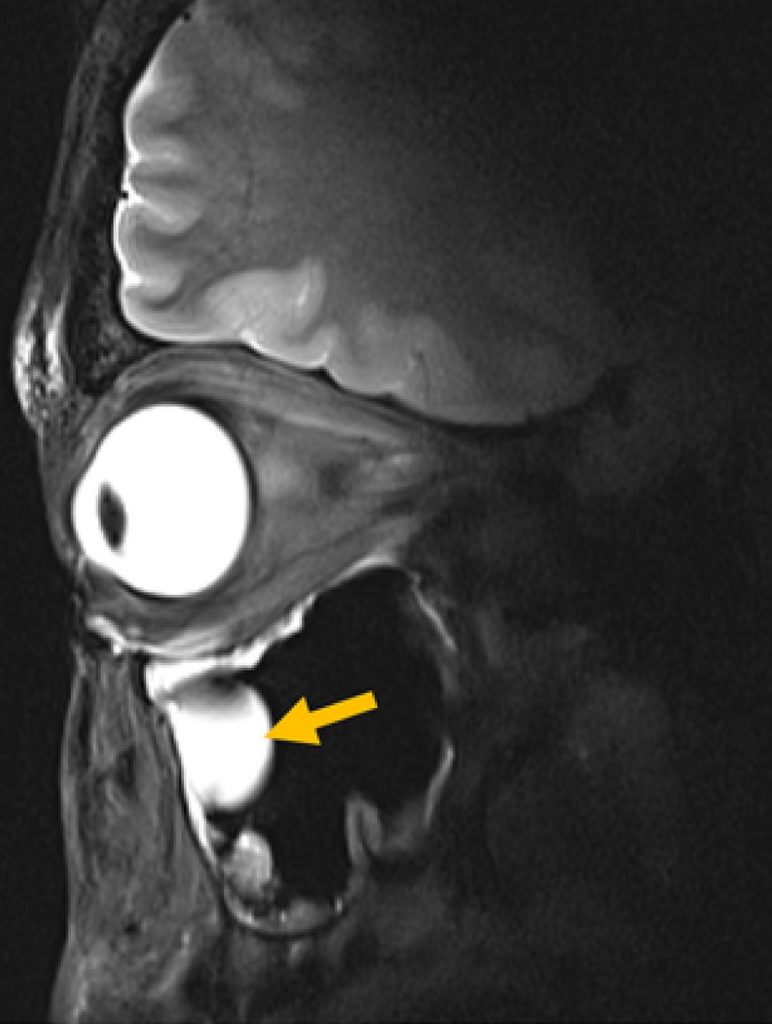
© AG Niendorf, Max Delbrück Center

“Our research demonstrates clear relevance for ophthalmological applications as it can facilitate anatomically detailed, high-spatial resolution MRI of the eye,” says Professor Oliver Stachs, a co-author of the paper at University Medicine Rostock. “It offers the potential to open a window into the eye and into (patho)physiological processes that in the past have been largely inaccessible.” 

“Our goal was to rethink MRI hardware from the modern physics of antenna design,” adds Saha. This technology can also be tuned to protect sensitive areas of the body during MRI, for example, to reduce unwanted heating around medical implants, she adds. It could also be used to focus RF energy more effectively for MRI guided therapies for various cancer treatments, such as gentle heating of tumors (hyperthermia) or thermal ablation of tissue. 

Better diagnostics

For patients, MRI scans can be uncomfortable and time-consuming – even more so when images need to be repeated because important details are hard to see. Faster scans mean patients spend less time inside scanners. Clearer images mean doctors can make diagnoses with greater confidence. And because the new antenna is lightweight and compact, it can also be designed to better fit specific parts of the body, improving comfort even further.

The technology could also be adapted to support MRI systems running at magnetic field strengths lower or higher than 7.0T, to image target anatomy other than the eye, orbit or the brain or to track metabolism or drug movement inside the body, says Niendorf. Special MRI scans that use other atoms, such as sodium or fluorine, could also benefit from this technology by producing clearer signals and better images, he adds. 

“Innovations in imaging hardware have the potential to transform diagnostics, and this study is an important step toward next-generation MRI technology,” says Dr Ebba Beller, a co-author of the paper at Rostock University Medical Center.

The researchers are already planning larger studies at multiple hospitals and adapting the design for other organs, such as the heart and kidneys. The collaboration will continue to be strengthened by long-standing reciprocal visiting scientist appointments of Stachs and Niendorf. 

Source: Max Delbrück Center for Molecular Medicine

Study Shows Low-Field MRI Is Feasible for Breast Screening

Mass General Brigham’s evaluation of low-field MRI performance lays potential groundwork for this technology to be a lower-cost, accessible option for breast imaging

Photo by National Cancer Institute on Unsplash

Researchers at Mass General Brigham have demonstrated the technical feasibility of using ultra-low field (ULF) magnetic resonance imaging (MRI) for breast imaging. With further refinement and evaluation, the technology could offer an alternative to existing breast cancer screening methods and may reduce barriers to screening. Results are published in Scientific Reports.

“These results are a very encouraging proof of principle, though larger studies are needed to establish diagnostic performance,” said project principal investigator and co-senior author Matthew Rosen, PhD, an associate professor of Radiology and director of the Low Field MRI laboratory in the Athinoula A. Martinos Center for Biomedical Imaging in the Mass General Brigham Department of Radiology. “They motivate our continued pursuit of safe, comfortable, lower-cost screening approaches that can expand access for patients.”

Current U.S. guidelines recommend screening mammography for women aged 40 to 74 years. Unlike mammography, ULF MRI doesn’t require breast compression, which many patients find uncomfortable. Another benefit of ULF MRI is that it doesn’t use ionizing radiation.

While higher risk patients may receive MRI screening for breast cancer, standard MRI machines are not used in routine breast cancer screening because they are expensive and not widely available. ULF MRI systems cost less than 5% of the price of standard MRI systems and have lower long-term operating costs.

In this study, ULF MRI scans were performed on 14 participants, including 11 women with no history of breast cancer, two women with a prior breast cancer diagnosis, and one woman with a benign mass.

When interpreting the ULF MRI scans, three radiologists could reliably identify essential breast features and distinguish fibroglandular tissue from adipose tissue. The authors note that discrepancies were likely related to the novelty of ULF MRI and may be reduced with additional training and experience.

“This early evidence suggests that ULF MRI can detect essential breast features and some abnormalities without radiation or injected contrast,” said co-first author Neha Koonjoo, PhD, an investigator at the Martinos Center. “These findings point to the potential for ULF MRI as an option that could complement existing screening tools in the future.”

“Even at very low magnetic field, the radiology team was able to make observations about the breast,” said co-principal investigator and co-senior author Kathryn E. Keenan, PhD, from the US National Institute of Standards and Technology. “We attempted this study in hopes that the breast features would be visible, but you don’t always have success. We’re very motivated by this study to continue our work on ultra-low-field MRI for breast screening.”

The researchers note that further study is needed to determine the diagnostic accuracy of ULF MRI for breast cancer screening, including studies in larger cohorts that include patients with benign and malignant lesions. They also emphasize that further refinements in ULF MRI technology are needed to meet clinical resolution standards for breast cancer screening.

“These results will guide the next engineering steps to improve image quality and enable a more comfortable exam and help bring screening to more settings and more patients,” said co-first author Sheng Shen, PhD, of the Martinos Center for Biomedical Imaging.

Source: Mass General Brigham

Doctors Complain About Choice of Equipment at Gauteng Hospital as Thousands Await Cancer Scans

Concern about decision to buy Chinese MRI machine from local company instead of one from Philips

Credit: Pixabay CC0

By Chris Bateman and Raymond Joseph

As thousands of cancer patients wait months for diagnostic scans, senior clinicians at Charlotte Maxeke Academic Hospital have questioned a decision by the Gauteng Health Department to override their choice of MRI machine.

In a letter to Gauteng Health Department’s acting chief financial officer, the head of supply chain management at the hospital, Solly Mokgoko, expressed a concern that a recommendation by the head of radiology and the acting clinical director to buy a Philips scanner had been overridden by the Gauteng health department’s central office. The letter is dated 31 October 2025.

Mokgoko said the doctors had preferred the Philips MRI scanner – at a cost of about R27.4-million – on the grounds of “technological advancement, operational sustainability, and clinical research potential”.

However, the department had chosen a machine from Mamello Clinical Solutions at R38.5-million, they said. The room in which the machine will be installed is currently being prepared.

The letter said the Philips unit’s cost “offers reduced lifecycle expenditure due to minimal helium dependency and extended operational uptime”. The Philips scanner used low-maintenance technology, “requiring minimal or no helium top-ups, thereby reducing lifecycle costs and mitigating downtime risks”.

The Mamello-proposed model, by contrast, “relies on traditional cryogenic technology, which entails higher running costs and environmental exposure”, they said.

They said the decision is inconsistent with value-for-money principles set out in the Public Finance Management Act (PFMA) and Treasury regulations.

The purchase of a Chinese MRI scanner from Mamello is part of a R304-million roll-out of eight scanners across Gauteng public hospitals, in which roughly R190-million has been awarded to Mamello Clinical Solutions (five machines) and the remainder to Philips SA.

The Gauteng Department of Health rejected any suggestion of irregularity, saying the purchase was made under a lawful, competitively awarded contract and that both suppliers met the required technical standards.

In this case, the original procurement contract was drawn up by the Limpopo Health Department, with the Gauteng department piggybacking on it.

Clinicians at Charlotte Maxeke who spoke to GroundUp say the procurement shift occurred without adequate consultation and against explicit technical recommendations — allegations the department disputes.

Approximately 2,600 oncology patients are awaiting MRI scans at Charlotte Maxeke alone, with outpatient bookings extending to December 2026. Similar waiting lists exist at Chris Hani Baragwanath Academic Hospital.

The letter said that besides the external patient scans waiting list, there are over 50 inpatients awaiting scans.

One department head said: “How can the hospital order an MRI that’s over R10-million more expensive in an environment where it can’t even provide decent food, [and where there is] widespread cost-cutting and a dire shortage of doctors?” Late last year, the hospital made headlines for shortages of adequate patient meals.

Mamello Clinical Solutions, a private company based in Polokwane, was established in December 2014, trading as Mamello Development until 2019 when it changed its name. Robert Makhubedu, its sole director, was appointed in June 2023 after two previous directors resigned, according to official company registration records.

Makhubedu previously worked as chief radiographer at Charlotte Maxeke Hospital in the early 1990s, then spent more than two decades as director of business development at Tecmed, before joining Mamello Clinical Solutions.

A Gauteng Health Department spokesperson “categorically” denied any irregular, inflated or non-compliant procurement.

He said the MRI acquisitions had been made under a lawful, competitively advertised contract which had been evaluated in line with constitutional, PFMA and Treasury requirements.

Philips Healthcare and Mamello Clinical Solutions had both met minimum safety, functional and performance specifications, he said.

While acknowledging that Charlotte Maxeke clinicians preferred the Philips MRI, the spokesperson said procurement decisions could not be driven by “brand preference or proprietary technology.” He said over the life of the machine the price difference between the two was about R1.07-million, not R11.1-million.

Treasury rules, he said, did not permit sole-supplier selection where multiple bidders meet approved specifications. Multi-supplier models were standard public-sector practice.

Makhubedu pointed out that the tender had not called for a “helium-free” scanner. He attributed the doctors’ complaints to a combination of “brand bias” and hostility towards emerging black-owned companies, compared to multinationals.

“Some black companies awarded these contracts in the past could not relate to the business and clinical profile of the projects,” he said. “The legacy of that is that you have to prove yourself all the time.”

Makhubedu said that provinces tried to strike a procurement balance between emerging and established companies. He said his scanner was in fact R300,000 cheaper than the Philips machine over the life of the machine, and Mamello was capturing market share because of scanner quality and price.

“We believe we were fairly, legally and transparently awarded the contract. And we were cheaper.”

Republished from GroundUp under a Creative Commons Licence.

Read the original article.

Diagnostic Breast MRI may be Unnecessary for Some Patients with Early-stage Breast Cancer

Adding breast magnetic resonance imaging (MRI) to a diagnostic mammogram did not reduce five-year cancer recurrence rates for patients with stage I/II hormone receptor (HR)-negative breast cancer, according to researchers at The University of Texas MD Anderson Cancer Center. 

The Phase III Alliance A011104/ACRIN6694 trial found that five-year locoregional recurrence rates were 6.8% in patients who received an MRI as part of a diagnostic work-up and 4.3% in those who did not. These data were presented today at the San Antonio Breast Cancer Symposium (SABCS) by principal investigator Isabelle Bedrosian, MD, professor of Breast Surgical Oncology (Abstract GS2-07).

“We have long assumed that finding more breast cancer on an MRI and removing it with surgery would help lower the chance of a patient’s cancer coming back,” Bedrosian said. “When you look at our findings alongside earlier trials, the message is clear: adding MRI before surgery doesn’t improve results for patients – and may not have to be used as a standard part of the diagnostic process.”

No additional MRI benefit in this group

The trial enrolled 319 patients between 2014 and 2018 with newly diagnosed stage I or II HR-negative breast cancer. These patients were eligible for lumpectomy and did not have germline BRCA1/2 mutations, bilateral breast cancer or a history of prior breast cancer. All patients had undergone diagnostic mammography with or without ultrasound prior to trial enrolment.  Patients were randomly assigned to undergo additional imaging by breast MRI (161 patients) or to receive no further imaging (158 patients).

Not only did breast MRI not impact five-year recurrence rates, but there were also not significant differences between groups for five-year distant recurrence-free survival nor overall survival. 

A small subset of patients with tumour subtypes (HR- HER2+ and HR-HER2-) and those over the age of 50 at diagnosis also showed no benefit to MRI.

Pre-op MRI not finding anything important

Breast MRI is a common part of the diagnostic evaluation because it can reveal cancer that mammography might not detect. However, the evidence that it improves surgical outcomes for patients has been limited.

“We believe the reason MRI did not reduce recurrence rates may be twofold,” Bedrosian said. “It is possible that MRI didn’t uncover many lesions that mammography hadn’t already found, or perhaps identifying and surgically removing those additional lesions was not important to reducing risk of the cancer coming back. It’s possible that in the group that did not receive MRI, radiation and chemotherapy effectively treated the occult areas of disease”. 

Experts are now analysing how often breast MRI identified additional lesions in the trial population to better understand why breast MRI did not impact oncologic outcomes.

Study limitations 

Limitations included that most patients involved in the trial had breast cancer that hadn’t spread to their lymph nodes, which may partly explain why recurrence rates were low overall. Despite being open to women of all ages, the study enrolled mostly older women who may have been less likely to benefit from breast MRI. 

Study Highlights the Limits of AI in Heart Care

Human heart. Credit: Scientific Animations CC4.0

There are limits in applying AI to images of the heart, a new study from the Smidt Heart Institute at Cedars-Sinai reveals. The findings were published in the Journal of the American Society of Echocardiography.

Investigators trained multiple artificial intelligence models to read images from echocardiograms, a type of ultrasound test that evaluates the structure and function of the heart. Their goal was to determine whether AI could use these images to calculate measurements like inflammation and scarring that are normally obtained through another, more costly test called cardiac magnetic resonance imaging (CMRI). By examining findings from 1453 patients who had undergone both tests, they found the AI models could not accomplish this task.

“As compared to echocardiograms, cardiac MRI machines are expensive and not available for many patients, especially those in rural areas, so we had hoped that AI could reduce the need for it,” said Alan Kwan, MD, assistant professor in the Department of Cardiology in the Smidt Heart Institute at Cedars-Sinai and co-senior author of the study. “Our results showed the limited powers of AI in this area.”

Source: Cedars-Sinai Medical Center

New Method Noninvasively Assesses Achilles Tendon in Dancers

Photo by Nihal Demirci on Unsplash

A study in the Journal of Orthopaedic Research uses a noninvasive, nonradioactive imaging-based method to measure the structure and function of the Achilles tendon in professional ballet dancers. The method could potentially be developed to help prevent injuries and improve rehabilitation efforts in athletes, as well as in the general public.

The study involved what is called multi-echo ultrashort echo time (UTE) magnetic resonance imaging (MRI) to assess collagen and other components of the Achilles tendon. These structural UTE MRI assessments were combined with functional assessments of the Achilles tendon based on sheer wave elastography (SWE) ultrasound, which measures tendon stiffness.

Professional dancers tended to have more tendon stiffness compared with non-dancers, consistent with prior observations of a training effect from repeated loading with exercise. UTE MRI measures corresponded with the degree of stiffness from SWE ultrasound.

“These findings highlight the potential of integrating UTE and SWE imaging to investigate tendon structure‐function relationships and adaptations to mechanical loading,” the authors write. “Enhanced structure‐function assessment of tendon health and injury status could improve rehabilitation protocols or injury prevention strategies for athletes, including professional dancers.”

Source: Wiley

Faster MRI Scans Offer New Hope for Dementia Diagnosis

Photo by Mart Production on Pexels

The time to carry out diagnostic MRI scans for dementia can be cut to one third of their standard length, according to a new study led by UCL researchers.

The findings, published in Alzheimer’s & Dementia, have been described as a step towards ending ‘the postcode lottery in dementia diagnosis’. Shorter scans would be easier and more comfortable for patients and also enable more people to be scanned at a lower cost. The team behind the study say this could at least double the number of dementia scans able to be done in one day.

Senior author Professor Nick Fox, Director of the UCL Dementia Research Centre at the UCL Queen Square Institute of Neurology, said: “As more treatments that can slow or change the course of dementia are being developed, it’s important to make sure MRI scans are available to everyone. This is because people living with dementia often need an MRI scan as part of their diagnosis before they can access these treatments.

“To help make this possible, our team carried out the first study looking at how new imaging techniques – called parallel imaging – could speed up MRI scans in clinics. Their goal is to move closer to a future where every person with dementia can get a diagnosis through a scan.”

MRI scans often play a key role in an accurate dementia diagnosis, including ruling out other causes of symptoms and assisting in diagnosing the type of dementia. Emerging disease-modifying treatments such as lecanemab and donanemab also require an MRI scan before starting treatment and for safety monitoring during the course of treatment. Reducing the cost of scanning would contribute to lowering the total cost of delivering for such treatments. 

The ADMIRA study (Accelerated Magnetic Resonance Imaging for Alzheimer’s disease), part funded by Alzheimer’s Society’s Heather Corrie Impact Fund, aimed to understand the reliability of fast MRI scans compared to standard-of-care clinical scans. The neurologists on the study were joined by co-authors from the UCL Hawkes Institute and the UCL Advanced Research Computing Centre in the faculty of Engineering.

The research team scanned 92 people in an outpatient setting where an MRI brain scan was planned as part of their routine clinical assessment. The accelerated scans were carried out and enhanced to increase the quality of the image using new scanning methods. Three neuroradiologists examined these scans, and weren’t aware if they were looking at fast or standard-of-care scans.

Co-author Professor Geoff Parker (UCL Hawkes Institute and UCL Medical Physics and Biomedical Engineering) said: “Our research has taken advantage of recent breakthroughs in scanner technology. Our task was to work out just how fast we could scan while maintaining image quality good enough for diagnosis.”

The team found that the quicker scans reduced time in the scanner by 63% and they were as reliable as the standard-of-care scans for diagnosis and visual ratings.

First author Dr Miguel Rosa-Grilo (UCL Queen Square Institute of Neurology) said: “We were confident that the new scan would prove non-inferior to the standard scan, given the high image quality – but it was remarkable how well it performed.”

Richard Oakley, Associate Director of Research and Innovation at Alzheimer’s Society, said: “Dementia is the UK’s biggest killer, but one in three people living with the condition haven’t had a diagnosis. An early and accurate diagnosis isn’t just a label, it’s the first step to getting vital care, support and treatment.

“While MRIs aren’t the only way to diagnosis dementia, very few people with concerns about their cognitive health are offered one as part of the diagnosis process, mainly because they are expensive and not widely available. These faster MRIs, which take less than half the time of standard scans, could help end this postcode lottery in dementia diagnosis, cut costs and potentially give more people access to them.

“MRI scans can be an uncomfortable and daunting experience for patients, so anything we can do to make it an easier process is really positive.

“So far, this shortened MRI scan has been tested at one specialist centre with one type of MRI scanner, so more research is needed to make sure this works across different types of scanners and a diverse range of people. We’re hugely encouraged by this progress and eager to see how it continues.”

The team will now build on their early results by making sure the approach works across different types of MRI machines, so it can benefit as many hospitals and clinics as possible.

Source: University College London

Gluten Free Diet Reduces Coeliac Symptoms – and ‘Good’ Gut Bacteria

Photo by Mariana Kurnyk: https://www.pexels.com/photo/two-baked-breads-1756062/

A research team led by the University of Nottingham has used magnetic resonance imaging (MRI) to better understand the impact a gluten free diet has on people with coeliac disease, which could be the first step towards finding new ways of treating the condition.

The MARCO study – MAgnetic Resonance Imaging in COliac disease is published in Clinical Gastroenterology and Hepatology (CGH) (link connects to BioRxiv copy).

Coeliac disease is a chronic condition affecting around one person in every 100 in the general population. When people with coeliac disease eat gluten, which is found in pasta and bread, their immune system produces an abnormal reaction that inflames and damages the gut tissue and causes symptoms such as abdominal pain and bloating.

The only treatment is a life- long commitment to a gluten free diet, which helps recovery of the gut tissue but still leaves many patients with gastrointestinal symptoms.

Luca Marciani, Professor of Gastrointestinal Imaging at the University, led the study. He said: “Despite being a common chronic condition, we still don’t precisely know how coeliac disease affects the basic physiological functioning of the gut and how the gluten free diet treatment may further change this.

“We launched the MARCO study to try and address this issue, by using MRI along with gut microbiome analysis to give us new insights into how a gluten-free diet affects people with coeliac disease.”

The team recruited 36 people who had just been diagnosed with coeliac disease and 36 healthy volunteers to participate in the study. Images were taken of their guts with MRI, along with blood and stool samples. The patients then followed a gluten free diet for one year and came back to repeat the study. The healthy participants came back one year later too and repeated the study, but they did not follow any diet treatment.

The study found that the newly diagnosed patients with coeliac disease had more gut symptoms, more fluid in the small bowel and that the transit of food in the bowel was slower than in the healthy controls.

The microbiota (the ‘bugs’ living in the colon) of the patients showed higher levels of ‘bad bugs’ such as E.coli. After one year of a gluten free diet, gut symptoms, bowel water and gut transit improved in the patients, but without returning to normal values. But the gluten free diet also reduced some of the ‘good bugs’ in the microbiota, such as Bifidobacteria associated with reduced intake of starch and wheat nutrients, due to the different diet.

The patient study was conducted by Radiographer Dr Carolyn Costigan, from Nottingham University Hospitals, as part of her PhD studies at the University of Nottingham.

It was particularly interesting to see how the imaging results on gut function correlated with changes in the ‘bugs’ in the colon microbiota. The findings increase our understanding of gut function and physiology in coeliac disease and open the possibility of developing prebiotic treatments to reverse the negative impact of the gluten free diet on the microbiome.”

Luca Marciani, Professor of Gastrointestinal Imaging

Dr Frederick Warren from the Quadram Institute, which contributed to the research, said: “This study is the result of an exciting and innovative research collaboration bringing together medical imaging technology and gut microbiome analysis. We provide important insights which pave the way for future studies which may identify novel approaches to alleviate long-term symptoms in coeliac patients.”

Source: University of Nottingham

MRI-guided Radiation Therapy Reduces Side Effects from Prostate Cancer Radiotherapy

A technique that uses MRI as a guide can make radiotherapy safer for prostate cancer patients by better aiming beams at the prostate while sparing nearby tissue in the bladder, urethra, and rectum. That is the finding of a thorough analysis of all published clinical trials of the technique, called magnetic resonance–guided daily adaptive stereotactic body radiotherapy (MRg-A-SBRT). The analysis is published in CANCER.

By providing detailed images, MRg-A-SBRT can be used to adjust a patient’s radiation plan every day to account for anatomical changes and to monitor the position of the prostate in real time while the radiation beam is on to ensure that treatment is being directed accurately to the prostate. Although MRg-A-SBRT is becoming more popular and multiple clinical trials have tested it, it is unclear whether the technique, which requires more time and resources than standard procedures, has an impact on clinical outcomes and side effects compared with other ways of delivering radiation.

To investigate, Jonathan E. Leeman, MD, of the Dana-Farber Cancer Institute and Brigham and Women’s Hospital, and his colleagues combined data from 29 clinical trials testing MRg-A-SBRT versus conventional CT-guided treatment, with a total of 2457 patients.

MRg-A-SBRT was associated with significantly fewer urinary and bowel side effects in the short term following radiation. Specifically, there was a 44% reduction in urinary side effects and a 60% reduction in bowel side effects.

“The study is the first to directly evaluate the benefits of MR-guided adaptive prostate radiation in comparison to another more standard and conventional form of radiation, and it provides support for use of this treatment in the management of prostate cancer,” said Dr Leeman.

Dr Leeman noted that the study also raises further questions regarding this type of treatment. For example, will the short-term benefits lead to long-term benefits, which are more impactful for patients? Longer follow-up will help answer this question because MRg-A-SBRT is a relatively new treatment. Also, which aspect of the technology is responsible for the improved outcomes seen in clinical trials? “It could potentially be the capability for imaging-based monitoring during the treatment or it could be related to the adaptive planning component. Further studies will be needed to disentangle this,” said Dr Leeman.

An accompanying editorial discusses the analysis’ findings, weighs the potential benefits and shortcomings of adopting this treatment strategy for patients, and questions the value of broad adoption.

Source: Wiley

Ultrasound Scans Proven Effective in Prostate Cancer Diagnosis

Photo by MART PRODUCTION from Pexels

Results from a clinical trial showed that ultrasound scans are effective in prostate cancer diagnosis, which would be a cheaper option than MRI for low- and middle income countries. The study is published in Lancet Oncology.

“MRI scans are one of the tests we use to diagnose prostate cancer,” said Professor Hashim Ahmed, lead author of the study and Chair of Urology at Imperial College London. “Although effective, these scans are expensive, take up to 40 minutes to perform and are not easily available to all. Also, there are some patients who are unable to have MRI scans such as those with hip replacements or claustrophobia fears.  As cancer waiting lists build as a result of the COVID pandemic, there is a real need to find more efficient and cheaper tests to diagnose prostate cancer.

“Our study is the first to show that a special type of ultrasound scan can be used as a potential test to detect clinically significant cases of prostate cancer.  They can detect most cases of prostate cancer with good accuracy, although MRI scans are slightly better.

“We believe that this test can be used in low and middle income settings where access to expensive MRI equipment is difficult and cases of prostate cancer are growing.”

Prostate cancer develops slowly and symptoms such as the blood in the urine do not appear until the disease has developed. It usually affects men over 50 and often men with a family history of the disease. Black men are disproportionately impacted by the disease and prostate cancer deaths now exceed those from breast cancer.

One of the principal means of prostate cancer diagnosis is a multi-parametric MRI (mpMRI) scan. However, the 40-minute scan costs £350–450 (R7000-9000).

This new study tested multiparametric ultrasound (mpUSS) to image the prostate. Elastography examines tissue hardness, doppler and contrast-enhancement with microbubbles measures blood flow. As cancers are denser and have greater blood supply, they show up more clearly.
While mpUSS is more widely available than mpMRI, no large-scale studies have been done thus far to validate its effectiveness as a test to detect prostate cancer cases.

For the trial the team recruited 370 men at risk of prostate cancer. They were identified following initial tests such as a prostate-specific antigen (PSA) test and/or an abnormal digital rectal examination.

The men underwent both mpUSS and mpMRI scans at separate visits. This was then followed by biopsies for 257 patients who had a positive mpUSS or mpMRI test result. Cancer was detected in 133 men, with 83 men diagnosed with clinically significant cancer.

Individually, mpUSS detected 66 cases of clinically significant cancer compared to mpMRI which detected 77 cases.

Although mpUSS detected 4.3% fewer clinically-important prostate cancers compared to mpMRI, this method would lead to 11.1% more patients being biopsied as a result of false positives from the mpUSS.

The researchers believe that mpUSS could be an alternative to mpMRI as a first test for patients at risk of prostate cancer, particularly where mpMRI cannot be carried out. As both imaging tests missed clinically-important cancers detected by the other, using both would increase the detection of clinically-important prostate cancers overall.

Source: Imperial College London